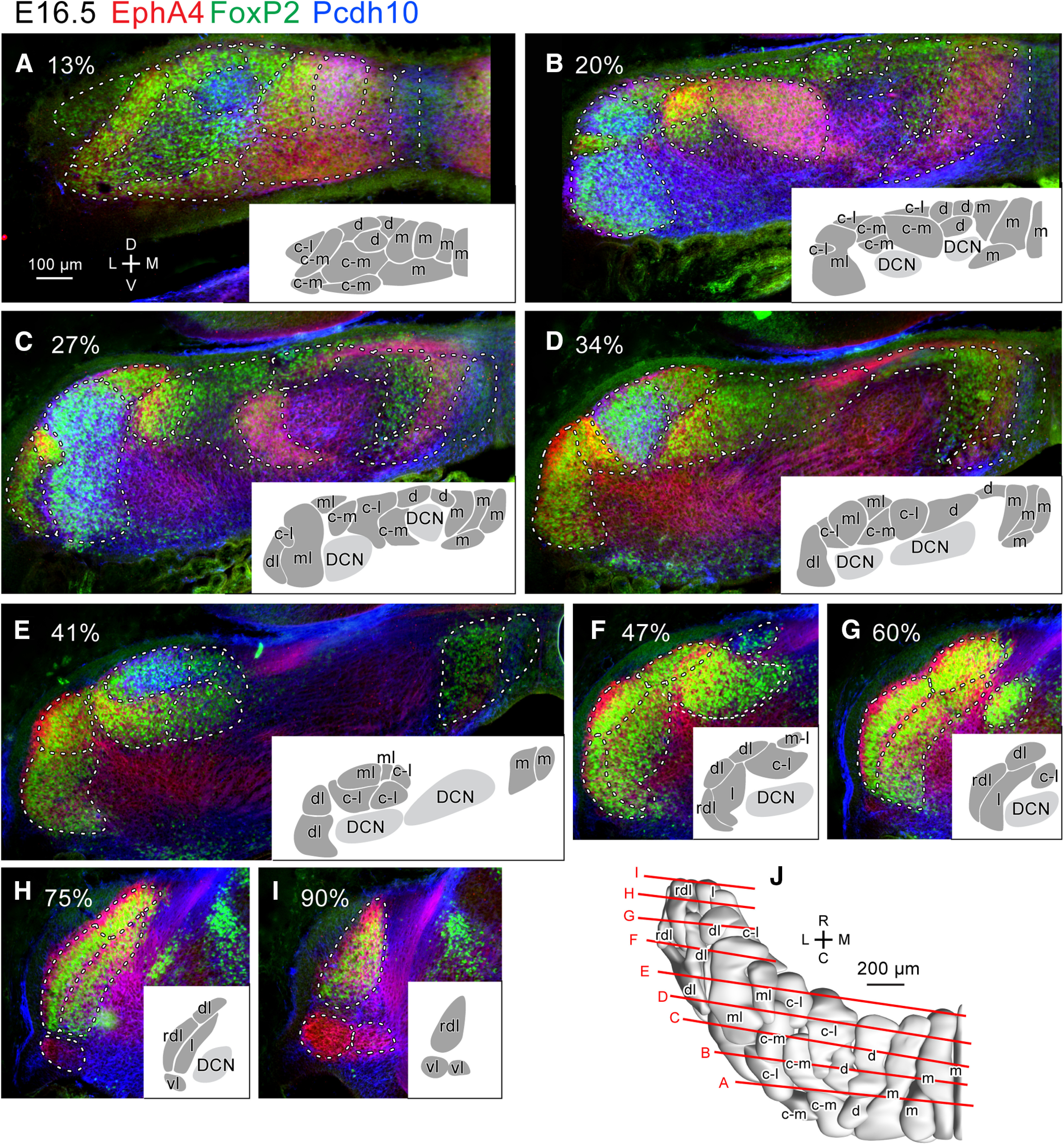
Figure 4.

Purkinje cell clusters in coronal sections of the left E16.5 mouse cerebellum identified by marker expression profiles. A–I, Sections at different caudorostral levels indicated by percentile. Fluorescent signals of immunostaining for EphA4 (red), Pcdh10 (blue) and FoxP2 (green) are merged. White dashed lines indicate the boundary of recognized clusters. Inset in each panel shows drawings of recognized clusters at half magnification. Names of each cluster were given later based on the lineage analysis (c.f. Fig. 7). J, Dorsal view of the 3D reconstruction of clusters. Red lines indicate the rostrocaudal level of the section in each panel. Scale bar in A (100 μm) applies to panels A–I. Abbreviations, c-l, c-m, d, dl, l, m, ml, rdl, vl, names of the lineage of clusters; C, caudal; D, dorsal; DCN, deep cerebellar nucleus; L, lateral; M, medial; R, rostral.V, ventral.
