

Abstract
Patient: Male, 42-year-old
Final Diagnosis: Ecthyma contagiosum • Orf
Symptoms: Nodular skin lesion • skin lesion
Medication: —
Clinical Procedure: N/A
Specialty: Dermatology • Infectious Diseases
Objective:
Rare disease
Background:
Orf, also known as ecthyma contagiosum, is a zoonotic disease caused by a parapox virus, which is endemic in goats and sheep but rare in camels. Orf is usually transmitted to humans who are in contact with infected animals. The clinical manifestation of the disease and a personal history of contact with an infected animal are sufficient to diagnose orf virus infection.
Case Report:
In this case report, we present a 42-year-old man with an unremarkable medical history who came into contact with an infected camel and developed a typical orf lesion. There were multiple erythematous, dome-shaped to round painless nodules on the right forearm of the patient. Some of them had coalesced, forming large plaques, and a few nodules had watery or yellowish discharge. The lesion was complicated by lymphadenopathy. The diagnosis of orf was made based upon clinical suspicion. The patient was treated with fusidic acid cream and observed until the lesion resolved spontaneously, leaving some post-inflammatory hyperpigmentation. We believe this is the first report of orf transmission from a camel to a human.
Conclusions:
All physicians should consider this disease as a differential diagnosis in any patient who has a history of contact with camels. Although orf is a self-limiting condition, its early clinical recognition is critical to avoid complications, unwarranted psychological stress, and unnecessary surgical intervention.
MeSH Keywords: Camels, Orf Virus, Parapoxvirus
Background
Orf virus is a zoonotic, large DNA virus belonging to the family of Poxviridae (genus Parapoxvirus) [1–5]. This virus is important because it is responsible for causing orf, also known as ecthyma contagiosum, a highly contagious dermatological disease [4,6,7]. The orf virus infects small ruminant species, mainly goats, sheep, and cattle, and is rarely reported in camels [2,4,7]. Orf is an endemic virus that can be transmitted to humans who are in contact with disease-ridden animals [4–6,8]. It causes solitary or several skin lesions that appear commonly on the fingers, hands, or forearms. The lesions start as small, firm, red-to-blue papules, then proliferate and progress to a severe ulcerative, pustular dermatitis [3,9,10]. The clinical manifestation of the disease and a personal history of contact with an infected animal are sufficient to diagnose orf virus infection [6,11]. When the diagnosis is in doubt, a real-time polymerase chain reaction test can be performed, as this method has a high level of sensitivity and specificity [6,12]. Other investigations include cell culture isolation, electron microscopy, and enzyme-linked immunosorbent assay [6,13]. In healthy people, conservative management of orf is likely warranted. Orf has no specific treatment since the disease is self-limited; however, giant orf lesions may be more problematic [6]. Some reports have demonstrated the efficacy of different therapeutic options in treating these lesions, including cidofovir, shave excision, topical imiquimod, cryotherapy, and electrocautery [11,14,15]. Although orf is a benign self-limiting disease, it may be complicated in immunocompromised patients [6,15]. The reported complications of orf virus infection include secondary bacterial infections, lymphangitis, lymphadenopathy, erythema multiforme, erysipelas, and Stevens-Johnson syndrome [6,16,17]. To the best of our knowledge, this report is the first case of orf transmitted to a human from an infected camel. This case will be of use for future studies in public health.
Case Report
A 42-year-old man, working as an academic staff member at a university, presented to the dermatology clinic with multiple large, erythematous, fluid-filled, dome-shaped to round nodules across the right forearm. Some of them had coalesced, forming large plaques, and a few nodules had watery or yellowish discharge (Figure 1). The nodules were painless but causing discomfort. He had developed them over a few weeks, and they were increasing in number. The patient denied any history of injury or exposure to a similar condition. He was not on any medications and had no allergies with negative constitutional symptoms. His past medical history was unremarkable. Upon examination, the patient had only 2 freely mobile lymph nodes at the right axilla. He was afebrile, and his other vital signs were within the normal ranges. No lesions were present elsewhere, and he was otherwise healthy. Upon verification of his history, it was revealed that he used to care for his camels and milk them occasionally. One of his camels had a rash around the mouth and both lips (Figure 2). Upon examining the camel, it was revealed that the animal had a rash typical of orf, which normally affects sheep and goats. The patient also said he used to see this rash on his camels and that it usually disappeared within 2 months without any problems. The affected camel’s mouth was in direct contact with the patient’s forearm while he was milking it a few weeks prior to the development of the rash. Therefore, the diagnosis of orf was made based upon clinical suspicion. The clinical manifestation of the orf and a personal history of contact with an infected camel were sufficient to diagnose orf virus infection [6,11]. The patient was given 2% fusidic acid cream to prevent bacterial superinfection and was followed for several weeks until all the nodules disappeared spontaneously, leaving some post-inflammatory hyperpigmentation (Figure 3).
Figure 1.
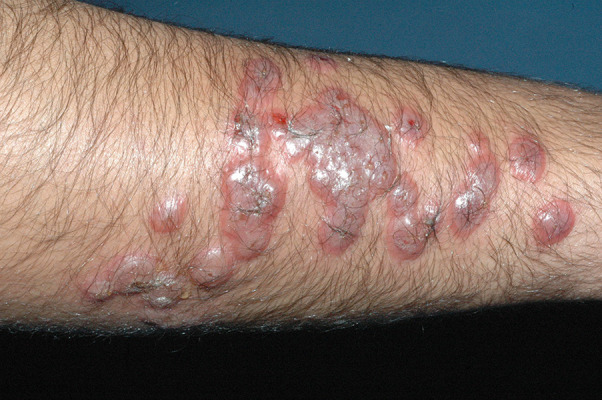
Multiple large, erythematous, fluid-filled, dome-shaped to round painless nodules across the right forearm of the patient. Some of them had coalesced, forming large plaques, and a few nodules had watery or yellowish discharge.
Figure 2.

The camel had a rash around the mouth and both lips. This rash is typical of orf, which affects sheep and goats.
Figure 3.
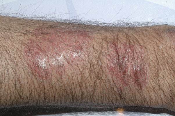
The patient was given 2% fusidic acid cream. After several weeks, all the nodules disappeared spontaneously, leaving some post-inflammatory hyper-pigmentation.
Discussion
Orf in camels is relatively rare, and transmission of the infection from camels to humans has not been previously reported. The lack of data on the incidence of humans contracting orf from infected camels could be due to different causes. The fact that camels rarely transfer orf virus to humans may lead to misdiagnosis by physicians or a lack of reporting infections owing to the virus’s self-limiting nature. Another probable explanation is that patients who are infected are often familiar with this condition and do not seek medical advice. Nevertheless, it is wise for dermatologists and general practitioners to consider orf in cases of such an angry-looking rash, in particular in the era of the Middle East respiratory syndrome-related coronavirus (MERS-CoV), which can also be transferred from camels. Clinical suspicion of the orf disease is crucial in diagnosis, especially when patients deal with animals, including sheep, goats, and camels. The differential diagnosis may include different types of fungal and bacterial infection. Skin biopsy for hematoxylin-eosin staining and cultures may be needed to make a definitive diagnosis. In the human literature, this is likely the first report of orf transferred from camel to human.
Conclusions
In conclusion, although orf is a self-limiting condition, early clinical recognition is critical to avoid complications, unwar-ranted psychological stress, and unnecessary surgical intervention. All physicians should consider this disease as a differential diagnosis in any patient who has a history of contact with camels. Taking the precise history of the patient is the correct way to reach a final diagnosis. We think that there is a need for more case reports of human orf transmitted by contact with infected camels to prevent the underdiagnosis and maltreatment of orf infection.
Acknowledgments
We would like to acknowledge the patient for his cooperation.
Footnotes
Department and institution where work was done
King Khalid University Hospital, King Saud University, Riyadh, Saudi Arabia
Conflict of interest
None.
References:
- 1.Demiraslan H, Dinc G, Doganay M. An overwiev of orf virus infection in humans and animals. Recent Pat Antiinfect Drug Discov. 2017;12(1):21–30. doi: 10.2174/1574891X12666170602080301. [DOI] [PubMed] [Google Scholar]
- 2.Wang R, Wang Y, Liu F, Luo S. Orf virus: A promising new therapeutic agent. Rev Med Virol. 2019;29(1):e2013. doi: 10.1002/rmv.2013. [DOI] [PubMed] [Google Scholar]
- 3.Haig DM, Mercer AA. Ovine diseases. Orf. Vet Res. 1998;29(3–4):311–26. [PubMed] [Google Scholar]
- 4.Caravaglio JV, Khachemoune A. Orf virus infection in humans: A review with a focus on advances in diagnosis and treatment. J Drugs Dermatol. 2017;16(7):684. [PubMed] [Google Scholar]
- 5.Veraldi S, Esposito L, Pontini P, et al. Feast of sacrifice and orf, Milan, Italy, 2015–2018. Emerg Infect Dis. 2019;25(8):1585–86. doi: 10.3201/eid2508.181063. [DOI] [PMC free article] [PubMed] [Google Scholar]
- 6.Bergqvist C, Kurban M, Abbas O. Orf virus infection. Rev Med Virol. 2017;27(4):e1932. doi: 10.1002/rmv.1932. [DOI] [PubMed] [Google Scholar]
- 7.Azwai SM, Carter SD, Woldehiwet Z. Immune responses of the camel (Camelus dromedarius) to contagious ecthyma (Orf) virus infection. Vet Microbiol. 1995;47(1–2):119–31. doi: 10.1016/0378-1135(95)00055-f. [DOI] [PubMed] [Google Scholar]
- 8.Veraldi S, Nazzaro G, Vaira F, Çuka E. Presentation of orf (ecthyma contagiosum) after sheep slaughtering for religious feasts. Infection. 2014;42(4):767–69. doi: 10.1007/s15010-014-0591-7. [DOI] [PubMed] [Google Scholar]
- 9.McKeever DJ, Jenkinson DM, Hutchison G, Reid HW. Studies of the pathogenesis of orf virus infection in sheep. J Comp Pathol. 1988;99(3):317–28. doi: 10.1016/0021-9975(88)90052-7. [DOI] [PubMed] [Google Scholar]
- 10.Guo J, Zhang Z, Edwards JF, et al. Characterization of a North American orf virus isolated from a goat with persistent, proliferative dermatitis. Virus Res. 2003;93(2):169–79. doi: 10.1016/s0168-1702(03)00095-9. [DOI] [PubMed] [Google Scholar]
- 11.Al-Qattan MM. Orf infection of the hand. J Hand Surg Am. 2011;36(11):1855–58. doi: 10.1016/j.jhsa.2011.08.019. [DOI] [PubMed] [Google Scholar]
- 12.Gallina L, Dal Pozzo F, Mc Innes CJ, et al. A real time PCR assay for the detection and quantification of orf virus. J Virol Methods. 2006;134(1–2):140–45. doi: 10.1016/j.jviromet.2005.12.014. [DOI] [PubMed] [Google Scholar]
- 13.Sanchez RL, Hebert A, Lucia H, Swedo J. Orf. A case report with histologic, electron microscopic, and immunoperoxidase studies. Arch Pathol Lab Med. 1985;109(2):166–70. [PubMed] [Google Scholar]
- 14.Kilic SS, Puel A, Casanova J-L. Orf infection in a patient with Stat1 gain-of-function. J Clin Immunol. 2015;35(1):80–83. doi: 10.1007/s10875-014-0111-7. [DOI] [PubMed] [Google Scholar]
- 15.Lederman ER, Green GM, DeGroot HE, et al. Progressive ORF virus infection in a patient with lymphoma: Successful treatment using imiquimod. Clin Infect Dis. 2007;44(11):e100–103. doi: 10.1086/517509. [DOI] [PubMed] [Google Scholar]
- 16.Wilkinson JD. Orf: A family with unusual complications. Br J Dermatol. 1977;97(4):447–50. doi: 10.1111/j.1365-2133.1977.tb14256.x. [DOI] [PubMed] [Google Scholar]
- 17.Joseph RH, Haddad FA, Matthews AL, et al. Erythema multiforme after orf virus infection: A report of two cases and literature review. Epidemiol Infect. 2015;143(2):385–90. doi: 10.1017/S0950268814000879. [DOI] [PMC free article] [PubMed] [Google Scholar]