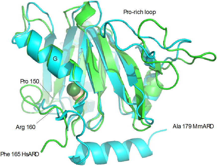
Figure 8.

Alignment of Fe-HsARD solution structure (green) with Ni-MmARD structure 5I91 (cyan). Fe is shown as yellow sphere, Ni as green. Features discussed in the text are labeled. Due to disorder, the HsARD structure is truncated at Phe 165 in the figure.
