PLOS ONE logoLink to PLOS ONE
. 2020 Dec 28;15(12):e0244397. doi: 10.1371/journal.pone.0244397

Non-invasive myocardial work is reduced during transient acute coronary occlusion

Jolanda Sabatino 1,2,3, Salvatore De Rosa 1,2,*, Isabella Leo 1, Carmen Spaccarotella 1,2, Annalisa Mongiardo 1, Alberto Polimeni 1,2, Sabato Sorrentino 1,2, Giovanni Di Salvo 3,4, Ciro Indolfi 1,2,5,*
Editor: Vincenzo Lionetti6
PMCID: PMC7769261  PMID: 33370359

Abstract

Background

During ischemia a close relationship exists between sub-endocardial blood flow and myocardial function. Strain parameters can capture an impairment of regional longitudinal function but are load dependent. Recently, a novel non-invasive method to calculate Myocardial Work (MW) showed a strong correlation with invasive work measurements.

Our aim was to investigate the ability of non-invasive MW indices to identify the ischaemic risk area during transient acute coronary occlusion (TACO).

Methods and results

The study population comprises 50 individuals with critical coronary stenosis (CCS). Echocardiography recordings were obtained before coronary angiography, during TACO and after revascularization to measure global longitudinal strain (GLS), Myocardial Work Index (MWI), Myocardial Constructive Work (MCW), Myocardial Wasted work (MWW), Myocardial work efficiency (MWE).

Compared to baseline, we found a significant reduction of GLS (p = 0.005), MWI, MCW and MWE (p<0.001) during TACO.

Conclusions

The non-invasive measurement of MW parameters is a sensitive and early marker of myocardial ischemia during TACO.

Introduction

The prompt recognition of acutely ischemic myocardium has crucial therapeutic and prognostic implications [1].

With the introduction of more sensitive cardiac biomarkers, the fourth universal definition of myocardial infarction [2] has been released, taking into account myocardial injury detected by necrosis biomarkers, together with clinical symptoms, ECG changes or new regional wall motion abnormalities.

However, regional wall motion abnormalities deserve special considerations in this setting, as they appear early after flow reduction in the temporal sequence of the ischemic cascade [3]. The routine evaluation of myocardial function by echocardiography in the acute setting is mainly based on the visual assessment of wall motion. Such qualitative method has well recognized limitations [4,5] and may fail to distinguish subtle ischemia-induced signs in regional mechanics.

Recent studies showed that two-dimensional speckle tracking echocardiography (2D-STE) might identify an impairment of longitudinal function downward of critical coronary stenosis [613], differentiating acutely ischemic segments from both normal and dysfunctional myocardium [14].

Nevertheless, since strain parameters are load dependent, they might not reflect systolic function accurately [15,16] in specific settings.

Myocardial Work Index (MWI), a non-invasive method to quantify myocardial work using segmental strain and a standardized LV pressure (LVP) curve has been recently introduced [1719].

Recently, Boe et al. [20] investigated the ability of regional MWI to identify acute coronary occlusion; however, their study was focused on patients with acute myocardial infarction (AMI).

Therefore, the aim of this study was to assess the impact of transient acute coronary occlusion on non-invasive myocardial work and 2D-STE-derived Longitudinal Strain (LS) to evaluate the impact of myocardial ischemia on these sensitive indices of LV function.

Materials and methods

Study population

We included 50 consecutive patients referred for coronary angiography in a single tertiary coronary care centre. Patients were included in the study population if they presented the following criteria:

  • ≥18 years of age;

  • clinical indication for coronary angiography;

  • critical coronary stenosis (single vessel disease) as diagnosed during coronary angiography;

  • gave their consent to participate.

Exclusion criteria were recent myocardial infarction (within 30 days), QRS-width of ≥120 ms, severe valvular disease, previous heart surgery, extensive comorbidity, or atrial fibrillation.

All patients were clinically and haemodynamically stable. The regional ethics committee (Comitato Etico Regione Calabria–Area Centro) approved the study, and all the patients provided written informed consent.

Study timeline, procedures and analysis plan

Echocardiography recordings with simultaneous measurement of both non-invasive (NINV) automatic oscillometric and invasive intra-arterial blood pressure (INV) were obtained in the catheterization laboratory immediately before coronary angiography, during transient acute coronary occlusion (TACO) and at the end of the procedure. The design of the study finds its parallel in studies previously performed by Edvardsen et al [21].

TACO was obtained by inflating a coronary balloon with a 1:1 balloon diameter-to-reference diameter ratio at the site of coronary stenosis at low pressure. Complete occlusion was verified injecting contrast medium, where TACO was defined as a TIMI flow of 0 (no perfusion) distal of the balloon inflation site. Pressure measurements and echocardiographic recordings during TACO were obtained starting at 60 seconds after balloon inflation.

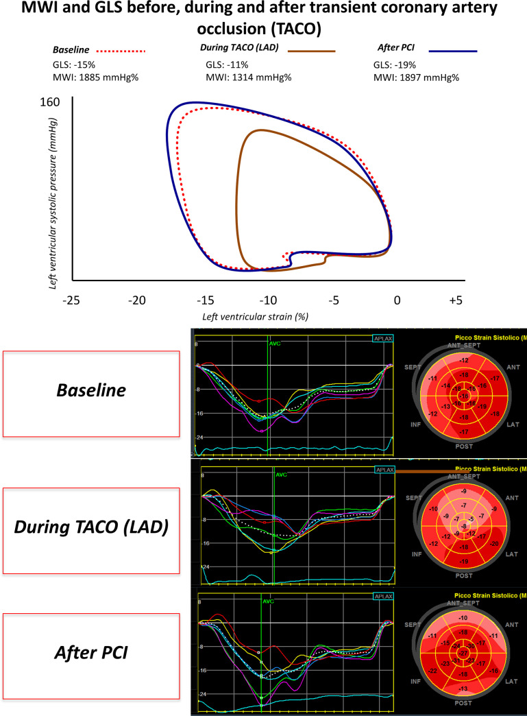
The present study consists of the assessment of the impact of TACO on Myocardial Strain and Myocardial Work parameters. To this regard, Fig 1 depicts a representative example showing the changes in the strain-pressure loops from baseline, through TACO, to recover (after PCI).

Fig 1. Strain-pressure loop and GLS changes during coronary occlusion.

Fig 1

Example of changes in Myocardial Work Index (MWI) and global longitudinal strain (GLS) at baseline, during transient coronary occlusion and after PCI. GLS: Global longitudinal strain. MWI: Myocardial work index. LAD: Left anterior descendent coronary artery. TACO: Transient acute coronary occlusion. PCI: Percutaneous coronary intervention.

Echocardiographic analyses

Two-dimensional (2D) 4-chambers, 3-chambers and 2-chambers apical views were acquired, as previously described [13,22], with a frame rate ≥60 frames/s and, then, transferred to a dedicated workstation for the offline analysis (EchoPAC, GE Healthcare). The recordings were processed using an acoustic-tracking software (EchoPAC version 112.99, Research Release, GE Healthcare), which allowed an offline semi-automated analysis of speckle-based strain [23]. To calculate LV peak systolic longitudinal strain values and Post-systolic Shortening Index (PSI), a line was traced along the LV endocardium's inner border in each of the three apical views, and a region of interest, between the endocardial and epicardial borders, was recognized by the EchoPAC software. The region of interest was, then, adjusted to ensure that the wall thickness was incorporated in the analysis, avoiding the pericardium and following myocardial motion, as recommended. Results of LV peak systolic longitudinal strain and PSI were then provided by the software and analysed by an 18-segment model. PSI was calculated according to the formula by Kulkuski et al (PSI = (peak systolic strain − end-systolic strain)/peak systolic strain) [14].

The timing of mitral and aortic valve closure and opening were obtained for Myocardial Work estimation.

Calculation of non-invasive myocardial work

MWI was calculated as the area of the LV pressure-strain loop (GE-Healthcare). Along with segmental and global values for myocardial work, a set of additional indices are also measured:

  • Myocardial Constructive work (MCW): work performed by a segment during shortening in systole adding negative work during lengthening in IVR;

  • Myocardial Wasted work (MWW): negative work performed by a segment during lengthening in systole adding work performed during shortening in IVR;

  • Myocardial work efficiency (MWE): constructive work divided by the sum of constructive and wasted work (0–100%).

Evaluation of regional function during transient coronary occlusion

The ischemic risk area (IRA), downward the coronary artery transient occluded, and the non-risk area (NRA), were selected taking into consideration the vascular distribution of each coronary vessel, as previously detailed by Kukulski et al [14]. In brief, during transient occlusion of the right coronary artery (RCA), we considered IRA segments the inferior mid- and basal segments in apical two-chamber view. Transient occlusion of coronary arteries for some minutes has been shown to be safe in different clinical context [24,25]. During transient occlusion of the left circumflex coronary artery (LCx), the lateral basal- and mid segments, imaged in the apical four chamber view, were selected as IRA [14]. Both the septal mid- and apical segments in apical four-chamber view, were considered “at-risk” in presence of a transient occlusion of the left anterior descending coronary artery (LAD), as previously earlier described [11,14,21].

Reproducibility study

Fifteen echocardiographic examinations were randomly selected to assess inter-rater and intra-rater variability. Two operators performed the echocardiographic assessment in a blinded fashion. In addition, one of the two operators had to analyze the same series of exams twice without knowing it. Inter- and intra-rater reliability was then assessed using the intraclass correlation coefficient (ICC).

Data analysis and statistics

Values are expressed as mean+SD or absolute numbers and/or percentages (%). Differences between groups were analysed with ANOVA. Comparison of continuous variables before versus during TACO were performed using the Wilcoxon U Test.

The intraclass correlation coefficient (ICC) was used to assess inter- and intra-rater reliability, as previously described [26].

Receiver operating characteristic (ROC) analysis was used to evaluate the diagnostic performance for each parameter. Significance of single ROC curves was assessed using the Hanley & McNeill method.

The statistical analyses were performed using SPSS v.21 (SPSS Inc., Chicago, IL, USA). A two-tailed P-value of 0.05 was considered significant.

Results

Study population

The study population comprises 50 individuals undergoing coronary angiography for clinical indication. All the patients included in the analysis had coronary artery disease (CAD) documented at invasive coronary angiography. Among CAD patients, seven were excluded for poor image quality, one patient was excluded from analysis for ongoing ventricular bigeminism during cardiac catheterization and one was excluded from the regional analysis for inadequate tracking. Patient characteristics, medication, and risk factors are listed in Table 1.

Table 1. Baseline patients characteristics.

PATIENTS (n = 41)
Age (years) 67 ± 9
Male, n (%) 32 (78)
Smoker, n (%) 11 (26)
Hypercolesterolemia, n (%) 30 (73)
Diabetes Mellitus, n (%) 11 (26)
Hypertension, n (%) 32 (78)
EF (%) 55±4
GLS (%) -17,2 ± 4,2
Target Vessel, n (%)
- LAD 25 (60)
- LCx 5 (12)
- RCA 11 (26)

Impact of acute coronary occlusion on global longitudinal strain and myocardial work indices

We found an impairment of LV systolic function during TACO, as demonstrated by reduced EF, GLS and MWI values. Six out 41 patients experienced chest pain above 6 (in a 1-to-10 analogic scale) during TACO; of those, 5 patients have undergone transient occlusion of the LAD and 1 of the RCA.

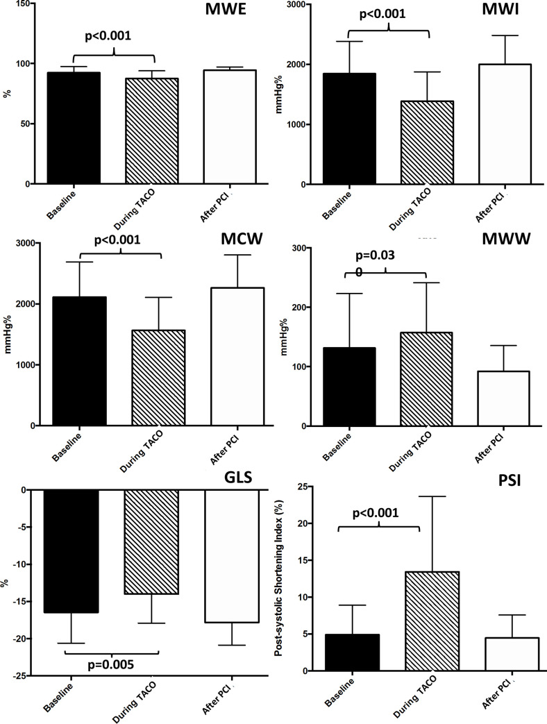
The average peak systolic GLS was significantly impaired during ischemia (Fig 1) compared to baseline (p = 0.005), with a significant reduction and a return to baseline values after reperfusion (p<0.001) (Fig 2). PSI was significantly increased during TACO (25.0±7.2%) compared to baseline (4.9±4%) (p<0.001) with a return to baseline values after PCI. Global MWI was significantly reduced during TACO compared to baseline (p<0.001). Similarly, global Myocardial Work Efficiency (MWE) index was significantly reduced during TACO (p<0.001), with a full recovery after PCI (p<0.001).

Fig 2. Myocardial work indices and global longitudinal strain during transient acute coronary occlusion.

Fig 2

The graph shows the impact of acute coronary occlusion on myocardial work indices and global longitudinal strain. GLS: Global longitudinal strain. TACO: Transient acute coronary occlusion. PCI: Percutaneous coronary intervention. MWI: Myocardial work index. MCW: Myocardial Constructive work. MWW: Myocardial Wasted work. MWE: Myocardial work efficiency.

In line with this last finding, we observed a significant increase of the global Myocardial Wasted Work index (MWW, p = 0.030) along with a significant reduction of the global Myocardial Constructive Work index (MCW, p<0.001) during TACO (Table 2).

Table 2. Echocardiographic data.

Baseline (n = 41) During TACO (n = 41) After PCI (n = 41)
LVEF (%) 55±11 52±14 59±7
GLS (%) -16.5±4.1 -14.0±3.9a -17.8±3.0
SBP 143.8±17 123.6±23a 139.8±21
DBP 81.2±9 74.8±15 76.2±13
SBP x Strain -2372±69 -1730±90 -2488±63
MWI (mmHg%) 1843±540 1387±488a 2000±480
MWE (%) 92.3±5.1 87.5±6.3a 94.5±2.5
MCW (mmHg%) 2112±577 1578±444a 2220±455
MWW (mmHg%) 131.6±91.5 157.3±84.1a 92.3±43.1

a = p<0.05 compared to baseline.

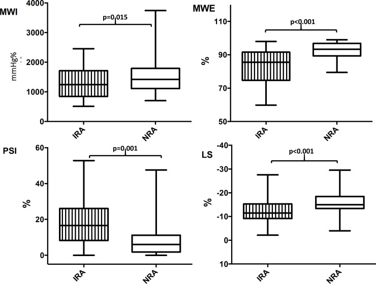
As concern results from segmental analyses, regional MWE, measured acutely within the IRA, underlying the target vessel during transient coronary occlusion, was significantly decreased by 10% (p<0.001), compared with the NRA of the same patients. Also, regional LS (p<0.001) and regional PSI, measured acutely within the IRA, were observed being significantly reduced the former, and significantly increased the latter, compared with the NRA (-12.0±5.6% VS -16±4.8%, p<0.001; 18.4±13.3% VS 9.0±10.9%, p = 0.001) (Fig 3).

Fig 3. Regional myocardial work and longitudinal strain parameters during transient acute coronary occlusion.

Fig 3

The chart shows regional MWE, MWI, LS and PSI, measured within the IRA (underlying the target vessel undergoing TACO), and compared to the NRA of the same patients. IRA = Ischemic risk area. NRA = Non-risk area. MWE = Myocardial work efficiency. MWI = Myocardial work index. PSI = Post systolic shortening index. LS = Longitudinal strain.

The diagnostic performance of those regional parameters measured acutely within the IRA, to ascertain the occurring of a transient acute coronary occlusion, were evaluated via ROC analyses. The area under the curve (AUC) was higher for regional MWE (AUC = 0.835, p<0.001) compared to both regional PSI (AUC = 0.792, p<0.001) and regional LS (AUC = 0.803, p<0.001) (Table 3).

Table 3. AUCs calculated by ROC curves for regional MWE, MWI, LS, PSI.

AUC 95% C.I. p
MWE 0.835 0.74–0.94 <0.001
MWI 0.766 0.67–0.87 <0.001
LS 0.803 0.71–0.90 <0.001
PSI 0.792 0.68–0.90 <0.001

AUC = Area under the curve. ROC = Receiver operating characteristic. MWE = Myocardial work efficiency. MWI = Myocardial work index. PSI = Post systolic shortening index. LS = Longitudinal strain.

Blood pressure during the study

Mean non-invasive blood pressure (BP) values are reported in Table 2. Invasive BP from a study subgroup are shown in Table 4.

Table 4. Invasive pressures.

Invasive pressures Baseline During TACO After PCI
SBP 141.2±23 118.2±24 a 140.1±21
DBP 76.1±10 70.5±16 75.7±12

a = p<0.05 compared to baseline.

The average NINV systolic (SBP) values at baseline, during TACO and post-PCI were 143.8±17 mmHg, 123.6±23 mmHg and 139.8±21 mmHg, respectively (Table 2), with a significant reduction during ischemia compared to baseline (p<0.001) and a return to baseline values after reperfusion (p<0.001).

Similar to non invasive pressure values, average INV SBP values at baseline, during TACO and post-PCI were 141.2±23 mmHg, 118.2±24 mmHg and 140.1±21mmHg, respectively (Table 4), with values obtained under ischemia being significantly lower compared to baseline (p<0.001).

There were no significant differences between NINV and INV systolic and diastolic blood pressure measurements at baseline (p = 0.68 and p = 0.06), during TACO (p = 0.48 and p = 0.35) and post-PCI (p = 0.92 and p = 0.89), respectively.

INV showed good correlation with NINV systolic (ρ = 0.904; p<0.001) and diastolic (ρ = 0.684; p<0.001) blood pressure measurements at baseline. The correlation between INV and NINV was maintained under TACO (SBP: ρ = 0.756; p<0.001 and DBP: ρ = 0.808; p<0.001) and post PCI (SBP: ρ = 0.980; p<0.001 and DBP: ρ = 0.993; p<0.001).

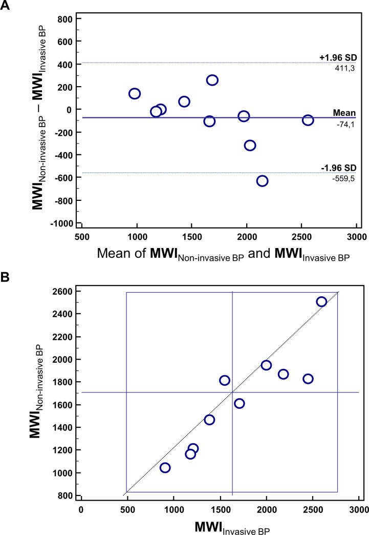
Finally, we demonstrated a strong correlation (R = 0.94, 95% CI = 0.75–0.96 p = 0.0001) between MWI calculated using INV vs NINV blood pressures by means of Bland-Altman and Youden plots (Fig 4).

Fig 4. Invasive vs non-invasive blood pressure measurement.

Fig 4

(A) Bland-Altman Plot comparing MWI obtained using invasive BP versus non invasive BP. (B) Youden Plot showing the scatter between MWI obtained using invasive BP versus non invasive BP.

Reproducibility analyses

Intraclass correlation coefficient was very good for global MWI (ICC = 0.977; 95% CI: 0.944–0.991).

Discussion

The main findings of our study are: 1) short and transient coronary occlusion results in an early reduction of non-invasive global myocardial work indices, as MWI, MCW, MWE, and in an increase of MWW; 2) MWI calculated using INV presents a strong correlation with MWI assessed using NINV blood pressures.

Impairment of myocardial function during acute ischemia

An acute reduction in coronary blood flow induces a regional contractile dysfunction within a few seconds, resulting in impairment of regional deformation indices [27].

During ischemia, the longitudinal and circumferential systolic shortening of the ischemic segment are decreased, together with the radial thickening.

Moreover, diastolic relaxation is markedly impaired during ischemia and, in turn, the physiologic early diastolic thinning and lengthening are substituted by ongoing post-systolic thickening and shortening [28].

Consistent changes in early diastolic deformation have been demonstrated in several studies and proposed as an early marker of regional ischemia [14,29]. In our study, indeed, deformation indices were impaired during coronary occlusion in patients with chronic coronary syndrome. Indeed, not only the average peak systolic GLS was significantly impaired during ischemia, but also, not surprisingly, PSI was significantly increased during TACO compared to baseline, as consequence of the increased amount of post-systolic thickening occurred in the ischemic myocardium.

In 1987, Lazar et al [30] demonstrated a significant decrease in stroke work index, assessed with invasive measurements of LV Pressure-Volume loops, in 17 dogs when the proximal left anterior descending coronary artery was occluded for 45 minutes.

We assessed for the first time in humans the impact of transient acute coronary occlusion, obtained by 60 minutes of balloon inflation, on a novel index that non-invasively estimate the myocardial work.

In agreement with findings by Lazar et al [30], obtained by means of invasive measurements in a dog model, we have observed that non-invasive MWI was significantly reduced during TACO compared to baseline values (p<0.001). Similarly, its derived indices as MCW and MWE index were significantly reduced during TACO (p<0.001).

Myocardial work and ventricular function

Global and regional LV function is dependent on sub-endocardial blood flow [31]. Indices of longitudinal deformation can be influenced by the loading conditions, which limits their accuracy. Invasively measured Myocardial Work was introduced as marker of ventricular contractility since the 1970s [3235]. It was later shown to provide similar physiological information to pressure/strain loops [36].

More recently, Russell et al [17] introduced a method for calculating non-invasive MW, on the basis of speckle tracking analysis with the estimation of LV pressure from brachial artery cuff pressure. The NORRE sub-study provided reference ranges for non-invasive MW, reporting a good reproducibility [37].

Most recently, Chan et al. [38] reported results of MW indices in three cardiovascular conditions, such as hypertension, ischaemic and not-ischaemic dilated cardiomyopathy. Particularly, they demonstrated a high impact of blood pressure on MW indices and a significant increase of MWI in hypertensive patients compared to controls, despite normal global longitudinal strain values.

In this regard, since myocardial work indices encompass multiple hemodynamic factors, they may—at least in part—complement and correct the estimation of systolic function compared to the sole strain measurements.

Application of non-invasive myocardial work in clinical practice

Our findings might have a relevant impact on clinical practice. The identification and quantitation of non-invasive myocardial work abnormalities could serve as a valuable adjunct to the conventional diagnostic approach to chest pain patients, as they might be helpful to recognize early LV impairment in a more sensitive fashion, allowing the quicker identification of myocardial ischemia, with relevant impact both on patient’s prognosis and clinical management workflow [39,40]. Furthermore, since data acquisition is totally non-invasive and safe and we have demonstrated MWI can be alternatively calculated both with INV or NINV, this novel method is appropriate for the monitoring of myocardial function even at short intervals after coronary revascularization. Early diagnosis or exclusion of critical coronary stenoses might be very useful both for the clinical management and for the prognostic impact [40]. In this regard, it should be pointed out that the echocardiographic assessment was performed in the catheterization laboratory on patients laying on the interventional table real-time during the procedure. This suggests that a rapid assessment to check for residual ischemia after initial PCI is ultimately possible. In this regard, the prognostic relevance of a timely complete coronary revascularization has been repeatedly demonstrated [4143]. Finally, the new non-invasive MWI might be a more sensitive and precise alternative to visual assessment to distinguish between segments that benefited and did not benefit from PCI. In this regard, our results confirm previous evidence on global noninvasive work parameters and extends those results to regional LV assessment [44].

Limitations

The design of the study with transient coronary occlusion may represent a study limitation, as the coronary occlusion has generally a longer duration in patients with acute myocardial infarction. However, the study design has also its strengths due to the controlled setting where each patient is his/her own control.

Echocardiographic exams in 16% of patients were not analysable due to insufficient image quality. Although these results are in line with recent studies [37], they could limit the applicability of our findings. MW analysis with contrast agents might improve its feasibility.

Conclusions

Assessment of non-invasive Myocardial Work Indices is able to detect an impairment of LV function very early during coronary occlusion.

Data Availability

The data underlying the results presented in the study are available from Open Science Framework: https://osf.io/bqnmh/.

Funding Statement

This study was partly supported through an ESC Training Grant 2019, awarded to JS. There was no additional external funding received for this study.

References

  • 1.Niccoli G, Montone RA, Ibanez B, Thiele H, Crea F, Heusch G, et al. Optimized Treatment of ST-Elevation Myocardial Infarction. Circulation Research. 2019;125(2):245–58. Available from: 10.1161/CIRCRESAHA.119.315344 [DOI] [PubMed] [Google Scholar]
  • 2.Thygesen K, Alpert JS, Jaffe AS, Chaitman BR, Bax JJ, Morrow DA, et al. Fourth Universal Definition of Myocardial Infarction (2018). Journal of the American College of Cardiology. 2018;72(18):2231–64. Available from: 10.1016/j.jacc.2018.08.1038 [DOI] [PubMed] [Google Scholar]
  • 3.Hauser AM, Gangadharan V, Ramos RG, Gordon S, Timmis GC, Dudlets P. Sequence of mechanical, electrocardiographic and clinical effects of repeated coronary artery occlusion in human beings: Echocardiographic observations during coronary angioplasty. Journal of the American College of Cardiology. 1985;5(2):193–7. Available from: 10.1016/s0735-1097(85)80036-x [DOI] [PubMed] [Google Scholar]
  • 4.Kvitting J-PE, Wigström L, Strotmann JM, Sutherland GR. How Accurate Is Visual Assessment of Synchronicity in Myocardial Motion? An In Vitro Study with Computer-Simulated Regional Delay in Myocardial Motion: Clinical Implications for Rest and Stress Echocardiography Studies. Journal of the American Society of Echocardiography. 1999;12(9):698–705. Available from: 10.1016/s0894-7317(99)70019-2 [DOI] [PubMed] [Google Scholar]
  • 5.Whitaker D, McGraw PV, Pearson S. Non-veridical size perception of expanding and contracting objects. Vision Research. 1999;39(18):2999–3009. Available from: 10.1016/s0042-6989(99)00010-3 [DOI] [PubMed] [Google Scholar]
  • 6.Sutherland GR, Di Salvo G, Claus P, D’hooge J, Bijnens B. Strain and strain rate imaging: a new clinical approach to quantifying regional myocardial function. Journal of the American Society of Echocardiography. 2004;17(7):788–802. Available from: 10.1016/j.echo.2004.03.027 [DOI] [PubMed] [Google Scholar]
  • 7.Liel-Cohen N, Tsadok Y, Beeri R, Lysyansky P, Agmon Y, Feinberg MS, et al. A New Tool for Automatic Assessment of Segmental Wall Motion Based on Longitudinal 2D Strain. Circulation: Cardiovascular Imaging. 2010;3(1):47–53. Available from: 10.1161/CIRCIMAGING.108.841874 [DOI] [PubMed] [Google Scholar]
  • 8.Reisner SA, Lysyansky P, Agmon Y, Mutlak D, Lessick J, Friedman Z. Global longitudinal strain: a novel index of left ventricular systolic function. Journal of the American Society of Echocardiography. 2004;17(6):630–3. Available from: 10.1016/j.echo.2004.02.011 [DOI] [PubMed] [Google Scholar]
  • 9.Jones CJ, Raposo L, Gibson DG. Functional importance of the long axis dynamics of the human left ventricle. Heart. 1990;63(4):215–20. Available from: 10.1136/hrt.63.4.215. [DOI] [PMC free article] [PubMed] [Google Scholar]
  • 10.Choi J-O, Cho SW, Song YB, Cho SJ, Song BG, Lee S-C, et al. Longitudinal 2D strain at rest predicts the presence of left main and three vessel coronary artery disease in patients without regional wall motion abnormality. European Journal of Echocardiography. 2009;10(5):695–701. Available from: 10.1093/ejechocard/jep041 [DOI] [PubMed] [Google Scholar]
  • 11.Dahlslett T, Karlsen S, Grenne B, Eek C, Sjøli B, Skulstad H, et al. Early Assessment of Strain Echocardiography Can Accurately Exclude Significant Coronary Artery Stenosis in Suspected Non–ST-Segment Elevation Acute Coronary Syndrome. Journal of the American Society of Echocardiography. 2014;27(5):512–9. Available from: 10.1016/j.echo.2014.01.019 [DOI] [PubMed] [Google Scholar]
  • 12.Bijnens B, Claus P, Weidemann F, Strotmann J, Sutherland GR. Investigating Cardiac Function Using Motion and Deformation Analysis in the Setting of Coronary Artery Disease. Circulation. 2007;116(21):2453–64. Available from: 10.1161/CIRCULATIONAHA.106.684357 [DOI] [PubMed] [Google Scholar]
  • 13.Mazzitelli M, Torti C, Sabatino J, D'Ascoli GL, Costa C, Pisani V, et al. Evaluation of cardiac function by global longitudinal strain before and after treatment with sofosbuvir-based regimens in HCV infected patients. BMC Infect Dis. 2018;18(1):518 Available from: 10.1186/s12879-018-3426-9 [DOI] [PMC free article] [PubMed] [Google Scholar]
  • 14.Kukulski T, Jamal F, Herbots L, D’hooge J, Bijnens B, Hatle L, et al. Identification of acutely ischemic myocardium using ultrasonic strain measurements. Journal of the American College of Cardiology. 2003;41(5):810–9. Available from: 10.1016/s0735-1097(02)02934-0 [DOI] [PubMed] [Google Scholar]
  • 15.Fredholm M, Jörgensen K, Houltz E, Ricksten S-E. Load-dependence of myocardial deformation variables—a clinical strain-echocardiographic study. Acta Anaesthesiologica Scandinavica. 2017;61(9):1155–65. Available from: 10.1111/aas.12954 [DOI] [PubMed] [Google Scholar]
  • 16.Murai D, Yamada S, Hayashi T, Okada K, Nishino H, Nakabachi M, et al. Relationships of left ventricular strain and strain rate to wall stress and their afterload dependency. Heart and Vessels. 2016;32(5):574–83. Available from: 10.1007/s00380-016-0900-4 [DOI] [PubMed] [Google Scholar]
  • 17.Russell K, Eriksen M, Aaberge L, Wilhelmsen N, Skulstad H, Remme EW, et al. A novel clinical method for quantification of regional left ventricular pressure–strain loop area: a non-invasive index of myocardial work. European Heart Journal. 2012;33(6):724–33. Available from: 10.1093/eurheartj/ehs016 [DOI] [PMC free article] [PubMed] [Google Scholar]
  • 18.Urheim S, Rabben SI, Skulstad H, Lyseggen E, Ihlen H, Smiseth OA. Regional myocardial work by strain Doppler echocardiography and LV pressure: a new method for quantifying myocardial function. American Journal of Physiology-Heart and Circulatory Physiology. 2005;288(5):H2375–80. Available from: 10.1152/ajpheart.00946.2004 [DOI] [PubMed] [Google Scholar]
  • 19.Russell K, Eriksen M, Aaberge L, Wilhelmsen N, Skulstad H, Gjesdal O, et al. Assessment of wasted myocardial work: a novel method to quantify energy loss due to uncoordinated left ventricular contractions. American Journal of Physiology-Heart and Circulatory Physiology. 2013;305(7):H996–1003. Available from: 10.1152/ajpheart.00191.2013 [DOI] [PubMed] [Google Scholar]
  • 20.Boe E, Russell K, Eek C, Eriksen M, Remme EW, Smiseth OA, et al. Non-invasive myocardial work index identifies acute coronary occlusion in patients with non-ST-segment elevation-acute coronary syndrome. European Heart Journal–Cardiovascular Imaging. 2015;16(11):1247–55. Available from: 10.1093/ehjci/jev078 [DOI] [PubMed] [Google Scholar]
  • 21.Edvardsen T, Skulstad H, Aakhus S, Urheim S, Ihlen H. Regional myocardial systolic function during acute myocardial ischemia assessed by strain Doppler echocardiography. J Am Coll Cardiol. 2001;37(3):726–730. Available from: 10.1016/s0735-1097(00)01160-8 [DOI] [PubMed] [Google Scholar]
  • 22.Sabatino J, Di Salvo G, Krupickova S, Fraisse A, Prota C, Bucciarelli V, et al. Left Ventricular Twist Mechanics to Identify Left Ventricular Noncompaction in Childhood. Circulation: Cardiovascular Imaging. 2019;12(4). Available from: 10.1161/CIRCIMAGING.118.007805 [DOI] [PubMed] [Google Scholar]
  • 23.Cutruzzolà A, Irace C, Frazzetto M, Sabatino J, Gullace R, De Rosa S, et al. Functional and morphological cardiovascular alterations associated with neurofibromatosis 1. Sci Rep 2020; 10:12070 10.1038/s41598-020-68908-0 [DOI] [PMC free article] [PubMed] [Google Scholar]
  • 24.De Rosa S, Seeger FH, Honold J, Fischer-Rasokat U, Lehmann R, Fichtlscherer S, et al. Procedural safety and predictors of acute outcome of intracoronary administration of progenitor cells in 775 consecutive procedures performed for acute myocardial infarction or chronic heart failure. Circ Cardiovasc Interv. 2013;6(1):44–51. 10.1161/CIRCINTERVENTIONS.112.971705 [DOI] [PubMed] [Google Scholar]
  • 25.Sorrentino S, De Rosa S, Ambrosio G, Mongiardo A, Spaccarotella C, Polimeni A, et al. The duration of balloon inflation affects the luminal diameter of coronary segments after bioresorbable vascular scaffolds deployment. BMC Cardiovasc Disord. 2015;15:169 10.1186/s12872-015-0163-5 [DOI] [PMC free article] [PubMed] [Google Scholar]
  • 26.Borrelli N, Di Salvo G, Sabatino J, Ibrahim A, Avesani M, Sirico D, et al. Serial changes in longitudinal strain are associated with outcome in children with hypoplastic left heart syndrome. Int J Cardiol. 2020; S0167-5273(20)30229-1. 10.1016/j.ijcard.2020.03.085 [DOI] [PubMed] [Google Scholar]
  • 27.Theroux P, Franklin D, Ross J Jr, Kemper Ws. Regional Myocardial Function during Acute Coronary Artery Occlusion and Its Modification by Pharmacologic Agents in the Dog. Circulation Research. 1974;35(6):896–908. Available from: 10.1161/01.res.35.6.896 [DOI] [PubMed] [Google Scholar]
  • 28.Takayama M, Norris RM, Brown MA, Armiger LC, Rivers JT, White HD. Postsystolic shortening of acutely ischemic canine myocardium predicts early and late recovery of function after coronary artery reperfusion. Circulation. 1988;78(4):994–1007. Available from: 10.1161/01.cir.78.4.994 [DOI] [PubMed] [Google Scholar]
  • 29.Safwat A, Leone BJ, Norris RM, Foëx P. Pressure-length loop area: Its components analyzed during graded myocardial ischemia. Journal of the American College of Cardiology. 1991;17(3):790–6. Available from: 10.1016/s0735-1097(10)80199-8 [DOI] [PubMed] [Google Scholar]
  • 30.Lazar HL, Wei J, Dirbas FM, Haasler GB, Spotnitz HM. Controlled Reperfusion Following Regional Ischemia. The Annals of Thoracic Surgery. 1987;44(4):350–5. Available from: 10.1016/s0003-4975(10)63789-7 [DOI] [PubMed] [Google Scholar]
  • 31.Indolfi C, Guth BD, Miura T, Miyazaki S, Schulz R, Ross J Jr. Mechanisms of improved ischemic regional dysfunction by bradycardia. Studies on UL-FS 49 in swine. Circulation. 1989;80(4):983–93. Available from: 10.1161/01.cir.80.4.983 [DOI] [PubMed] [Google Scholar]
  • 32.Sagawa K, Suga H, Shoukas AA, Bakalar KM. End-systolic pressure/volume ratio: A new index of ventricular contractility. The American Journal of Cardiology. 1977;40(5):748–53. Available from: 10.1016/0002-9149(77)90192-8 [DOI] [PubMed] [Google Scholar]
  • 33.Suga H. Total mechanical energy of a ventricle model and cardiac oxygen consumption. American Journal of Physiology-Heart and Circulatory Physiology. 1979;236(3):H498–505. Available from: 10.1152/ajpheart.1979.236.3.H498 [DOI] [PubMed] [Google Scholar]
  • 34.Miyazaki S, Goto Y, Guth BD, Miura T, Indolfi C, Ross J Jr. Changes in regional myocardial function and external work in exercising dogs with ischemia. American Journal of Physiology-Heart and Circulatory Physiology. 1993;264(1):H110–6. Available from: 10.1152/ajpheart.1993.264.1.H110 [DOI] [PubMed] [Google Scholar]
  • 35.Hisano R, Cooper G 4th. Correlation of force-length area with oxygen consumption in ferret papillary muscle. Circulation Research. 1987;61(3):318–28. Available from: 10.1161/01.res.61.3.318 [DOI] [PubMed] [Google Scholar]
  • 36.Delhaas T, Arts T, Prinzen FW, Reneman RS. Regional fibre stress-fibre strain area as an estimate of regional blood flow and oxygen demand in the canine heart. The Journal of Physiology. 1994;477(3):481–96. Available from: 10.1113/jphysiol.1994.sp020209 [DOI] [PMC free article] [PubMed] [Google Scholar]
  • 37.Manganaro R, Marchetta S, Dulgheru R, Ilardi F, Sugimoto T, Robinet S, et al. Echocardiographic reference ranges for normal non-invasive myocardial work indices: results from the EACVI NORRE study. European Heart Journal—Cardiovascular Imaging. 2018;20(5):582–90. Available from: 10.1093/ehjci/jey188. [DOI] [PubMed] [Google Scholar]
  • 38.Chan J, Edwards NFA, Khandheria BK, Shiino K, Sabapathy S, Anderson B, et al. A new approach to assess myocardial work by non-invasive left ventricular pressure–strain relations in hypertension and dilated cardiomyopathy. European Heart Journal—Cardiovascular Imaging. 2018;20(1):31–9. Available from: 10.1093/ehjci/jey131. [DOI] [PubMed] [Google Scholar]
  • 39.Wang C, Smedby O. Integrating automatic and interactive methods for coronary artery segmentation: let the PACS workstation think ahead. Int J Comput Assist Radiol Surg. 2010;5(3):275–85. 10.1007/s11548-009-0393-z [DOI] [PubMed] [Google Scholar]
  • 40.Irace C, De Rosa S, Tripolino C, Ambrosio G, Covello C, Abramo E, et al. Delayed flow-mediated vasodilation and critical coronary stenosis. J Investig Med. 2018;66(5):1–7. 10.1136/jim-2017-000644 [DOI] [PubMed] [Google Scholar]
  • 41.Pavasini R, Biscaglia S, Barbato E, Tebaldi M, Dudek D, Escaned J, et al. Complete revascularization reduces cardiovascular death in patients with ST-segment elevation myocardial infarction and multivessel disease: systematic review and meta-analysis of randomized clinical trials. Eur Heart J. 2019:ehz896 10.1093/eurheartj/ehz896 [DOI] [PubMed] [Google Scholar]
  • 42.De Rosa S, Polimeni A, Sabatino J, Indolfi C. Long-term outcomes of coronary artery bypass grafting versus stent-PCI for unprotected left main disease: a meta-analysis. BMC Cardiovasc Disord. 2017;17(1):240 10.1186/s12872-017-0664-5 [DOI] [PMC free article] [PubMed] [Google Scholar]
  • 43.Schwietz T, Spyridopoulos I, Pfeiffer S, Laskowski R, Palm S, DE Rosa S, et al. Risk stratification following complex PCI: clinical versus anatomical risk stratification including "post PCI residual SYNTAX-score" as quantification of incomplete revascularization J Interv Cardiol. 2013;26(1):29–37. 10.1111/j.1540-8183.2013.12014.x [DOI] [PubMed] [Google Scholar]
  • 44.Edwards NFA, Scalia GM, Shiino K, Sabapathy S, Anderson B, Chamberlain R, et al. Global Myocardial Work Is Superior to Global Longitudinal Strain to Predict Significant Coronary Artery Disease in Patients With Normal Left Ventricular Function and Wall Motion. J Am Soc Echocardiogr. 2019;32(8):947–957. 10.1016/j.echo.2019.02.014 [DOI] [PubMed] [Google Scholar]

Decision Letter 0

Vincenzo Lionetti

27 Jul 2020

PONE-D-20-18937

Non-invasive myocardial work is reduced during transient acute coronary occlusion.

PLOS ONE

Dear Dr. De Rosa,

Thank you for submitting your manuscript to PLOS ONE. After careful consideration, we feel that it has merit but does not fully meet PLOS ONE’s publication criteria as it currently stands. Therefore, we invite you to submit a revised version of the manuscript that addresses the points raised during the review process.

==============================

ACADEMIC EDITOR: All issues raised by expert reviewers are required.

==============================

Please submit your revised manuscript by Sep 10 2020 11:59PM. If you will need more time than this to complete your revisions, please reply to this message or contact the journal office at plosone@plos.org. When you're ready to submit your revision, log on to https://www.editorialmanager.com/pone/ and select the 'Submissions Needing Revision' folder to locate your manuscript file.

Please include the following items when submitting your revised manuscript:

  • A rebuttal letter that responds to each point raised by the academic editor and reviewer(s). You should upload this letter as a separate file labeled 'Response to Reviewers'.

  • A marked-up copy of your manuscript that highlights changes made to the original version. You should upload this as a separate file labeled 'Revised Manuscript with Track Changes'.

  • An unmarked version of your revised paper without tracked changes. You should upload this as a separate file labeled 'Manuscript'.

If you would like to make changes to your financial disclosure, please include your updated statement in your cover letter. Guidelines for resubmitting your figure files are available below the reviewer comments at the end of this letter.

If applicable, we recommend that you deposit your laboratory protocols in protocols.io to enhance the reproducibility of your results. Protocols.io assigns your protocol its own identifier (DOI) so that it can be cited independently in the future. For instructions see: http://journals.plos.org/plosone/s/submission-guidelines#loc-laboratory-protocols

We look forward to receiving your revised manuscript.

Kind regards,

Vincenzo Lionetti, M.D., PhD

Academic Editor

PLOS ONE

Journal Requirements:

When submitting your revision, we need you to address these additional requirements.

1. Please ensure that your manuscript meets PLOS ONE's style requirements, including those for file naming. The PLOS ONE style templates can be found at

https://journals.plos.org/plosone/s/file?id=wjVg/PLOSOne_formatting_sample_main_body.pdf and

https://journals.plos.org/plosone/s/file?id=ba62/PLOSOne_formatting_sample_title_authors_affiliations.pdf

2.  Thank you for including your ethics statement:  "The ethics review board approved the study, and all the patients provided written informed consent.".   

i) Please amend your current ethics statement to include the full name of the ethics committee/institutional review board(s) that approved your specific study.

ii) Once you have amended this/these statement(s) in the Methods section of the manuscript, please add the same text to the “Ethics Statement” field of the submission form (via “Edit Submission”).

For additional information about PLOS ONE ethical requirements for human subjects research, please refer to http://journals.plos.org/plosone/s/submission-guidelines#loc-human-subjects-research.

3.  Thank you for stating in your Funding Statement:

"This study was partly supported through an ESC Training Grant 2019, awarded to Jolanda Ssabatino. The funders had no role in study design, data collection and analysis, decision to publish, or preparation of the manuscript".

i) Please provide an amended statement that declares *all* the funding or sources of support (whether external or internal to your organization) received during this study, as detailed online in our guide for authors at http://journals.plos.org/plosone/s/submit-now.  Please also include the statement “There was no additional external funding received for this study.” in your updated Funding Statement.

ii) Please include your amended Funding Statement within your cover letter. We will change the online submission form on your behalf.

4. Please include captions for your Supporting Information files at the end of your manuscript, and update any in-text citations to match accordingly. Please see our Supporting Information guidelines for more information: http://journals.plos.org/plosone/s/supporting-information.

[Note: HTML markup is below. Please do not edit.]

Reviewers' comments:

Reviewer's Responses to Questions

Comments to the Author

1. Is the manuscript technically sound, and do the data support the conclusions?

The manuscript must describe a technically sound piece of scientific research with data that supports the conclusions. Experiments must have been conducted rigorously, with appropriate controls, replication, and sample sizes. The conclusions must be drawn appropriately based on the data presented.

Reviewer #1: Yes

Reviewer #2: Yes

**********

2. Has the statistical analysis been performed appropriately and rigorously?

Reviewer #1: Yes

Reviewer #2: Yes

**********

3. Have the authors made all data underlying the findings in their manuscript fully available?

The PLOS Data policy requires authors to make all data underlying the findings described in their manuscript fully available without restriction, with rare exception (please refer to the Data Availability Statement in the manuscript PDF file). The data should be provided as part of the manuscript or its supporting information, or deposited to a public repository. For example, in addition to summary statistics, the data points behind means, medians and variance measures should be available. If there are restrictions on publicly sharing data—e.g. participant privacy or use of data from a third party—those must be specified.

Reviewer #1: Yes

Reviewer #2: Yes

**********

4. Is the manuscript presented in an intelligible fashion and written in standard English?

PLOS ONE does not copyedit accepted manuscripts, so the language in submitted articles must be clear, correct, and unambiguous. Any typographical or grammatical errors should be corrected at revision, so please note any specific errors here.

Reviewer #1: Yes

Reviewer #2: Yes

**********

5. Review Comments to the Author

Please use the space provided to explain your answers to the questions above. You may also include additional comments for the author, including concerns about dual publication, research ethics, or publication ethics. (Please upload your review as an attachment if it exceeds 20,000 characters)

Reviewer #1: In everyday clinical practice the afterload dependency of all means of systolic ventricular function is still a challenge and unfortunately also often ignored. The noninvasive estimation of myocardial work invented by Prof Smiseth and his group in Oslo has moved the field a significant step forward as the afterload can be accounted for in the noninvasive work estimation to measure myocardial function. The method is now clinical available and the authors should be applauded in their work to validate this method. Thus, this paper by Sabation et al increases the knowledge in the field and well written.

Major comments:

• Why is no data on regional dysfunction included? I would have been very interesting to see the data from the different regions during the transient occlusion of the corresponding artery and probably increase the sensitivity of the method.

• The authors should use their data to demonstrate the possible increased or additional value of myocardial work vs strain.

Minor comments:

• In fact, the design of the study finds its parallel in some studies performed by Edvardsen and Skulstad in the early days of strain validation (for instance JACC, 2001). This should be addressed in the methods and included in the reference list.

• The authors use the term acute coronary occlusion and the abbreviation ACO. This confused this reviewer as none of the patients presented acute coronary occlusion, but critical coronary stenosis (Methods line 79). I suggest that this abbreviation should be solely used according to clinically acute occlusion as a thrombotic event like in an acute infarction. Why not stick to the term “transient acute coronary occlusion” as used in the heading also in the text and the figures?

• How many of the patients expressed chest pain during the angioplasty? This may have impact on their finding and should be discussed.

• The data in the supplementary tables should preferably be included in the main manuscript, either in the text or as tables.

• Please use SI units and replace “sec” by “s”.

• I suggest simplifying page 6 line 136 to “Two operators performed….”

• The design of the study with transient coronary occlusion should be discussed as a limitation as the occlusion has a longer duration in patients with acute infarction. However, the design also has its strengths due to the controlled situation where each patient is his/her own control.

• Include “n=41” also in table 2?

• Why is the term PTCA and not PCI used in the figures ? Replace ?

Reviewer #2: The manuscript treats an interesting topic, has a good structure, and is well written.

Major issues:

• Methods: if the presence of a critical stenosis is an inclusion criterion and every patient signed a written consent, it is licit to presume that coronary angiography and PCI (with the study-related measurements) happened at different times; how can a "planned coronary angiography" be an inclusion criterion as well? please clarify.

• the "Data analysis and statistics" section needs revision and expansion.

Minor issues:

• Potential clinical applications of an innovative diagnostic method is crucial and authors opportunely added an apposite section; nevertheless if the idea is that the method could be potentially useful in the setting of ACS and have a "relevant clinical impact both on patient's prognosis and clinical management" because more sensitive and able to detect functional abnormalities earlier than standard echographic methods, all these statements need to be supported by evidences in the literature. No references for the potential timesaving of the method, for the impact on prognosis of earlier diagnosis, for the sensitivity comparison among echographic techniques are present in the section. Please add some references (if already cited in the introduction, some can be re-cited).

• Data analysis and statistics: some parameters are also expressed as percentages (see table 1).

• Few language errors might be corrected; e.g.:

1. line 75: probably the past tense for "present" would be more correct

2. line 293: "eventually" is probably not the most appropriate word

**********

6. PLOS authors have the option to publish the peer review history of their article (what does this mean?). If published, this will include your full peer review and any attached files.

If you choose “no”, your identity will remain anonymous but your review may still be made public.

Do you want your identity to be public for this peer review? For information about this choice, including consent withdrawal, please see our Privacy Policy.

Reviewer #1: Yes: Helge Skulstad

Reviewer #2: No

[NOTE: If reviewer comments were submitted as an attachment file, they will be attached to this email and accessible via the submission site. Please log into your account, locate the manuscript record, and check for the action link "View Attachments". If this link does not appear, there are no attachment files.]

While revising your submission, please upload your figure files to the Preflight Analysis and Conversion Engine (PACE) digital diagnostic tool, https://pacev2.apexcovantage.com/. PACE helps ensure that figures meet PLOS requirements. To use PACE, you must first register as a user. Registration is free. Then, login and navigate to the UPLOAD tab, where you will find detailed instructions on how to use the tool. If you encounter any issues or have any questions when using PACE, please email PLOS at figures@plos.org. Please note that Supporting Information files do not need this step.

PLoS One. 2020 Dec 28;15(12):e0244397. doi: 10.1371/journal.pone.0244397.r002

Author response to Decision Letter 0


14 Oct 2020

Editorial requirements:

1. Please ensure that your manuscript meets PLOS ONE's style requirements, including those for file naming. The PLOS ONE style templates can be found at

https://journals.plos.org/plosone/s/file?id=wjVg/PLOSOne_formatting_sample_main_body.pdf and

https://journals.plos.org/plosone/s/file?id=ba62/PLOSOne_formatting_sample_title_authors_affiliations.pdf

Authors’ response: we revised the manuscript according to the journal’s style requirements.

2. Thank you for including your ethics statement: "The ethics review board approved the study, and all the patients provided written informed consent.".

i) Please amend your current ethics statement to include the full name of the ethics committee/institutional review board(s) that approved your specific study.

ii) Once you have amended this/these statement(s) in the Methods section of the manuscript, please add the same text to the “Ethics Statement” field of the submission form (via “Edit Submission”).

For additional information about PLOS ONE ethical requirements for human subjects research, please refer to http://journals.plos.org/plosone/s/submission-guidelines#loc-human-subjects-research.

Authors’ response: we amended the Ethics Statement as requested, adding the full name of the Ethics Review Board both in the revised manuscript and in the “Ethics Statement field” of the online submission form.

3. Thank you for stating in your Funding Statement:

"This study was partly supported through an ESC Training Grant 2019, awarded to Jolanda Sabatino. The funders had no role in study design, data collection and analysis, decision to publish, or preparation of the manuscript".

i) Please provide an amended statement that declares *all* the funding or sources of support (whether external or internal to your organization) received during this study, as detailed online in our guide for authors at http://journals.plos.org/plosone/s/submit-now. Please also include the statement “There was no additional external funding received for this study.” in your updated Funding Statement.

ii) Please include your amended Funding Statement within your cover letter. We will change the online submission form on your behalf.

Authors’ response: we amended the Funding statement as requested in the revised manuscript.

4. Please include captions for your Supporting Information files at the end of your manuscript, and update any in-text citations to match accordingly. Please see our Supporting Information guidelines for more information: http://journals.plos.org/plosone/s/supporting-information.

Authors’ response: please notice that we have no Supporting Information files anymore, as the only files we had in the original version of the manuscript were added to the main content upon request by the external reviewers.

Response to Reviewers' comments:

5. Review Comments to the Author

Reviewer #1: In everyday clinical practice the afterload dependency of all means of systolic ventricular function is still a challenge and unfortunately also often ignored. The noninvasive estimation of myocardial work invented by Prof Smiseth and his group in Oslo has moved the field a significant step forward as the afterload can be accounted for in the noninvasive work estimation to measure myocardial function. The method is now clinical available and the authors should be applauded in their work to validate this method. Thus, this paper by Sabatino et al increases the knowledge in the field and well written.

Authors’ response: we thank the Reviewer for the general good comment about our manuscript.

Major comments:

• Why is no data on regional dysfunction included? I would have been very interesting to see the data from the different regions during the transient occlusion of the corresponding artery and probably increase the sensitivity of the method.

Authors’ response: we thank the Reviewer for this useful suggestion. Accordingly, we have now included in the manuscript results on regional dysfunction of myocardial work parameters, obtained acutely during transient coronary occlusion of the corresponding artery (page 6 and 9, lines 136-148 and 196-201, Fig 3).

• The authors should use their data to demonstrate the possible increased or additional value of myocardial work vs strain.

Authors’ response: we thank the Reviewer for this observation. The diagnostic performance of those regional parameters measured acutely within the IRA, to ascertain the occurring of a transient acute coronary occlusion, were evaluated via ROC analysis. The area under the curve (AUC) was higher for regional MWE (AUC=0.835, p<0.001) compared to both regional PSI (AUC=0.792, p<0.001) and regional LS (AUC=0.803, p<0.001) (Table 3).

We added these results and a table (Table 3) in our manuscript (page 9, lines 202-206).

Minor comments:

• In fact, the design of the study finds its parallel in some studies performed by Edvardsen and Skulstad in the early days of strain validation (for instance JACC, 2001). This should be addressed in the methods and included in the reference list.

Authors’ response: we thank the Reviewer and referenced this study (ref. 21) as correctly suggested.

• The authors use the term acute coronary occlusion and the abbreviation ACO. This confused this reviewer as none of the patients presented acute coronary occlusion, but critical coronary stenosis (Methods line 79). I suggest that this abbreviation should be solely used according to clinically acute occlusion as a thrombotic event like in an acute infarction. Why not stick to the term “transient acute coronary occlusion” as used in the heading also in the text and the figures?

Authors’ response: we thank the Reviewer and replaced the wording “acute coronary occlusion” (AC0) with “transient acute coronary occlusion” (TACO) throughout the manuscript.

• How many of the patients expressed chest pain during the angioplasty? This may have impact on their finding and should be discussed.

Authors’ response: we thank the Reviewer and we added these data in Results Section (page 9, lines 183-185). “Six out 41 patients experienced chest pain over 6 (in a 1-to-10 analogic scale) during TACO; of those, 5 patients have undergone transient occlusion of the LAD and 1 of the RCA vessel.”

• The data in the supplementary tables should preferably be included in the main manuscript, either in the text or as tables.

Authors’ response: according to the Reviewer’s suggestion Supplemental Table 1 is now Table 4 and the content of Supplemental Table 2 has been included in the text of the main manuscript.

• Please use SI units and replace “sec” by “s”.

Authors’ response: we replaced “sec” by “s.

• I suggest simplifying page 6 line 136 to “Two operators performed….”

Authors’ response: we simplified the sentence as requested.

• The design of the study with transient coronary occlusion should be discussed as a limitation as the occlusion has a longer duration in patients with acute infarction. However, the design also has its strengths due to the controlled situation where each patient is his/her own control.

Authors’ response: we thank the Reviewer for the suggestion, and we included this argument in the Study Limitation Section (page 16, lines 332-335).

• Include “n=41” also in table 2?

Authors’ response: we included “n=41” also in table 2, as requested.

• Why is the term PTCA and not PCI used in the figures? Replace?

Authors’ response: we replaced PTCA with PCI.

Reviewer #2: The manuscript treats an interesting topic, has a good structure, and is well written.

Major issues:

• Methods: if the presence of a critical stenosis is an inclusion criterion and every patient signed a written consent, it is licit to presume that coronary angiography and PCI (with the study-related measurements) happened at different times; how can a "planned coronary angiography" be an inclusion criterion as well? please clarify.

Authors’ response: we apologize for the lack of clarity here. The sentence has been revised (page 4) to better reflect what was really meant:

“-clinical indication for coronary angiography;”.

• the "Data analysis and statistics" section needs revision and expansion.

Authors’ response: we thank the Reviewer for this suggestion. We reviewed and expanded the "Data analysis and statistics" Section, as requested.

Minor issues:

• Potential clinical applications of an innovative diagnostic method is crucial and authors opportunely added an apposite section; nevertheless if the idea is that the method could be potentially useful in the setting of ACS and have a "relevant clinical impact both on patient's prognosis and clinical management" because more sensitive and able to detect functional abnormalities earlier than standard echographic methods, all these statements need to be supported by evidences in the literature. No references for the potential timesaving of the method, for the impact on prognosis of earlier diagnosis, for the sensitivity comparison among echographic techniques are present in the section. Please add some references (if already cited in the introduction, some can be re-cited).

Authors’ response: we thank the Reviewer for this comment. As suggested we revised the paragraph on potential clinical applications and provided the necessary supporting evidence (page 16, lines 311-330):

“Our findings might have a relevant impact on clinical practice. The identification and quantitation of non-invasive myocardial work abnormalities could serve as a valuable adjunct to the conventional diagnostic approach to chest pain patients, as they might be helpful to recognize early LV impairment in a more sensitive fashion, allowing the quicker identification of myocardial ischemia, with relevant impact both on patient’s prognosis and clinical management workflow (39-40). Furthermore, since data acquisition is totally non-invasive and safe and we have demonstrated MWI can be alternatively calculated both with INV or NINV, this novel method is appropriate for the monitoring of myocardial function even at short intervals after coronary revascularization. Early diagnosis or exclusion of critical coronary stenoses might be very useful both for the clinical management and for the prognostic impact (40). In this regard, it should be pointed out that the echocardiographic assessment was performed in the catheterization laboratory on patients laying on the interventional table real-time during the procedure. This suggests that a rapid assessment to check for residual ischemia after initial PCI is ultimately possible. In this regard, the prognostic relevance of a timely complete coronary revascularization has been repeatedly demonstrated (41-43). Finally, the new non-invasive MWI might be a more sensitive and precise alternative to visual assessment to distinguish between segments that benefited and did not benefit from PCI. In this regard, our results confirm previous evidence on global non-invasive work parameters and extends those results to regional LV assessment (44).”

• Data analysis and statistics: some parameters are also expressed as percentages (see table 1).

Authors’ response: we thank the Reviewer and corrected it as requested.

• Few language errors might be corrected; e.g.:

1. line 75: probably the past tense for "present" would be more correct

2. line 293: "eventually" is probably not the most appropriate word

Authors’ response: we apologise for the errors and corrected them as suggested.

Attachment

Submitted filename: Reviewer response - Sabatino J final.docx

Decision Letter 1

Vincenzo Lionetti

25 Nov 2020

PONE-D-20-18937R1

Non-invasive myocardial work is reduced during transient acute coronary occlusion.

PLOS ONE

Dear Dr. De Rosa,

Thank you for submitting your manuscript to PLOS ONE. After careful consideration, we feel that it has merit but does not fully meet PLOS ONE’s publication criteria as it currently stands. Therefore, we invite you to submit a revised version of the manuscript that addresses the points raised during the review process.

==============================

ACADEMIC EDITOR: All issues raised by expert reviewers are required in order to support the conclusions.

==============================

Please submit your revised manuscript by Jan 09 2021 11:59PM. If you will need more time than this to complete your revisions, please reply to this message or contact the journal office at plosone@plos.org. When you're ready to submit your revision, log on to https://www.editorialmanager.com/pone/ and select the 'Submissions Needing Revision' folder to locate your manuscript file.

Please include the following items when submitting your revised manuscript:

  • A rebuttal letter that responds to each point raised by the academic editor and reviewer(s). You should upload this letter as a separate file labeled 'Response to Reviewers'.

  • A marked-up copy of your manuscript that highlights changes made to the original version. You should upload this as a separate file labeled 'Revised Manuscript with Track Changes'.

  • An unmarked version of your revised paper without tracked changes. You should upload this as a separate file labeled 'Manuscript'.

If you would like to make changes to your financial disclosure, please include your updated statement in your cover letter. Guidelines for resubmitting your figure files are available below the reviewer comments at the end of this letter.

If applicable, we recommend that you deposit your laboratory protocols in protocols.io to enhance the reproducibility of your results. Protocols.io assigns your protocol its own identifier (DOI) so that it can be cited independently in the future. For instructions see: http://journals.plos.org/plosone/s/submission-guidelines#loc-laboratory-protocols

We look forward to receiving your revised manuscript.

Kind regards,

Vincenzo Lionetti, M.D., PhD

Academic Editor

PLOS ONE

[Note: HTML markup is below. Please do not edit.]

Reviewers' comments:

Reviewer's Responses to Questions

Comments to the Author

1. If the authors have adequately addressed your comments raised in a previous round of review and you feel that this manuscript is now acceptable for publication, you may indicate that here to bypass the “Comments to the Author” section, enter your conflict of interest statement in the “Confidential to Editor” section, and submit your "Accept" recommendation.

Reviewer #1: (No Response)

Reviewer #2: (No Response)

**********

2. Is the manuscript technically sound, and do the data support the conclusions?

The manuscript must describe a technically sound piece of scientific research with data that supports the conclusions. Experiments must have been conducted rigorously, with appropriate controls, replication, and sample sizes. The conclusions must be drawn appropriately based on the data presented.

Reviewer #1: No

Reviewer #2: (No Response)

**********

3. Has the statistical analysis been performed appropriately and rigorously?

Reviewer #1: I Don't Know

Reviewer #2: (No Response)

**********

4. Have the authors made all data underlying the findings in their manuscript fully available?

The PLOS Data policy requires authors to make all data underlying the findings described in their manuscript fully available without restriction, with rare exception (please refer to the Data Availability Statement in the manuscript PDF file). The data should be provided as part of the manuscript or its supporting information, or deposited to a public repository. For example, in addition to summary statistics, the data points behind means, medians and variance measures should be available. If there are restrictions on publicly sharing data—e.g. participant privacy or use of data from a third party—those must be specified.

Reviewer #1: No

Reviewer #2: (No Response)

**********

5. Is the manuscript presented in an intelligible fashion and written in standard English?

PLOS ONE does not copyedit accepted manuscripts, so the language in submitted articles must be clear, correct, and unambiguous. Any typographical or grammatical errors should be corrected at revision, so please note any specific errors here.

Reviewer #1: No

Reviewer #2: (No Response)

**********

6. Review Comments to the Author

Please use the space provided to explain your answers to the questions above. You may also include additional comments for the author, including concerns about dual publication, research ethics, or publication ethics. (Please upload your review as an attachment if it exceeds 20,000 characters)

Reviewer #1: No new version is added, only two old versions.

I guess the authors have done something wrong. They should receive help from the editorial office.

Reviewer #2: The version PONE-D-20-18937_R1_reviewer contains two identical versions of the manuscript without any of the changes the authors have described in their responses. This issue was communicate to the editor in a previous email. If it is my fault I do apologize in advance. At the present state of things my review cannot be completed.

**********

7. PLOS authors have the option to publish the peer review history of their article (what does this mean?). If published, this will include your full peer review and any attached files.

If you choose “no”, your identity will remain anonymous but your review may still be made public.

Do you want your identity to be public for this peer review? For information about this choice, including consent withdrawal, please see our Privacy Policy.

Reviewer #1: No

Reviewer #2: No

[NOTE: If reviewer comments were submitted as an attachment file, they will be attached to this email and accessible via the submission site. Please log into your account, locate the manuscript record, and check for the action link "View Attachments". If this link does not appear, there are no attachment files.]

While revising your submission, please upload your figure files to the Preflight Analysis and Conversion Engine (PACE) digital diagnostic tool, https://pacev2.apexcovantage.com/. PACE helps ensure that figures meet PLOS requirements. To use PACE, you must first register as a user. Registration is free. Then, login and navigate to the UPLOAD tab, where you will find detailed instructions on how to use the tool. If you encounter any issues or have any questions when using PACE, please email PLOS at figures@plos.org. Please note that Supporting Information files do not need this step.

PLoS One. 2020 Dec 28;15(12):e0244397. doi: 10.1371/journal.pone.0244397.r004

Author response to Decision Letter 1


27 Nov 2020

Editorial requirements:

1. Please ensure that your manuscript meets PLOS ONE's style requirements, including those for file naming. The PLOS ONE style templates can be found at

https://journals.plos.org/plosone/s/file?id=wjVg/PLOSOne_formatting_sample_main_body.pdf and

https://journals.plos.org/plosone/s/file?id=ba62/PLOSOne_formatting_sample_title_authors_affiliations.pdf

Authors’ response: we revised the manuscript according to the journal’s style requirements.

2. Thank you for including your ethics statement: "The ethics review board approved the study, and all the patients provided written informed consent.".

i) Please amend your current ethics statement to include the full name of the ethics committee/institutional review board(s) that approved your specific study.

ii) Once you have amended this/these statement(s) in the Methods section of the manuscript, please add the same text to the “Ethics Statement” field of the submission form (via “Edit Submission”).

For additional information about PLOS ONE ethical requirements for human subjects research, please refer to http://journals.plos.org/plosone/s/submission-guidelines#loc-human-subjects-research.

Authors’ response: we amended the Ethics Statement as requested, adding the full name of the Ethics Review Board both in the revised manuscript and in the “Ethics Statement field” of the online submission form.

3. Thank you for stating in your Funding Statement:

"This study was partly supported through an ESC Training Grant 2019, awarded to Jolanda Sabatino. The funders had no role in study design, data collection and analysis, decision to publish, or preparation of the manuscript".

i) Please provide an amended statement that declares *all* the funding or sources of support (whether external or internal to your organization) received during this study, as detailed online in our guide for authors at http://journals.plos.org/plosone/s/submit-now. Please also include the statement “There was no additional external funding received for this study.” in your updated Funding Statement.

ii) Please include your amended Funding Statement within your cover letter. We will change the online submission form on your behalf.

Authors’ response: we amended the Funding statement as requested in the revised manuscript.

4. Please include captions for your Supporting Information files at the end of your manuscript, and update any in-text citations to match accordingly. Please see our Supporting Information guidelines for more information: http://journals.plos.org/plosone/s/supporting-information.

Authors’ response: please notice that we have no Supporting Information files anymore, as the only files we had in the original version of the manuscript were added to the main content upon request by the external reviewers.

Original Response to Reviewers' comments:

5. Review Comments to the Author

Reviewer #1: In everyday clinical practice the afterload dependency of all means of systolic ventricular function is still a challenge and unfortunately also often ignored. The noninvasive estimation of myocardial work invented by Prof Smiseth and his group in Oslo has moved the field a significant step forward as the afterload can be accounted for in the noninvasive work estimation to measure myocardial function. The method is now clinical available and the authors should be applauded in their work to validate this method. Thus, this paper by Sabatino et al increases the knowledge in the field and well written.

Authors’ response: we thank the Reviewer for the general good comment about our manuscript.

Major comments:

• Why is no data on regional dysfunction included? I would have been very interesting to see the data from the different regions during the transient occlusion of the corresponding artery and probably increase the sensitivity of the method.

Authors’ response: we thank the Reviewer for this useful suggestion. Accordingly, we have now included in the manuscript results on regional dysfunction of myocardial work parameters, obtained acutely during transient coronary occlusion of the corresponding artery (page 6 and 9, lines 136-148 and 196-201, Fig 3).

• The authors should use their data to demonstrate the possible increased or additional value of myocardial work vs strain.

Authors’ response: we thank the Reviewer for this observation. The diagnostic performance of those regional parameters measured acutely within the IRA, to ascertain the occurring of a transient acute coronary occlusion, were evaluated via ROC analysis. The area under the curve (AUC) was higher for regional MWE (AUC=0.835, p<0.001) compared to both regional PSI (AUC=0.792, p<0.001) and regional LS (AUC=0.803, p<0.001) (Table 3).

We added these results and a table (Table 3) in our manuscript (page 9, lines 202-206).

Minor comments:

• In fact, the design of the study finds its parallel in some studies performed by Edvardsen and Skulstad in the early days of strain validation (for instance JACC, 2001). This should be addressed in the methods and included in the reference list.

Authors’ response: we thank the Reviewer and referenced this study (ref. 21) as correctly suggested.

• The authors use the term acute coronary occlusion and the abbreviation ACO. This confused this reviewer as none of the patients presented acute coronary occlusion, but critical coronary stenosis (Methods line 79). I suggest that this abbreviation should be solely used according to clinically acute occlusion as a thrombotic event like in an acute infarction. Why not stick to the term “transient acute coronary occlusion” as used in the heading also in the text and the figures?

Authors’ response: we thank the Reviewer and replaced the wording “acute coronary occlusion” (AC0) with “transient acute coronary occlusion” (TACO) throughout the manuscript.

• How many of the patients expressed chest pain during the angioplasty? This may have impact on their finding and should be discussed.

Authors’ response: we thank the Reviewer and we added these data in Results Section (page 9, lines 183-185). “Six out 41 patients experienced chest pain over 6 (in a 1-to-10 analogic scale) during TACO; of those, 5 patients have undergone transient occlusion of the LAD and 1 of the RCA vessel.”

• The data in the supplementary tables should preferably be included in the main manuscript, either in the text or as tables.

Authors’ response: according to the Reviewer’s suggestion Supplemental Table 1 is now Table 4 and the content of Supplemental Table 2 has been included in the text of the main manuscript.

• Please use SI units and replace “sec” by “s”.

Authors’ response: we replaced “sec” by “s.

• I suggest simplifying page 6 line 136 to “Two operators performed….”

Authors’ response: we simplified the sentence as requested.

• The design of the study with transient coronary occlusion should be discussed as a limitation as the occlusion has a longer duration in patients with acute infarction. However, the design also has its strengths due to the controlled situation where each patient is his/her own control.

Authors’ response: we thank the Reviewer for the suggestion, and we included this argument in the Study Limitation Section (page 16, lines 332-335).

• Include “n=41” also in table 2?

Authors’ response: we included “n=41” also in table 2, as requested.

• Why is the term PTCA and not PCI used in the figures? Replace?

Authors’ response: we replaced PTCA with PCI.

Reviewer #2: The manuscript treats an interesting topic, has a good structure, and is well written.

Major issues:

• Methods: if the presence of a critical stenosis is an inclusion criterion and every patient signed a written consent, it is licit to presume that coronary angiography and PCI (with the study-related measurements) happened at different times; how can a "planned coronary angiography" be an inclusion criterion as well? please clarify.

Authors’ response: we apologize for the lack of clarity here. The sentence has been revised (page 4) to better reflect what was really meant:

“-clinical indication for coronary angiography;”.

• the "Data analysis and statistics" section needs revision and expansion.

Authors’ response: we thank the Reviewer for this suggestion. We reviewed and expanded the "Data analysis and statistics" Section, as requested.

Minor issues:

• Potential clinical applications of an innovative diagnostic method is crucial and authors opportunely added an apposite section; nevertheless if the idea is that the method could be potentially useful in the setting of ACS and have a "relevant clinical impact both on patient's prognosis and clinical management" because more sensitive and able to detect functional abnormalities earlier than standard echographic methods, all these statements need to be supported by evidences in the literature. No references for the potential timesaving of the method, for the impact on prognosis of earlier diagnosis, for the sensitivity comparison among echographic techniques are present in the section. Please add some references (if already cited in the introduction, some can be re-cited).

Authors’ response: we thank the Reviewer for this comment. As suggested we revised the paragraph on potential clinical applications and provided the necessary supporting evidence (page 16, lines 311-330):

“Our findings might have a relevant impact on clinical practice. The identification and quantitation of non-invasive myocardial work abnormalities could serve as a valuable adjunct to the conventional diagnostic approach to chest pain patients, as they might be helpful to recognize early LV impairment in a more sensitive fashion, allowing the quicker identification of myocardial ischemia, with relevant impact both on patient’s prognosis and clinical management workflow (39-40). Furthermore, since data acquisition is totally non-invasive and safe and we have demonstrated MWI can be alternatively calculated both with INV or NINV, this novel method is appropriate for the monitoring of myocardial function even at short intervals after coronary revascularization. Early diagnosis or exclusion of critical coronary stenoses might be very useful both for the clinical management and for the prognostic impact (40). In this regard, it should be pointed out that the echocardiographic assessment was performed in the catheterization laboratory on patients laying on the interventional table real-time during the procedure. This suggests that a rapid assessment to check for residual ischemia after initial PCI is ultimately possible. In this regard, the prognostic relevance of a timely complete coronary revascularization has been repeatedly demonstrated (41-43). Finally, the new non-invasive MWI might be a more sensitive and precise alternative to visual assessment to distinguish between segments that benefited and did not benefit from PCI. In this regard, our results confirm previous evidence on global non-invasive work parameters and extends those results to regional LV assessment (44).”

• Data analysis and statistics: some parameters are also expressed as percentages (see table 1).

Authors’ response: we thank the Reviewer and corrected it as requested.

• Few language errors might be corrected; e.g.:

1. line 75: probably the past tense for "present" would be more correct

2. line 293: "eventually" is probably not the most appropriate word

Authors’ response: we apologise for the errors and corrected them as suggested.

Response to the additional Reviewers' comments:

Reviewer #1: No new version is added, only two old versions.

I guess the authors have done something wrong. They should receive help from the editorial office.

Authors’ response: we apologise for the mishap. We are sorry for the time you lost around this revision. We are not able to explain what was the issue why you didn’t receive the revised manuscript file. All files have been now newly uploaded and the previous ones deleted from the editorial manager to avoid any further issues.

Reviewer #2: The version PONE-D-20-18937_R1_reviewer contains two identical versions of the manuscript without any of the changes the authors have described in their responses. This issue was communicate to the editor in a previous email. If it is my fault I do apologize in advance. At the present state of things my review cannot be completed.

Authors’ response: we apologise for the mishap, which we were aware of only upon receipt of the decision. We are sorry for the time you lost around this revision. We are not able to explain what was the issue why you didn’t receive the revised manuscript file. All files have been now newly uploaded and the previous ones deleted from the editorial manager to avoid any further issues.

Attachment

Submitted filename: Reviewer response - 20201127.docx

Decision Letter 2

Vincenzo Lionetti

9 Dec 2020

Non-invasive myocardial work is reduced during transient acute coronary occlusion.

PONE-D-20-18937R2

Dear Dr. De Rosa,

We’re pleased to inform you that your manuscript has been judged scientifically suitable for publication and will be formally accepted for publication once it meets all outstanding technical requirements.

Within one week, you’ll receive an e-mail detailing the required amendments. When these have been addressed, you’ll receive a formal acceptance letter and your manuscript will be scheduled for publication.

An invoice for payment will follow shortly after the formal acceptance. To ensure an efficient process, please log into Editorial Manager at http://www.editorialmanager.com/pone/, click the 'Update My Information' link at the top of the page, and double check that your user information is up-to-date. If you have any billing related questions, please contact our Author Billing department directly at authorbilling@plos.org.

If your institution or institutions have a press office, please notify them about your upcoming paper to help maximize its impact. If they’ll be preparing press materials, please inform our press team as soon as possible -- no later than 48 hours after receiving the formal acceptance. Your manuscript will remain under strict press embargo until 2 pm Eastern Time on the date of publication. For more information, please contact onepress@plos.org.

Kind regards,

Vincenzo Lionetti, M.D., PhD

Academic Editor

PLOS ONE

Additional Editor Comments (optional):

Reviewers' comments:

Reviewer's Responses to Questions

Comments to the Author

1. If the authors have adequately addressed your comments raised in a previous round of review and you feel that this manuscript is now acceptable for publication, you may indicate that here to bypass the “Comments to the Author” section, enter your conflict of interest statement in the “Confidential to Editor” section, and submit your "Accept" recommendation.

Reviewer #1: All comments have been addressed

**********

2. Is the manuscript technically sound, and do the data support the conclusions?

The manuscript must describe a technically sound piece of scientific research with data that supports the conclusions. Experiments must have been conducted rigorously, with appropriate controls, replication, and sample sizes. The conclusions must be drawn appropriately based on the data presented.

Reviewer #1: Yes

**********

3. Has the statistical analysis been performed appropriately and rigorously?

Reviewer #1: Yes

**********

4. Have the authors made all data underlying the findings in their manuscript fully available?

The PLOS Data policy requires authors to make all data underlying the findings described in their manuscript fully available without restriction, with rare exception (please refer to the Data Availability Statement in the manuscript PDF file). The data should be provided as part of the manuscript or its supporting information, or deposited to a public repository. For example, in addition to summary statistics, the data points behind means, medians and variance measures should be available. If there are restrictions on publicly sharing data—e.g. participant privacy or use of data from a third party—those must be specified.

Reviewer #1: Yes

**********

5. Is the manuscript presented in an intelligible fashion and written in standard English?

PLOS ONE does not copyedit accepted manuscripts, so the language in submitted articles must be clear, correct, and unambiguous. Any typographical or grammatical errors should be corrected at revision, so please note any specific errors here.

Reviewer #1: Yes

**********

6. Review Comments to the Author

Please use the space provided to explain your answers to the questions above. You may also include additional comments for the author, including concerns about dual publication, research ethics, or publication ethics. (Please upload your review as an attachment if it exceeds 20,000 characters)

Reviewer #1: No further comments. The paper fulfill the criteria needed for publication. They have addressed all the comment properly.

**********

7. PLOS authors have the option to publish the peer review history of their article (what does this mean?). If published, this will include your full peer review and any attached files.

If you choose “no”, your identity will remain anonymous but your review may still be made public.

Do you want your identity to be public for this peer review? For information about this choice, including consent withdrawal, please see our Privacy Policy.

Reviewer #1: Yes: Helge Skulstad

Acceptance letter

Vincenzo Lionetti

15 Dec 2020

PONE-D-20-18937R2

Non-invasive myocardial work is reduced during transient acute coronary occlusion.

Dear Dr. De Rosa:

I'm pleased to inform you that your manuscript has been deemed suitable for publication in PLOS ONE. Congratulations! Your manuscript is now with our production department.

If your institution or institutions have a press office, please let them know about your upcoming paper now to help maximize its impact. If they'll be preparing press materials, please inform our press team within the next 48 hours. Your manuscript will remain under strict press embargo until 2 pm Eastern Time on the date of publication. For more information please contact onepress@plos.org.

If we can help with anything else, please email us at plosone@plos.org.

Thank you for submitting your work to PLOS ONE and supporting open access.

Kind regards,

PLOS ONE Editorial Office Staff

on behalf of

Prof. Vincenzo Lionetti

Academic Editor

PLOS ONE

Associated Data

    This section collects any data citations, data availability statements, or supplementary materials included in this article.

    Supplementary Materials

    Attachment

    Submitted filename: Reviewer response - Sabatino J final.docx

    Attachment

    Submitted filename: Reviewer response - 20201127.docx

    Data Availability Statement

    The data underlying the results presented in the study are available from Open Science Framework: https://osf.io/bqnmh/.


    Articles from PLoS ONE are provided here courtesy of PLOS

    RESOURCES