PLOS ONE logoLink to PLOS ONE
. 2020 Dec 28;15(12):e0237122. doi: 10.1371/journal.pone.0237122

The protective mutation A673T in amyloid precursor protein gene decreases Aβ peptides production for 14 forms of Familial Alzheimer’s Disease in SH-SY5Y cells

Antoine Guyon 1,2,*, Joël Rousseau 1,2, Gabriel Lamothe 1,2, Jacques P Tremblay 1,2
Editor: Hemant K Paudel3
PMCID: PMC7769289  PMID: 33370284

Abstract

The deposition of Aβ plaques in the brain leads to the onset and development of Alzheimer’s disease. The Amyloid precursor protein (APP) is cleaved by α-secretase (non-amyloidogenic processing of APP), however increased cleavage by β-secretase (BACE1) leads to the accumulation of Aβ peptides, which forms plaques. APP mutations mapping to exons 16 and 17 favor plaque accumulation and cause Familial Alzheimer Disease (FAD). However, a variant of the APP gene (A673T) originally found in an Icelandic population reduces BACE1 cleavage by 40%. A series of plasmids containing the APP gene, each with one of 29 different FAD mutations mapping to exon 16 and exon 17 was created. These plasmids were then replicated with the addition of the A673T mutation. Combined these formed the library of plasmids that was used in this study. The plasmids were transfected in neuroblastomas to assess the effect of this mutation on Aβ peptide production. The production of Aβ peptides was decreased for some FAD mutations due to the presence of the co-dominant A673T mutation. The reduction of Aβ peptide concentrations for the London mutation (V717I) even reached the same level as for A673T control in SH-SY5Y cells. These preliminary results suggest that the insertion of A673T in APP genes containing FAD mutations might confer a clinical benefit in preventing or delaying the onset of some FADs.

Introduction

The ageing population in the Western world is of grave importance as both a socioeconomic issue and a strain on the medical system [1]. More than 5% of the population above 60 years old is affected by dementia; of these, two thirds are the result of Alzheimer’s disease (AD) [24]. After the age of 65, the prevalence of AD almost doubles every five years. As a result, over 10.3% of individuals aged 90 years or older have AD [4, 5]. As baby boomers, one of the most populous generational groups in American history, enter retirement, AD is becoming an increasingly heavy burden on the medical system [1, 6]. As such, an increased focus on the diagnostic and treatment of this disease has become critical.

AD diagnosis is confirmed by two major histopathologic hallmarks: neurofibrillary tangles and senile plaques. The former are intracellular inclusions of the microtubule-associated tau protein while the latter are comprised of extracellular deposits of amyloid β (Aβ) peptides. These plaques, comprised mostly of β-amyloid peptides, are a central pathological feature of AD [7, 8]. The β-amyloid peptides are the result of sequential proteolytic processing of the amyloid-β precursor protein (APP) by β- and γ-secretases [9]. APP is a membrane protein expressed in many tissues but mostly in neuron synapses.

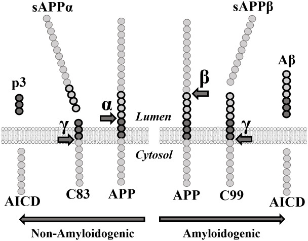
β-secretase, also known as aspartyl protease β-site APP cleaving enzyme 1 (BACE1), preferentially cleaves APP at the β-site in exon 16 (between Met671 and Asp672) [10, 11]. Subsequent cleavage by γ-secretase in exon 17 releases the β-amyloid peptides (40–42 amino acids long). In elderly people without Alzheimer’s disease, APP is preferentially processed by α-secretase prior to the cleavage by γ-secretase. The α-secretase enzyme targets the α-site located within the β-amyloid peptide sequence (Fig 1) and prevents the formation of the Aβ peptides thus reducing the formation of insoluble oligomers and protofibrils resulting from the aggregation of these peptides. These oligomers and protofibrils accumulate to form neurotoxic senile and neuritic plaques [12].

Fig 1. APP proteolytic pathways.

Fig 1

α: α-secretase. sAPPα: soluble APPα fragment. β: β-secretase. sAPPβ: soluble APPβ fragment. γ: γ-secretase. AICD: APP Intracellular domain.

Many pharmaceutical companies attempted to inhibit BACE1 to decrease Aβ peptide concentration. However, numerous clinical studies targeting BACE1 failed due to notable side effects [13, 14]. Indeed, BACE1 was found to be significantly implicated in several other pathways necessary for synaptic transmission [1517]. The soluble APPβ fragment (sAPPβ) generated by the cleavage of APP by BACE1 is also involved in axonal generation and neuronal death mediation [18] (Fig 1). This implies that an effective treatment for AD must decrease Aβ peptide concentrations without eliminating either sAPPβ or BACE1. Since eliminating the enzyme responsible for the excessive cleavage is unrealistic as a treatment, targeting the APP gene itself must become the focus of many future gene-based therapies.

Many APP mutations cause early-onset Familial Alzheimer’s disease (FAD). However, rather than causing FAD, the point mutation A673T decreases the incidence of this disease. In 2012, Jonsson et al. published a study in which they searched for APP coding variants in a sample of 1,795 Icelanders using whole-genome sequencing [19]. Their goal was to find low-frequency variants of the APP gene, which significantly reduced the risk of AD. Ultimately, they found a point mutation in the APP gene wherein the alanine at position 673 was substituted for a threonine (A673T). This mutation protects against AD and is adjacent to the β-site in exon 16 of the APP gene. The amino acid in question is located at position 2 in the ensuing β-amyloid peptide [17, 19, 20]. Due to the proximity of the A673T mutation to the β-site, the authors proposed that A673T specifically impairs the cleavage of the APP protein by β-secretase. In fact, the A673T mutation was shown to reduce the formation of β-amyloid peptides in wildtype APP by about 40% in vitro [1921].

The strong protective effect of the A673T mutation against AD serves as a proof of principle that reducing the β-cleavage of APP may protect against the disease. Moreover, the A673T mutation may also help to prolong the lifespan of its carriers. Indeed, individuals with this mutation were reported to have 1.47 times greater chances of reaching the age of 85 when compared to non-carriers [19]. Jonsson et al. concluded that the A673T mutation confers a strong protection against AD [19]. Later, Kero et al. found the A673T variant in a deceased individual aged 104.8 years whose brain presented little β-amyloid pathology [21]. This report supports the hypothesis that A673T protects the brain against β-amyloid accumulation and AD.

Although the A673T mutation is protective for people with an otherwise wild type APP gene, it is not known whether the A673T mutation will reduce the formation of β-amyloid peptides when the APP gene contains a deleterious FAD mutation. We thus aimed to study the interaction between the A673T mutation and 29 FAD mutations to determine to what extent it can provide a protective effect in patients. If the A673T is protective for FAD, this modification could eventually be introduced in an FAD patient genome with the CRISPR/Cas9 derived gene editing technology [22].

We report here that the presence of the Icelandic mutation (A673T) in several APP genes, each containing a FAD mutation, reduces the production of the Aβ40 and Aβ42 peptides. The introduction of the A673T mutation could thus eventually be an effective treatment for heritable FAD and perhaps even for sporadic AD.

Results

Amyloid-β peptide quantification

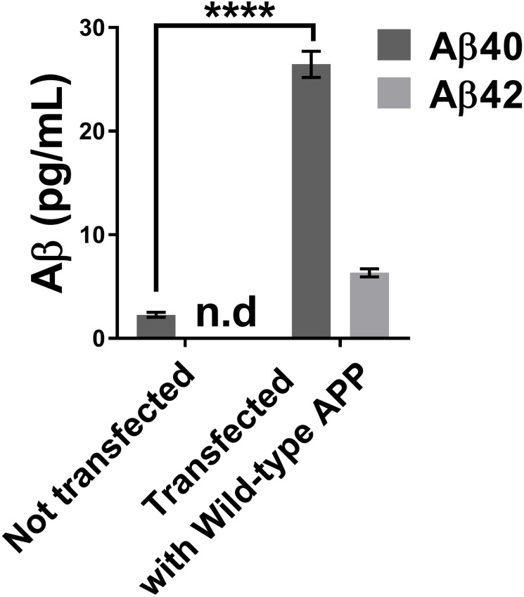
We first measured the concentration of Aβ peptides in the culture medium of neuroblastomas that were transfected or not with a plasmid overexpressing wild-type APP (Fig 2). The Aβ42 concentration of cells not transfected with the plasmid was so low that it was below the detection range of the MSD kit. There was, however, a clear increase of Aβ40 and Aβ42 peptide concentrations for the cells transfected with the wild-type APP plasmid.

Fig 2. Concentrations of Aβ40 and Aβ42 in the neuroblastoma supernatant using the MSD Elisa test.

Fig 2

The neuroblastoma transfection with a wild APP plasmid increased the concentrations of Aβ40 and Aβ42 peptides in the cell culture medium. Statistic test: Two-way ANOVA Sidak’s multiple comparisons test. P value style **** p<0.0001. n.d: not detectable.

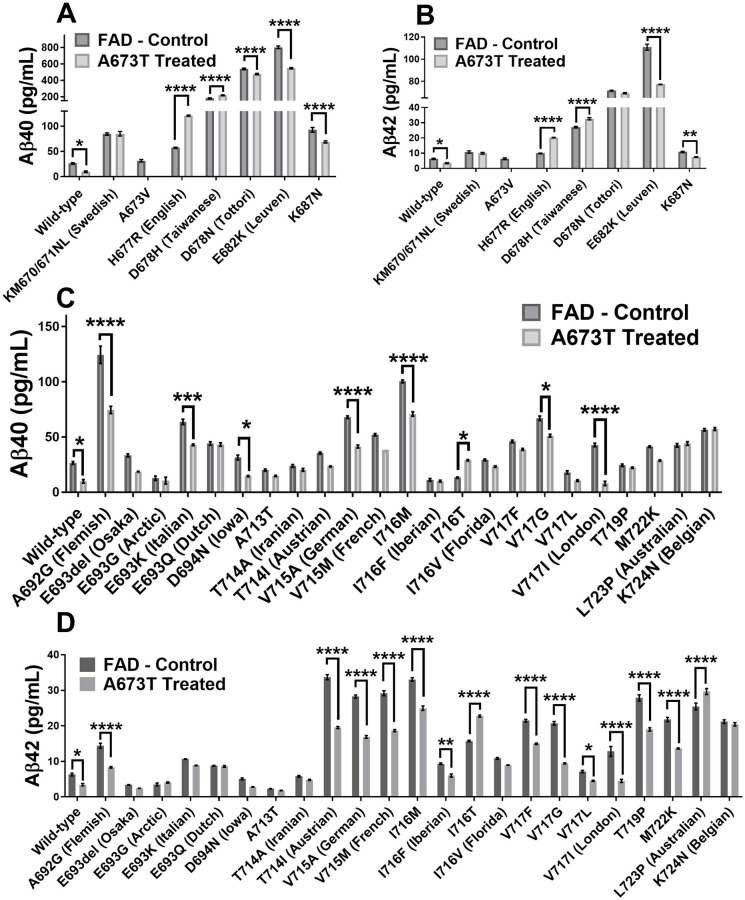
Two plasmid libraries were then tested. The first library was comprised of the APP plasmid with a unique FAD mutation in each plasmid. The second library was composed of the same APP/FAD plasmids but with the additional A673T mutation. We first analyzed the effect of the different FAD mutations on the Aβ peptide concentrations in the cell culture medium (Fig 3). Aβ40 and Aβ42 peptide concentrations were decreased in 10/29 (34%) and 14/29 (48%) FAD mutations respectively. The results obtained were consistent with the literature with some exceptions such as the H677R (English) and D678N (Tottori) mutation, which were reported to only enhance aggregation and not Aβ peptide accumulation [23].

Fig 3. Aβ peptide concentrations in culture medium.

Fig 3

Neuroblastomas were transfected with plasmids coding for 30 FAD mutations with or without an additional Icelandic (A673T) mutation (except for A673V). Aβ40 concentration (A) and Aβ42 concentration (B) when various FAD mutations are present in exon 16. Aβ40 concentration (C) and Aβ42 concentration (D) when various FAD mutations are present in exon 17. Statistical test: two-way ANOVA Sidak’s multiple comparisons test (n = 6). P value style * p<0.0332, ** p<0.0021, *** p<0.0002, **** p<0.0001.

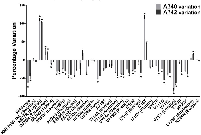
The insertion of the A673T Islandic mutation reduced the production of Aβ40 and Aβ42 peptides in the wild-type APP gene as well as 7/29 FAD mutations (Fig 4). It was further shown that the A673T mutation has a greater effect against FAD mutations in exon 17 than exon 16. The addition of the A673T mutation provided a statistically significant decrease in the Aβ40 concentration for 10 out of 29 FAD plasmids (34%). In addition, Aβ42 concentrations were also decreased in 14 out of 29 FAD plasmids (48%). However, the addition of the A673T mutation increased the Aβ40 concentrations in the presence of the D678H, I716T, and H677R FAD mutations (10%). Aβ42 concentrations were likewise increased in the L723P, D678H, I716T, and H677R FAD mutations (14%).

Fig 4. Variations of Aβ40 and Aβ42 peptide concentrations with A673T mutation.

Fig 4

Percentage changes of Aβ40 and Aβ42 peptide concentrations induced by the addition of the A673T mutation to plasmids containing or not an FAD mutation. * indicates this variation was statistically significant with the two-way ANOVA Sidak’s multiple comparisons test.

Aβ42/Aβ40 ratio

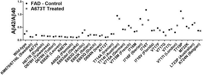
The Aβ42/Aβ40 ratios were calculated for all FAD mutations with and without the additional Icelandic mutation (Fig 5). However, since the Icelandic mutation also decreased the Aβ40 concentration, sometimes this ratio was increased by the Icelandic mutation insertion even though both Aβ peptide concentrations were significantly reduced. This was particularly the case for the London (V717I) mutation [24]. We thus ranked the different FAD mutations by their capacity to reduce the concentration of Aβ42 in Table 1. Such a ranking will be useful for choosing the right mouse model or patients should one attempt to design an FAD-specific therapy.

Fig 5. Aβ42/Aβ40 ratios.

Fig 5

Culture medium of cells transfected with plasmids coding for 30 FAD mutations with or without an additional Icelandic (A673T) mutation (except for A673V).

Table 1. Percentage variations in Aβ40 and Aβ42 peptide concentrations due to the A673T insertion.

EOAD mutation Aβ42 Concentration (%) Significance Aβ40 Concentration (%) Significance Exon
Wild-type -46 * -63 * na
V717I (London) -65 **** -81 **** 17
V717G -54 **** -24 * 17
D694N (Iowa) -44 ns -53 * 17
A692G (Flemish) -42 **** -40 **** 17
T714I (Austrian) -42 **** -34 ns 17
V715A (German) -40 **** -39 **** 17
M722K -37 **** -30 ns 17
V717L -36 * -41 ns 17
V715M (French) -36 **** -26 ns 17
I716F (Iberian) -36 ** -10 ns 17
T719P -32 **** -9 ns 17
K687N -31 ** -26 **** 16
V717F -30 **** -16 ns 17
E682K (Leuven) -30 **** -32 **** 16
E693del (Osaka) -28 ns -44 ns 17
I716M -24 **** -29 **** 17
A713T -21 ns -27 ns 17
I716V (Florida) -17 ns -21 ns 17
T714A (Iranian) -17 ns -15 ns 17
E693K (Italian) -17 ns -32 *** 17
KM670/671NL (Swedish) -8 ns 1 ns 16
K724N (Belgian) -3 ns 1 ns 17
D678N (Tottori) -3 ns -12 **** 16
E693Q (Dutch) -2 ns -2 ns 17
E693G (Arctic) 15 ns -17 ns 17
L723P (Australian) 16 **** 4 ns 17
D678H (Taiwanese) 20 **** 22 **** 16
I716T 44 **** 117 * 17
H677R (English) 103 **** 110 **** 16

FAD mutations were ranked from the highest decrease in Aβ42 concentration to the highest increase. The significance was determined by a Sidak’s ANOVA test. P value style * p<0.0332, ** p<0.0021, *** p<0.0002, **** p<0.0001.

The London mutation (APP V717I)

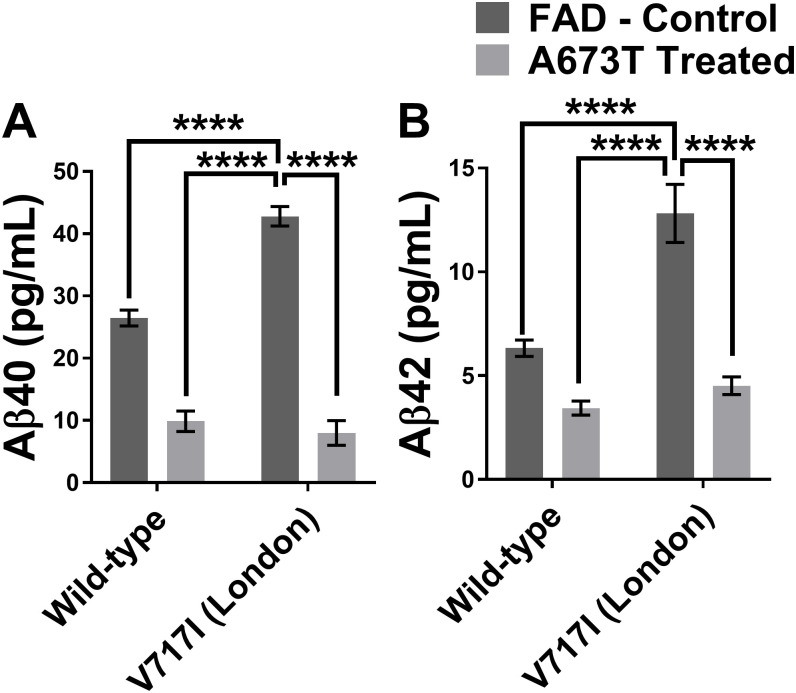
Among all the FAD mutations tested in the APP gene, the V717I (London mutation), located at the γ-secretase cutting site, presented very interesting results following the insertion of the A673T mutation. This mutation was able to diminish the Aβ42 percentage in the extracellular environment by 63% and Aβ40 by 80% (Table 1). The Aβ42 and Aβ40 peptide concentrations were actually reduced to levels that were similar to those observed in the A673T carrier without an FAD mutation (Fig 6).

Fig 6. Aβ peptide concentrations with wild type or London APP gene with or without A673T mutation.

Fig 6

Aβ40 (in A) and Aβ42 (in B) concentrations for neuroblastomas transfected with plasmids coding either for a wild type APP gene or an APP gene with the London mutation (V717I) with and without the Icelandic (A673T) mutation. Statistic test: Two-way ANOVA Sidak’s multiple comparison test (n = 6). P value style * p<0.0332, ** p<0.0021, *** p<0.0002, **** p<0.0001.

Discussion

The A673T mutation has been shown to provide protective effects against AD onset and development [19]. A lower β-secretase cleavage efficiency results in the carriers of this mutation into a 28% lower level of Aβ42 peptides monomer in plasma [25]. However, until now, the effects of this mutation when in the presence of FAD mutations has never been tested. Until now patients with a family history of Alzheimer’s disease have no effective treatment to prevent the loss of their mental faculties starting as early as their late 40’s [26]. In this study, we have shown that the addition of the A673T mutation in an APP cDNA can reduce Aβ42 peptide secretion in 14 of the 29 FAD mutations (48%) that we have studied and a reduction of Aβ40 peptide secretion in 10 of the 29 FAD mutations (34%) investigated in SH-SY5Y cells.

One notable observation of this study was that the A673T mutation generally had stronger protective effects against FAD mutations located in exon 17 compared to those located in exon 16 (Fig 3). This may be because some of the FAD mutations in exon 16 are located close to the BACE1 cut site and interfere with the A673T mutation’s capacity to reduce cutting of the APP protein by this enzyme. Each combination of A673T with one FAD mutation creates a unique structural conformation which will either favor or interfere with BACE1 cleavage and subsequent fibril formation.

Interestingly, plasmids with different mutations in the same codon of the APP gene demonstrated different Aβ peptide secretions when in conjunction with A673T. It was noted that A673T has a strong protective effect for the I716M mutation. Its presence resulted in a reduction in Aβ42 of 24% and Aβ40 of 29%. However, when A673T was in the presence of I716T, Aβ42 levels were instead increased by 44% while Aβ40 was increased by 117%.

Indeed, for some FAD mutations, the addition of the A673T mutation resulted in an increase rather than a decrease in the concentration of Aβ peptides (Fig 4). H677R (English), D678H (Taiwanese), I716T and L723P (Australian) FAD mutations increased the severity of Aβ peptide accumulation when in conjunction with A673T (Table 1). The unequal effects of the A673T mutation on the Aβ42 and Aβ40 concentrations indicates that the Aβ42/Aβ40 ratio is not a good measure of the effects of each mutation. The Aβ42/Aβ40 ratio has previously been measured in cerebrospinal fluid (CSF) and used to diagnose Alzheimer’s disease [27, 28]. In this study however, L723P (Australian) demonstrated four-fold increase in its Aβ42/Aβ40 ratio. The CSF Aβ42/Aβ40 ratio would state that this is a beneficial effect. However, the Aβ42 and Aβ40 concentrations were increased by 16% and 4% respectively. Given that the Aβ42 peptide is primarily responsible for the plaque, an increase in its concentration is detrimental [29]. The Aβ42/Aβ40 ratio is therefore not an appropriate measurement in this study. As such, the effects of each mutation have been evaluated by observing the concentration of Aβ42 and Aβ40.

The reduction of Aβ peptides in the extracellular environment following the introduction of the A673T mutation was especially encouraging in the case of the V717I London mutation. Not only did the percentages of Aβ42 and Aβ40 demonstrate the greatest reductions with this mutation (Table 1) but the London mutation is also one of the most common FAD mutations in the world (ALZ.org). Our results seem to indicate that the addition of the A673T mutation in a London patient has the potential to strongly reduce their production of Aβ peptides (Fig 6) and serve as a promising avenue for a gene therapy. Attempting a clinical trial for the addition of the A673T mutation in pre-symptomatic London patients would also likely be much easier than in patients lacking an FAD mutation. When weighing the perceived notion of risk versus the potential benefits in taking part of the trial, these individuals who are at risk for AD are more likely to join. Individuals lacking an FAD mutation are less likely to be willing to join a clinical trial for sporadic AD as the notion of risk will likely outweigh the benefits [30]. Our results could also be used to support the development of other gene therapies that are predicated on the addition of a protective mutation as opposed to the return of the gene to the wildtype.

In certain cases, the percentage decrease of Aβ40 and Aβ42 is insufficient to determine whether an FAD mutation is an optimal candidate for A673T treatment. For example, the Leuven E682K mutation demonstrated a considerable reduction of Aβ40 and Aβ42 concentrations (Table 1). However, this reduction was incapable of bringing the peptide concentrations to acceptable levels. Rather, the peptides were still present in a concentration that was severalfold that of the wildtype APP gene (Fig 3A and 3B). Ultimately, a gene therapy for this FAD mutation using A673T may slow down the progression of the disease as the concentration of the Aβ peptides is diminished; however, the data does not suggest that this treatment alone has the potential to prevent the onset of and development of AD based on Aβ peptide secretion.

Our report has solely studied the secretion of the Aβ peptides. It must be remembered that the aggregation of these peptides is also a very important parameter as it plays an essential role in the protective effect of A673T [31]. Most FAD mutations are considered pathogenic as they alter the aggregation of the Aβ peptides leading to detrimental outcomes. Adding A673T, which is also known to reduce aggregation, may create some competition with the pathogenic mutation and reduce total aggregation. However, the results are hard to predict without direct experimentation. Many teams working on aggregation have shown that A673T Aβ42 proteins aggregate more slowly than wild-type proteins [20, 3134]. Most recently, Limegrover et al. showed A673T inhibits oligomer formation and lowers binding affinity to the synaptic receptors, limiting neuronal degradation [32]. It is possible that some FAD mutations showing only a moderate reduction of Aβ peptide production following the insertion of the A673T mutation may experience a significant reduction in overall aggregation.

The next step will be to determine the extent to which the A673T mutation conveys beneficial effects to cells derived from V717I London FAD patients using base editing [22] or PRIME editing [35]. This will need to be performed first in vitro and subsequently in mouse models in vivo. This will demonstrate the feasibility of providing a protective effect against Alzheimer’s disease using the A673T Icelandic mutation and set the groundwork for an eventual gene therapy. A few teams have attempted to use the CRISPR/Cas9 system to target and disrupt AD-related genes [36]. However, the requirements for the original CRISPR system limits the number of FAD mutations that can be targeted. In addition, a gene therapy will need to be developed for each FAD they intend to target. Our proposed approach is novel in that we will attempt to leverage the protective effects of A673T to create a gene therapy that will be applicable to more than one FAD.

This study has demonstrated that the insertion of the A673T mutation decreases Aβ40 and Aβ42 production in SH-SY5Y cells in 10 and 14 FAD mutations respectively and may lead to potential benefits for these forms of Familial Alzheimer’s Disease. The decrease in Aβ42 production is especially encouraging as it is this protein that is primarily responsible for the formation of plaque. Our future experiments will attempt to verify whether the A673T mutation in APP affects other genes in trans. More specifically, genes that have been related to AD such as PSEN1 or PSEN2. A673T might also compensate for a weak clearing system which has been associated with sporadic AD cases by changing the conformation of the Aβ peptides [37]. This mutation may one day be the simplest and safest way to treat this disease.

Methods

Construction of plasmid libraries containing an FAD mutation

The backbone plasmid pcDNA6/V5-His was purchased from Invitrogen Inc. (Carlsbad, CA). The APP695 cDNA (courtesy of Dr. G. Levesque, CHUQ, Quebec) was inserted by ligation between Kpn1 and Xba1 cut sites. The ensuing plasmid was then mutated using the New England Biolabs (NEB, Ipswich, MA) mutagenesis Q5 kit in 29 different reactions. The 29 new plasmids each represented a form of FAD and served as a “normal” library version of each FAD. The mutations were located in exons 16 and 17 to better demonstrate the protective effects of A673T. Another “mutated” library was created by adding an additional A673T mutation to each FAD plasmid. Prior to the start of the experiments, the plasmids underwent Sanger sequencing to ensure that the only mutations present were those under study.

Transfection in SH-SY5Y of plasmid libraries

The transfection reagent (Lipofectamine 2000TM) and Opti-MEM-1 culture media were purchased from Life Technologies Inc. (Carlsbad, CA). The day before the transfection, 100,000 SH-SY5Y cells were seeded per well in 24 well plates in DMEM/F12 supplemented with 10% Fetal Bovine Serum (FBS) and antibiotics (penicillin/streptomycin 100 μg/mL). The following morning, the culture medium was changed for 500 μl of DMEM/F12 medium supplemented with 10% FBS without antibiotics. The plate was maintained at 37°C for the time required to prepare the transfection solution. For the transfection, solutions A and B were first prepared. Solution A contained 48 μl of Opti-MEM-1 and 2 μl of Lipofectamine 2000 for a final volume of 50 μl. Solution B was prepared as follows: a volume of DNA solution containing 800 ng of DNA was mixed with a volume of Opti-MEM-1 to obtain a final volume of 50 μl. Solutions A and B were then mixed together by up and down movements and incubated at room temperature for 20 minutes. 100 μl of the ensuing solution were then added to each well. The plate was left in the CO2 incubator for a period of 4 to 6 hours. The medium was replaced by 500 μl of DMEM F12 supplemented with 10% FBS and antibiotics. The plate was kept for 72 hours in the CO2 incubator before extraction of genomic DNA. The culture medium was harvested and protease inhibitors (1 mM PMSF + 1X complete tabs from Roche) were added. The media were then stored at -80°C.

Culture medium analysis

The concentrations of Aβ40 and Aβ42 peptides were measured with Meso Scale Discovery Inc. (MSD, Rockville, MA) Neurodegenerative Disease Assay 6E10 kit. Standards and samples were prepared according to the manufacturer’s protocols and tested in triplicate for each experiment.

Statistical analysis

All statistic tests and graphs were performed as recommended by GraphPad Prism 7.0. Two-way ANOVA Sidak’s multiple comparisons test was used to test significance with three biological replicas (two technical replicas each) for Figs 2, 3 and 6. P value style * p<0.0332, ** p<0.0021, *** p<0.0002, **** p<0.0001.

Supporting information

S1 Data

(XLSX)

Acknowledgments

We would like to thank Dr. G. Levesque (CHUQ, Quebec) for giving us the APP695 cDNA plasmid as well as Dr R. Lapointe (CHUM, Montréal) for allowing us to use the MSD equipment in his laboratory. We also would like to thank The Cell Network for their support.

Data Availability

All relevant data are within the manuscript and its Supporting information files.

Funding Statement

This project was funded by the Weston Brain Institute (WBI). AG holds a Bourse de la Fondation du CHU de Québec. GL holds a Training award from the CIHR. https://westonbraininstitute.ca/home/https://fondationduchudequebec.org/?gclid=Cj0KCQjw9b_4BRCMARIsADMUIyrETbZQ7IdT9XBUtrTNt8ivE7VxeTfFOz33hLRR1h8MizZH8NoGnwoaAjS1EALw_wcBhttps://cihr-irsc.gc.ca/e/193.html The funders had no role in study design, data collection and analysis, decision to publish, or preparation of the manuscript.

References

  • 1.Organization WH. Dementia 2020 [https://www.who.int/news-room/fact-sheets/detail/dementia.
  • 2.Ferri CP, Prince M, Brayne C, Brodaty H, Fratiglioni L, Ganguli M, et al. Global prevalence of dementia: a Delphi consensus study. Lancet. 2005;366(9503):2112–7. 10.1016/S0140-6736(05)67889-0 [DOI] [PMC free article] [PubMed] [Google Scholar]
  • 3.Plassman BL, Langa KM, Fisher GG, Heeringa SG, Weir DR, Ofstedal MB, et al. Prevalence of dementia in the United States: the aging, demographics, and memory study. Neuroepidemiology. 2007;29(1–2):125–32. 10.1159/000109998 [DOI] [PMC free article] [PubMed] [Google Scholar]
  • 4.Qiu C, Kivipelto M, von Strauss E. Epidemiology of Alzheimer’s disease: occurrence, determinants, and strategies toward intervention. Dialogues in clinical neuroscience. 2009;11(2):111–28. 10.31887/DCNS.2009.11.2/cqiu [DOI] [PMC free article] [PubMed] [Google Scholar]
  • 5.Alzheimer’s Disease I. World Alzheimer Report 2015. 2015.
  • 6.Mohr NM, Moreno-Walton L, Mills AM, Brunett PH, Promes SB, Society for Academic Emergency Medicine A, et al. Generational influences in academic emergency medicine: teaching and learning, mentoring, and technology (part I). Acad Emerg Med. 2011;18(2):190–9. 10.1111/j.1553-2712.2010.00985.x [DOI] [PMC free article] [PubMed] [Google Scholar]
  • 7.Masters CL, Multhaup G, Simms G, Pottgiesser J, Martins RN, Beyreuther K. Neuronal origin of a cerebral amyloid: neurofibrillary tangles of Alzheimer’s disease contain the same protein as the amyloid of plaque cores and blood vessels. The EMBO journal. 1985;4(11):2757–63. [DOI] [PMC free article] [PubMed] [Google Scholar]
  • 8.Alonso Vilatela ME, Lopez-Lopez M, Yescas-Gomez P. Genetics of Alzheimer’s disease. Archives of medical research. 2012;43(8):622–31. 10.1016/j.arcmed.2012.10.017 [DOI] [PubMed] [Google Scholar]
  • 9.Zhang YW, Thompson R, Zhang H, Xu H. APP processing in Alzheimer’s disease. Molecular brain. 2011;4:3 10.1186/1756-6606-4-3 [DOI] [PMC free article] [PubMed] [Google Scholar]
  • 10.Hussain I, Powell D, Howlett DR, Tew DG, Meek TD, Chapman C, et al. Identification of a novel aspartic protease (Asp 2) as beta-secretase. Molecular and cellular neurosciences. 1999;14(6):419–27. 10.1006/mcne.1999.0811 [DOI] [PubMed] [Google Scholar]
  • 11.Sinha S, Anderson JP, Barbour R, Basi GS, Caccavello R, Davis D, et al. Purification and cloning of amyloid precursor protein beta-secretase from human brain. Nature. 1999;402(6761):537–40. 10.1038/990114 [DOI] [PubMed] [Google Scholar]
  • 12.Urbanc B, Cruz L, Ding F, Sammond D, Khare S, Buldyrev SV, et al. Molecular dynamics simulation of amyloid beta dimer formation. Biophys J. 2004;87(4):2310–21. 10.1529/biophysj.104.040980 [DOI] [PMC free article] [PubMed] [Google Scholar]
  • 13.Merck. Merck Announces EPOCH Study of Verubecestat for the Treatment of People with Mild to Moderate Alzheimer’s Disease to Stop for Lack of Efficacy 2017 [https://investors.merck.com/news/press-release-details/2017/Merck-Announces-EPOCH-Study-of-Verubecestat-for-the-Treatment-of-People-with-Mild-to-Moderate-Alzheimers-Disease-to-Stop-for-Lack-of-Efficacy/default.aspx.
  • 14.Hu X, Fan Q, Hou H, Yan R. Neurological dysfunctions associated with altered BACE1-dependent Neuregulin-1 signaling. J Neurochem. 2016;136(2):234–49. 10.1111/jnc.13395 [DOI] [PMC free article] [PubMed] [Google Scholar]
  • 15.Laird FM, Cai H, Savonenko AV, Farah MH, He K, Melnikova T, et al. BACE1, a major determinant of selective vulnerability of the brain to amyloid-beta amyloidogenesis, is essential for cognitive, emotional, and synaptic functions. J Neurosci. 2005;25(50):11693–709. 10.1523/JNEUROSCI.2766-05.2005 [DOI] [PMC free article] [PubMed] [Google Scholar]
  • 16.Hartmann S, Zheng F, Kyncl MC, Karch S, Voelkl K, Zott B, et al. beta-Secretase BACE1 Promotes Surface Expression and Function of Kv3.4 at Hippocampal Mossy Fiber Synapses. J Neurosci. 2018;38(14):3480–94. 10.1523/JNEUROSCI.2643-17.2018 [DOI] [PMC free article] [PubMed] [Google Scholar]
  • 17.Das B, Yan R. Role of BACE1 in Alzheimer’s synaptic function. Transl Neurodegener. 2017;6:23 10.1186/s40035-017-0093-5 [DOI] [PMC free article] [PubMed] [Google Scholar]
  • 18.Furukawa K, Sopher BL, Rydel RE, Begley JG, Pham DG, Martin GM, et al. Increased activity-regulating and neuroprotective efficacy of alpha-secretase-derived secreted amyloid precursor protein conferred by a C-terminal heparin-binding domain. J Neurochem. 1996;67(5):1882–96. 10.1046/j.1471-4159.1996.67051882.x [DOI] [PubMed] [Google Scholar]
  • 19.Jonsson T, Atwal JK, Steinberg S, Snaedal J, Jonsson PV, Bjornsson S, et al. A mutation in APP protects against Alzheimer’s disease and age-related cognitive decline. Nature. 2012;488(7409):96–9. 10.1038/nature11283 [DOI] [PubMed] [Google Scholar]
  • 20.Maloney JA, Bainbridge T, Gustafson A, Zhang S, Kyauk R, Steiner P, et al. Molecular mechanisms of Alzheimer disease protection by the A673T allele of amyloid precursor protein. J Biol Chem. 2014;289(45):30990–1000. 10.1074/jbc.M114.589069 [DOI] [PMC free article] [PubMed] [Google Scholar]
  • 21.Kero M, Paetau A, Polvikoski T, Tanskanen M, Sulkava R, Jansson L, et al. Amyloid precursor protein (APP) A673T mutation in the elderly Finnish population. Neurobiol Aging. 2013;34(5):1518 e1–3. 10.1016/j.neurobiolaging.2012.09.017 [DOI] [PubMed] [Google Scholar]
  • 22.Komor AC, Kim YB, Packer MS, Zuris JA, Liu DR. Programmable editing of a target base in genomic DNA without double-stranded DNA cleavage. Nature. 2016;533(7603):420–4. 10.1038/nature17946 [DOI] [PMC free article] [PubMed] [Google Scholar]
  • 23.Hori Y, Hashimoto T, Wakutani Y, Urakami K, Nakashima K, Condron MM, et al. The Tottori (D7N) and English (H6R) familial Alzheimer disease mutations accelerate Abeta fibril formation without increasing protofibril formation. J Biol Chem. 2007;282(7):4916–23. 10.1074/jbc.M608220200 [DOI] [PubMed] [Google Scholar]
  • 24.Goate A, Chartier-Harlin MC, Mullan M, Brown J, Crawford F, Fidani L, et al. Segregation of a missense mutation in the amyloid precursor protein gene with familial Alzheimer’s disease. Nature. 1991;349(6311):704–6. 10.1038/349704a0 [DOI] [PubMed] [Google Scholar]
  • 25.Martiskainen H, Herukka SK, Stancakova A, Paananen J, Soininen H, Kuusisto J, et al. Decreased plasma beta-amyloid in the Alzheimer’s disease APP A673T variant carriers. Ann Neurol. 2017;82(1):128–32. 10.1002/ana.24969 [DOI] [PubMed] [Google Scholar]
  • 26.Maurer K, Volk S, Gerbaldo H. Auguste D and Alzheimer’s disease. Lancet. 1997;349(9064):1546–9. 10.1016/S0140-6736(96)10203-8 [DOI] [PubMed] [Google Scholar]
  • 27.Lewczuk P, Esselmann H, Otto M, Maler JM, Henkel AW, Henkel MK, et al. Neurochemical diagnosis of Alzheimer’s dementia by CSF Abeta42, Abeta42/Abeta40 ratio and total tau. Neurobiol Aging. 2004;25(3):273–81. 10.1016/S0197-4580(03)00086-1 [DOI] [PubMed] [Google Scholar]
  • 28.Lehmann S, Delaby C, Boursier G, Catteau C, Ginestet N, Tiers L, et al. Relevance of Abeta42/40 Ratio for Detection of Alzheimer Disease Pathology in Clinical Routine: The PLMR Scale. Front Aging Neurosci. 2018;10:138 10.3389/fnagi.2018.00138 [DOI] [PMC free article] [PubMed] [Google Scholar]
  • 29.Mann DM, Iwatsubo T, Ihara Y, Cairns NJ, Lantos PL, Bogdanovic N, et al. Predominant deposition of amyloid-beta 42(43) in plaques in cases of Alzheimer’s disease and hereditary cerebral hemorrhage associated with mutations in the amyloid precursor protein gene. Am J Pathol. 1996;148(4):1257–66. [PMC free article] [PubMed] [Google Scholar]
  • 30.Fogel DB. Factors associated with clinical trials that fail and opportunities for improving the likelihood of success: A review. Contemp Clin Trials Commun. 2018;11:156–64. 10.1016/j.conctc.2018.08.001 [DOI] [PMC free article] [PubMed] [Google Scholar]
  • 31.Benilova I, Gallardo R, Ungureanu AA, Castillo Cano V, Snellinx A, Ramakers M, et al. The Alzheimer disease protective mutation A2T modulates kinetic and thermodynamic properties of amyloid-beta (Abeta) aggregation. J Biol Chem. 2014;289(45):30977–89. 10.1074/jbc.M114.599027 [DOI] [PMC free article] [PubMed] [Google Scholar]
  • 32.Limegrover CS, LeVine H 3rd, Izzo NJ, Yurko R, Mozzoni K, Rehak C, et al. Alzheimer’s Protection Effect of A673T Mutation May Be Driven by Lower Abeta Oligomer Binding Affinity. J Neurochem. 2020. 10.1111/jnc.15212 [DOI] [PMC free article] [PubMed] [Google Scholar]
  • 33.Lin TW, Chang CF, Chang YJ, Liao YH, Yu HM, Chen YR. Alzheimer’s amyloid-beta A2T variant and its N-terminal peptides inhibit amyloid-beta fibrillization and rescue the induced cytotoxicity. PLoS One. 2017;12(3):e0174561 10.1371/journal.pone.0174561 [DOI] [PMC free article] [PubMed] [Google Scholar]
  • 34.Murray B, Sorci M, Rosenthal J, Lippens J, Isaacson D, Das P, et al. A2T and A2V Abeta peptides exhibit different aggregation kinetics, primary nucleation, morphology, structure, and LTP inhibition. Proteins. 2016;84(4):488–500. 10.1002/prot.24995 [DOI] [PubMed] [Google Scholar]
  • 35.Anzalone AV, Randolph PB, Davis JR, Sousa AA, Koblan LW, Levy JM, et al. Search-and-replace genome editing without double-strand breaks or donor DNA. Nature. 2019. 10.1038/s41586-019-1711-4 [DOI] [PMC free article] [PubMed] [Google Scholar]
  • 36.Gyorgy B, Loov C, Zaborowski MP, Takeda S, Kleinstiver BP, Commins C, et al. CRISPR/Cas9 Mediated Disruption of the Swedish APP Allele as a Therapeutic Approach for Early-Onset Alzheimer’s Disease. Mol Ther Nucleic Acids. 2018;11:429–40. 10.1016/j.omtn.2018.03.007 [DOI] [PMC free article] [PubMed] [Google Scholar]
  • 37.Castellano JM, Kim J, Stewart FR, Jiang H, DeMattos RB, Patterson BW, et al. Human apoE isoforms differentially regulate brain amyloid-beta peptide clearance. Sci Transl Med. 2011;3(89):89ra57 10.1126/scitranslmed.3002156 [DOI] [PMC free article] [PubMed] [Google Scholar]

Decision Letter 0

Hemant K Paudel

2 Sep 2020

PONE-D-20-22117

Protective mutation A673T as a potential gene therapy for most forms of APP Familial Alzheimer’s Disease

PLOS ONE

Dear Dr. Guyon:

Thank you for submitting your manuscript to PLOS ONE. Reviewers found your manuscript interesting. However, they also raised a number of issues. The results are thought to be exaggerated and conclusions drawn to far. It was also felt that data obtained from non neural cells must be cautiously interpreted.  We invite you to submit a revised version of the manuscript that addresses the points raised during the review process.

Please submit your revised manuscript within 60 days. If you will need more time than this to complete your revisions, please reply to this message or contact the journal office at plosone@plos.org. When you're ready to submit your revision, log on to https://www.editorialmanager.com/pone/ and select the 'Submissions Needing Revision' folder to locate your manuscript file.

Please include the following items when submitting your revised manuscript:

  • A rebuttal letter that responds to each point raised by the academic editor and reviewer(s). You should upload this letter as a separate file labeled 'Response to Reviewers'.

  • A marked-up copy of your manuscript that highlights changes made to the original version. You should upload this as a separate file labeled 'Revised Manuscript with Track Changes'.

  • An unmarked version of your revised paper without tracked changes. You should upload this as a separate file labeled 'Manuscript'.

If you would like to make changes to your financial disclosure, please include your updated statement in your cover letter. Guidelines for resubmitting your figure files are available below the reviewer comments at the end of this letter.

If applicable, we recommend that you deposit your laboratory protocols in protocols.io to enhance the reproducibility of your results. Protocols.io assigns your protocol its own identifier (DOI) so that it can be cited independently in the future. For instructions see: http://journals.plos.org/plosone/s/submission-guidelines#loc-laboratory-protocols

We look forward to receiving your revised manuscript.

Kind regards,

Hemant K. Paudel

Academic Editor

PLOS ONE

Journal Requirements:

When submitting your revision, we need you to address these additional requirements.

1. Please ensure that your manuscript meets PLOS ONE's style requirements, including those for file naming. The PLOS ONE style templates can be found at

https://journals.plos.org/plosone/s/file?id=wjVg/PLOSOne_formatting_sample_main_body.pdf and

https://journals.plos.org/plosone/s/file?id=ba62/PLOSOne_formatting_sample_title_authors_affiliations.pdf

[Note: HTML markup is below. Please do not edit.]

Reviewers' comments:

Reviewer's Responses to Questions

Comments to the Author

1. Is the manuscript technically sound, and do the data support the conclusions?

The manuscript must describe a technically sound piece of scientific research with data that supports the conclusions. Experiments must have been conducted rigorously, with appropriate controls, replication, and sample sizes. The conclusions must be drawn appropriately based on the data presented.

Reviewer #1: Partly

Reviewer #2: Partly

**********

2. Has the statistical analysis been performed appropriately and rigorously?

Reviewer #1: Yes

Reviewer #2: I Don't Know

**********

3. Have the authors made all data underlying the findings in their manuscript fully available?

The PLOS Data policy requires authors to make all data underlying the findings described in their manuscript fully available without restriction, with rare exception (please refer to the Data Availability Statement in the manuscript PDF file). The data should be provided as part of the manuscript or its supporting information, or deposited to a public repository. For example, in addition to summary statistics, the data points behind means, medians and variance measures should be available. If there are restrictions on publicly sharing data—e.g. participant privacy or use of data from a third party—those must be specified.

Reviewer #1: Yes

Reviewer #2: Yes

**********

4. Is the manuscript presented in an intelligible fashion and written in standard English?

PLOS ONE does not copyedit accepted manuscripts, so the language in submitted articles must be clear, correct, and unambiguous. Any typographical or grammatical errors should be corrected at revision, so please note any specific errors here.

Reviewer #1: Yes

Reviewer #2: Yes

**********

5. Review Comments to the Author

Please use the space provided to explain your answers to the questions above. You may also include additional comments for the author, including concerns about dual publication, research ethics, or publication ethics. (Please upload your review as an attachment if it exceeds 20,000 characters)

Reviewer #1: In this manuscript, the authors propose a novel potential gene therapy in Alzheimer’s disease by inserting/adding the protective Icelandic mutation A673T into carriers of other harmful APP mutations leading to familial, early onset AD. The idea is compelling, and the method and results presented in this study are simple and straight forward. However, the authors exaggerate the positive results as well as the potential benefit of the results.

A general concern in that the therapeutic benefit, focusing on FAD with APP mutations is very slim. The Early onset familial form of AD is at most 5% of the AD cases. Out of those APP mutations makes up 10-15 % resulting in 0.5% of all AD cases at the most.

My main concern for this manuscript is that the results are exaggerated and conclusions drawn to far.

TITLE

The title states that introducing the A673T rescue mutation could be “a potential gene therapy for most APP familial cases”.

- The results show that introducing the A673T mutation significantly decreases Abeta 42 in 14/29 mutations and Abeta 40 in 10/29. That should be the main results. Not “most” mutations are rescued.

- The study is performed on a cancerous cell line SH-SY5Y and much more studies needs to be performed before suggesting this to be a potential gene therapy.

The effect in carriers of the London mutation might be beneficial, but needs much further studies to make such a claim

A title that describes the results is more appropriate.

ABSTRACT

- Beta secretase cleavage is not an abnormal cleavage pattern. Abeta is produced in healthy individuals throughout life and Abeta has been suggested to have endogenous function.

- The A673T mutation is not only found in Icelanders, but mainly located to the Scandinavian countries.

- “In most cases the production of Abeta peptides was decreased by the co-dominant A673T mutation” is not true (as goes for the title and results), 14 out of 29 mutation variants are reduced for Abeta 42, which is not “most”. Looking at a significant reduction of both Abeta 40 and 42, 10 out of 29 were reduced.

RESULTS

- Row 135. The authors state that introducing the A673T mutation reduces Abeta 40 in 23/29 FAD plasmids. There is only a statistically significant decrease in 10 out of 29 FAD plasmids. Additionally, on row 137, it is stated that the A673T mutation reduces Abeta 42 in 24 out of 29 plasmids, but only 14 of those are statistically significant. It is the statically significant reduction’s that should be reported as the main finding!

- Figure 4, where the change in Abeta secretion is presented as percentage of control is lacking error bars. Also, as in figure 3, the main result should be the number of plasmids that resulted in a SIGNIFICANT decrease in Abeta production (10 when taking both 40 and 42 into account).

DISCUSSION

The discussion is lacking a solid comparison with earlier work, it only contains four references. References are specifically lacking on line 198, 208, 222 and 225, but I would like to see a broader discussion taking earlier work into comparison.

Also, the conclusions from this small study performed on a simple neuroblastoma cell line are drawn too far. The use of SH-SY5Y cells are an easily accessible and easy transfectable cell system. However, there are many things that differ this cell line from human neurons. SH-SY5Y cell produce relatively low levels of Abeta, requiring over transfection to even give measurable levels. Investigating the effects of introducing the A673T mutation, ie by CRISPR technique, in human iPSC derived neurons carrying the London mutation and other relevant mutations would give a better idea of the possibility to take this further.

Much more work is needed using human neurons (iPSC-derived or directly converted) as well as animal studies before knowing if inserting the A673T mutation using CRISPR technique results in the same changes in Abeta production.

It is also very speculative to suggest that introducing this mutation into the APP gene of sporadic AD cases may compensate for most genetic risk factors.

Reviewer #2: Guyon and colleagues investigated the hypothesis that A673T mutation may influence the production of Aβ 40 and Aβ 42 in 29 different FAD mutations mapping to APP exon 16 and 17 in an in vitro system, therefore exerting a protective role on AD development.

Although the study is interesting and presents a promising therapeutic approach that deserves further investigation, there are some points that need to be addressed:

-is there a correlation between the overproduction of Aβ 40 and 42 in the FAD mutation studied and the degree of Aβ 40 and 42 reduction exerted by the A673T mutation?

Please explain why the effect of decreasing Aβ 40 and 42 production exerted by A673T mutation does not apply to all the FAD mutations. And why for some mutations (please list them) there are an increased production?

Minor Comments

Title

‘most forms of APP Familial Alzheimer’s Disease’, please change to 'most forms of Familial Alzheimer’s Disease caused by APP mutations'.

• Abstract

‘The accumulation of plaque in the brain leads’, please change to ‘the deposition of plaques in the brain’

‘Numerous APP gene mutations’, please change to ‘APP mutations mapping to exon 16 and 17’

‘29 FAD mutations’, please change to ‘29 FAD mutations mapping to exon 16 and exon 17

‘prevent the onset of, slow down’, please change ‘to prevent or delay the onset’

• Introduction

‘somatic inclusions’, please change to ‘intracellular inclusions’

‘In AD-free individuals’ please change to ‘in elderly people without Alzheimer’s disease’

‘Initially, many big pharma companies’ please change to ‘many pharmaceutical companies’

‘not all mutations are made equal’ please delete it

• Results

Please always specify to what extent the A673T mutation decreased the Aβ40 and Aβ42 concentration

‘have been stuck without an effective’, please reformulate the sentence.

• Discussion

'AD-free', please change the expression

‘said peptides’, please reformulate the sentence.

**********

6. PLOS authors have the option to publish the peer review history of their article (what does this mean?). If published, this will include your full peer review and any attached files.

If you choose “no”, your identity will remain anonymous but your review may still be made public.

Do you want your identity to be public for this peer review? For information about this choice, including consent withdrawal, please see our Privacy Policy.

Reviewer #1: No

Reviewer #2: No

[NOTE: If reviewer comments were submitted as an attachment file, they will be attached to this email and accessible via the submission site. Please log into your account, locate the manuscript record, and check for the action link "View Attachments". If this link does not appear, there are no attachment files.]

While revising your submission, please upload your figure files to the Preflight Analysis and Conversion Engine (PACE) digital diagnostic tool, https://pacev2.apexcovantage.com/. PACE helps ensure that figures meet PLOS requirements. To use PACE, you must first register as a user. Registration is free. Then, login and navigate to the UPLOAD tab, where you will find detailed instructions on how to use the tool. If you encounter any issues or have any questions when using PACE, please email PLOS at figures@plos.org. Please note that Supporting Information files do not need this step.

PLoS One. 2020 Dec 28;15(12):e0237122. doi: 10.1371/journal.pone.0237122.r002

Author response to Decision Letter 0


18 Oct 2020

Response to reviewers

Journal Requirements:

When submitting your revision, we need you to address these additional requirements.

1. Please ensure that your manuscript meets PLOS ONE's style requirements, including those for file naming.

We have checked every point of the two guidelines. We have the impression that all requirements are respected.

Reviewer #1: In this manuscript, the authors propose a novel potential gene therapy in Alzheimer’s disease by inserting/adding the protective Icelandic mutation A673T into carriers of other harmful APP mutations leading to familial, early onset AD. The idea is compelling, and the method and results presented in this study are simple and straight forward. However, the authors exaggerate the positive results as well as the potential benefit of the results.

A general concern in that the therapeutic benefit, focusing on FAD with APP mutations is very slim. The Early onset familial form of AD is at most 5% of the AD cases. Out of those APP mutations makes up 10-15 % resulting in 0.5% of all AD cases at the most. My main concern for this manuscript is that the results are exaggerated and conclusions drawn to far.

We have reformulated the title, abstract, discussion, and conclusion to better represent the scope of our findings. The study’s main aim was to verify whether one or several FAD mutations responded well to the co-domination of the A673T mutation. It was our plan to continue this vein of research with other cell models/ CRISPR techniques. This is actually the first article pertaining to our Alzheimer project; since submitting this manuscript, we have submitted another in which we detailed a method of introducing the A673T mutation using base editing. Additionally, as previous studies have shown, individuals carrying the A673T mutation without a FAD do not develop Alzheimer’s Disease. This leads us to believe that the addition of this mutation in a wild-type patient would protect them against sporadic forms of Alzheimer. It was previously demonstrated that A673T protects wild-type patients, this paper therefore sought to determine the effects of this mutation when in conjunction with several APP FAD mutations.

TITLE

The title states that introducing the A673T rescue mutation could be “a potential gene therapy for most APP familial cases”.

-The results show that introducing the A673T mutation significantly decreases Abeta 42 in 14/29 mutations and Abeta 40 in 10/29. That should be the main results. Not “most” mutations are rescued.

We changed the title accordingly. It is important to note that in the case of Alzheimer’s, the reduction of Abeta 42 is the most important parameter.

-The study is performed on a cancerous cell line SH-SY5Y and much more studies need to be performed before suggesting this to be a potential gene therapy.

This is true, it will indeed be necessary to perform many more tests in the future before this conclusion can be made.

Thus presently we can not suggest that this is a potential gene therapy. We have replaced this erroneous statement with one that stipulates that the results are encouraging for future studies on the development of a gene therapy for Alzheimer’s Disease.

The effect in carriers of the London mutation might be beneficial, but needs much further studies to make such a claim

You are right.

We will change it by something along the lines of: “the results are encouraging for carriers of the London mutation; however, more studies will need to be performed before we can assess its potential clinical benefit for these patients.”

A title that describes the results is more appropriate.

We changed the title accordingly.

ABSTRACT

- Beta secretase cleavage is not an abnormal cleavage pattern. Abeta is produced in healthy individuals throughout life and Abeta has been suggested to have endogenous function.

This was poorly explained on our part, we meant that the cleavage was normal but was aggravated in familial or sporadic forms. This would in term trigger the creation of plaque.

- The A673T mutation is not only found in Icelanders, but mainly located to the Scandinavian countries.

That is correct. It was first found in an Icelandic population and it is thus named the Islandic mutation.

- “In most cases the production of Abeta peptides was decreased by the co-dominant A673T mutation” is not true (as goes for the title and results), 14 out of 29 mutation variants are reduced for Abeta 42, which is not “most”. Looking at a significant reduction of both Abeta 40 and 42, 10 out of 29 were reduced.

We corrected that.

RESULTS

- Row 135. The authors state that introducing the A673T mutation reduces Abeta 40 in 23/29 FAD plasmids. There is only a statistically significant decrease in 10 out of 29 FAD plasmids. Additionally, on row 137, it is stated that the A673T mutation reduces Abeta 42 in 24 out of 29 plasmids, but only 14 of those are statistically significant. It is the statically significant reduction’s that should be reported as the main finding!

We made the correction.

- Figure 4, where the change in Abeta secretion is presented as percentage of control is lacking error bars.

It was an oversight. We corrected it.

Also, as in figure 3, the main result should be the number of plasmids that resulted in a SIGNIFICANT decrease in Abeta production (10 when taking both 40 and 42 into account).

We understand your comment and your perspective, however, we thought it was relevant to show the other mutations. Our results demonstrate that a given locus may have significantly varying results depending on the amino acid substitution, which is present. For example, V717L and V717I (London mutation) gave different Aβ40 and Aβ42 concentrations in spite of being on the same locus.

We also sought to illustrate that the mutations at the beginning of exon 17 induced very little changes (from E693Del Osaka to T714A Iranian) in Abeta 42 concentrations when the co-dominant A673T is also present. Interestingly, this pattern was not necessarily always shown with ABeta 40.

This figure was intended to provide an illustration of the effects of each mutation according to their order in the exons. This was thought to demonstrate the overall efficiency of the A673T mutation and its "hot spots", especially in the case of exon 16 and the end of exon 17. We think it is also important to present negative results to show the importance of the good results.

DISCUSSION

The discussion is lacking a solid comparison with earlier work; it only contains four references. References are specifically lacking on line 198, 208, 222 and 225, but I would like to see a broader discussion taking earlier work into comparison.

We have added some additional references to the discussion, but there are not a lot of articles available. It is important to note that this study has never been done before. No one has ever tried to determine the effects of codominance between A673T and another FAD mutation. There have only been a few articles dealing with the A673T mutation since its discovery in 2012. On October 6, 2020 Limegrover et al. (https://doi.org/10.1111/jnc.15212) reported another beneficial effect of this mutation. They suggested that the mutation could potentially decrease the ABeta oligomer binding affinity to synapses.

Line 198: One notable observation in this study was that the A673T mutation generally had stronger protective effects against FAD mutations in exon 17 compared to exon 16.

This finding comes from this study and in particular from Figure 3 (see previous commentary).

Line 208: Indeed, for some FAD mutations, the addition of the A673T mutation resulted in an increase rather than a decrease in the concentration of Aβ peptides (Fig 4).

No one has ever observed this before us.

Line 222: . It would probably be difficult or even impossible to obtain ethical approval for a Phase I clinical trial for sporadic Alzheimer patients in a preclinical state, i.e., before symptom development.

We have reformulated this section. It was difficult to find a proper citation that could fully support our claims so we took a different approach. We are now stating that enrolling a sufficient number of patients to obtain statistical significance is unlikely. This is due to the large number of patients that would be required as well as the perceived notion of risk associated to clinical trials weighed against the uncertain reward of protection against Alzheimer’s disease. Here we have cited an article discussing the difficulty of obtaining patients due to the perceived notion of risk as well as an article discussing the low incidence of sporadic Alzheimer’s disease.

Line 225: It would make the development of a Phase I clinical trial much simpler since we would be able to know the genotype of the patients several years before the symptom apparition.

The sentence was reworded slightly. While the trial would be simpler, it wouldn’t necessarily be “much simpler”. We now discuss that due to the perceived notion of risk associated to their genotype, patient enrollment would likely be higher. Though obtaining enough patients for statistical significance would remain a challenge given that this form of FAD only represents a small fraction of the total population with Alzheimer patients.

Also, the conclusions from this small study performed on a simple neuroblastoma cell line are drawn too far. The use of SH-SY5Y cells are an easily accessible and easy transfectable cell system. However, there are many things that differ this cell line from human neurons. SH-SY5Y cell produce relatively low levels of Abeta, requiring over transfection to even give measurable levels. Investigating the effects of introducing the A673T mutation, i.e., by CRISPR technique, in human iPSC derived neurons carrying the London mutation and other relevant mutations would give a better idea of the possibility to take this further.

You are absolutely right. SH-SY5Y is not the best model but its ABeta secretion profile of FAD mutations although lower corresponds to the profiles found in other cell models (Li et al. Mutations of beta-amyloid precursor protein alter the consequence of Alzheimer's disease pathogenesis. Neural Regen Res. 2019 Apr;14(4):658-665. doi: 10.4103/1673-5374.247469. PMID: 30632506; PMCID: PMC6352587).

Much more work is needed using human neurons (iPSC-derived or directly converted) as well as animal studies before knowing if inserting the A673T mutation using CRISPR technique results in the same changes in Abeta production.

Those studies are ongoing in our laboratory. We have changed our phrasing to reflect that these studies have yet to be performed.

It is also very speculative to suggest that introducing this mutation into the APP gene of sporadic AD cases may compensate for most genetic risk factors.

You are right, it is a simple hypothesis, however, the person with the Islandic mutation are protected from becoming Alzheimer.

Reviewer #2: Guyon and colleagues investigated the hypothesis that A673T mutation may influence the production of Aβ 40 and Aβ 42 in 29 different FAD mutations mapping to APP exon 16 and 17 in an in vitro system, therefore exerting a protective role on AD development. Although the study is interesting and presents a promising therapeutic approach that deserves further investigation, there are some points that need to be addressed:

Is there a correlation between the over-production of Aβ 40 and 42 in the FAD mutation studied and the degree of Aβ 40 and 42 reduction exerted by the A673T mutation?

Please explain why the effect of decreasing Aβ 40 and 42 production exerted by A673T mutation does not apply to all the FAD mutations. And why for some mutations (please list them) there are an increased production?

If this correlation exists it has not yet been demonstrated by anyone. Indeed, the interest of our manuscript is to show that the Abeta peptide that will be produced by the addition of the A673T mutation will have a unique interaction with mutations found at the same locus. The amino acid responsible for FAD will cause different effects with each peptide as it will change its special conformation in different ways and therefore some mutation like H677R, D678H, I716T and L723P showed an increased production of Abeta peptides. (It is assumed that BACE1 has a greater affinity with the peptide produced). For example: please note that the decrease in ABeta peptide production after adding the A673T mutation is starkly different for the V717G and V717I mutations. While they share the same locus, the results were noticeably different; we could not anticipate this before performing the experiment.

On October 6, 2020 (a few days ago), Limegrover et al. (https://doi.org/10.1111/jnc.15212) demonstrated another beneficial effect of the A673T mutation. It is potentially linked to a decrease of the ABeta oligomer binding affinity for synapses.

Minor Comments

Title

‘most forms of APP Familial Alzheimer’s Disease’, please change to 'most forms of Familial Alzheimer’s Disease caused by APP mutations'.

We corrected it.

• Abstract

The accumulation of plaque in the brain leads’, please change to ‘the deposition of plaques in the brain ‘Numerous APP gene mutations’, please change to ‘APP mutations mapping to exon 16 and 17 ‘29 FAD mutations’, please change to ‘29 FAD mutations mapping to exon 16 and exon 17 ‘prevent the onset of, slow down’, please change ‘to prevent or delay the onset’.

We have made those changes.

• Introduction

somatic inclusions’, please change to ‘intracellular inclusions’

We have made that change.

‘In AD-free individuals’ please change to ‘in elderly people without Alzheimer’s disease’

We have made that change.

‘Initially, many big pharma companies’ please change to ‘many pharmaceutical companies’

We have made that change.

‘not all mutations are made equal’ please delete it

We have made that change.

• Results

Please always specify to what extent the A673T mutation decreased the Aβ40 and Aβ42 concentration

We have made these specifications.

‘have been stuck without an effective’, please reformulate the sentence.

This sentence has been reformulated.

• Discussion

'AD-free', please change the expression

‘said peptides’, please reformulate the sentence.

We changed all the sentences, thank you.

Attachment

Submitted filename: Response to Reviewers.docx

Decision Letter 1

Hemant K Paudel

12 Nov 2020

PONE-D-20-22117R1

The protective mutation A673T in Amyloid Precursor Protein gene decreases Aβ40 and Aβ42 production for some forms of Familial Alzheimer’s Disease in SH-SY5Y cells

PLOS ONE

Dear Dr. Guyon:

Thank you for submitting your manuscript to PLOS ONE. After careful consideration, we feel that it has merit but does not fully meet PLOS ONE’s publication criteria as it currently stands. Therefore, we invite you to submit a revised version of the manuscript that addresses the points raised during the review process.

 More clarity about FTD mutations is required. There are several other minor points need to be addressed.

Please submit your revised manuscript within one month. If you will need more time than this to complete your revisions, please reply to this message or contact the journal office at plosone@plos.org. When you're ready to submit your revision, log on to https://www.editorialmanager.com/pone/ and select the 'Submissions Needing Revision' folder to locate your manuscript file.

Please include the following items when submitting your revised manuscript:

  • A rebuttal letter that responds to each point raised by the academic editor and reviewer(s). You should upload this letter as a separate file labeled 'Response to Reviewers'.

  • A marked-up copy of your manuscript that highlights changes made to the original version. You should upload this as a separate file labeled 'Revised Manuscript with Track Changes'.

  • An unmarked version of your revised paper without tracked changes. You should upload this as a separate file labeled 'Manuscript'.

If you would like to make changes to your financial disclosure, please include your updated statement in your cover letter. Guidelines for resubmitting your figure files are available below the reviewer comments at the end of this letter.

If applicable, we recommend that you deposit your laboratory protocols in protocols.io to enhance the reproducibility of your results. Protocols.io assigns your protocol its own identifier (DOI) so that it can be cited independently in the future. For instructions see: http://journals.plos.org/plosone/s/submission-guidelines#loc-laboratory-protocols

We look forward to receiving your revised manuscript.

Kind regards,

Hemant K. Paudel

Academic Editor

PLOS ONE

[Note: HTML markup is below. Please do not edit.]

Reviewers' comments:

Reviewer's Responses to Questions

Comments to the Author

1. If the authors have adequately addressed your comments raised in a previous round of review and you feel that this manuscript is now acceptable for publication, you may indicate that here to bypass the “Comments to the Author” section, enter your conflict of interest statement in the “Confidential to Editor” section, and submit your "Accept" recommendation.

Reviewer #1: All comments have been addressed

Reviewer #2: All comments have been addressed

**********

2. Is the manuscript technically sound, and do the data support the conclusions?

The manuscript must describe a technically sound piece of scientific research with data that supports the conclusions. Experiments must have been conducted rigorously, with appropriate controls, replication, and sample sizes. The conclusions must be drawn appropriately based on the data presented.

Reviewer #1: Yes

Reviewer #2: Partly

**********

3. Has the statistical analysis been performed appropriately and rigorously?

Reviewer #1: Yes

Reviewer #2: N/A

**********

4. Have the authors made all data underlying the findings in their manuscript fully available?

The PLOS Data policy requires authors to make all data underlying the findings described in their manuscript fully available without restriction, with rare exception (please refer to the Data Availability Statement in the manuscript PDF file). The data should be provided as part of the manuscript or its supporting information, or deposited to a public repository. For example, in addition to summary statistics, the data points behind means, medians and variance measures should be available. If there are restrictions on publicly sharing data—e.g. participant privacy or use of data from a third party—those must be specified.

Reviewer #1: Yes

Reviewer #2: Yes

**********

5. Is the manuscript presented in an intelligible fashion and written in standard English?

PLOS ONE does not copyedit accepted manuscripts, so the language in submitted articles must be clear, correct, and unambiguous. Any typographical or grammatical errors should be corrected at revision, so please note any specific errors here.

Reviewer #1: Yes

Reviewer #2: Yes

**********

6. Review Comments to the Author

Please use the space provided to explain your answers to the questions above. You may also include additional comments for the author, including concerns about dual publication, research ethics, or publication ethics. (Please upload your review as an attachment if it exceeds 20,000 characters)

Reviewer #1: The Authors have adequately responded to all my comments.

Reviewer #2: The manuscript has improved. However, there are still some minor issues that need to be addressed.

Overall, the description of the mutations remains imprecise. Terms like ‘some’, ‘most of’ ecc..have to be replaced.

Moreover, it is not clear whether 29 or 30 FAD mutations have been tested:

Figure 3 A-C : overall 30 mutations tested

Figure 4: 29 mutations tested

Figure 5: 30 mutations tested

Minor comments

Title

production for some forms of Familial Alzheimer’s Disease in SH-SY5Y cells

Please replace ‘some’ with a more accurate term

Abstract

Line 17. The deposition of plaques in the brain leads to the onset and development of Alzheimer’s disease.

Please change to: the deposition of Aβ plaques in the brain

Line 18. The Amyloid precursor protein (APP) is usually cut by alpha-secretase

I would write: the Amyloid precursor protein (APP) is cleaved by alpha-secretase (non-amyloidogenic processing of APP)

Line 22-23.containing APP genes with 29 FAD mutations

Please change it to ‘APP gene’

Introduction

Line 72. one APP mutation decreases

Please refer to the specific mutation: one APP mutation (p.A673T)

Line 89. ‘passed away’, please change to ‘deceased’

Line 94. mutation and various FAD mutations: please change to 29 FAD mutations

Results

Line 106. wild-type APP plasmid (Fig 2). Please specify.

Line 132-134. The reduction of Aβ40 and Aβ42 peptide production by the insertion of the additional A673T Islandic mutation is clear not only for the wild-type APP control gene but also for several FAD mutations.

Please reformulate it

Line 120-123. Nearly all of the FAD mutations increased the Aβ40 and Aβ42 concentrations.

The results obtained were consistent with the literature with some exceptions such as the

H677R (English) and D678N (Tottori) mutation, which were reported to only enhance

aggregation and not Aβ peptide accumulation

'Nearly all', please state exactly how many.

Line 126. Fig 3. Plasmids coding for various FAD mutations, please state exactly how many

Line 133-134. The reduction of Aβ40 and Aβ42 peptide production by the insertion of the additional A673T Islandic mutation is clear not only for the wild-type APP control gene but also for several FAD mutations.

Please reformulate it: clear? Significant? several FAD mutations, how many (%)?

Line 138-139. However, the addition of the A673T mutation increased the Aβ40 concentrations for 3 FAD plasmids (10%) and the Aβ42 concentrations for 4 FAD plasmids (14%) FAD mutations

Please list the APP mutations for these 3 and 4 FAD Plasmids

Line 167-168. The most encouraging mutation among all the FAD mutations of the APP

Please change the title and write a paragraph only about ‘The London mutation (APP p. V717I)’

Line 168. all the FAD mutations please change it to all the FAD mutations tested

Discussion

Line 183-184. The A673T mutation has been theorized to provide protective effects against AD onset and development [18].

Please, change to ‘The A673T mutation has been shown’

Line 190-191. 14 of the 29 FAD .. 10 of the 29 FAD mutations investigated

Please report (%)

Line 199-200. Another interesting observation of this study was the drastic difference in the presence of Aβ peptides, especially when different mutations of the same codon.

This sentence is not clear, please reformulate it.

Line 202-203. However, the presence of A673T actually increased the formation of both peptides in presence of the I716T mutation.

Please comment on it.

Line 212. All said, for the purposes,

Please reformulate it

Line 219. to strongly reduce of their Aβ peptide levels

Please rephrase it.

Line 220-225. Enrolling pre-symptomatic patients lacking FAD mutations

for clinical trials targeting sporadic Alzheimer patients would likely prove difficult. The large

sample size required for statistical significance in the face of patient reluctance when weighing the risk against the odds of developing Alzheimer’s disease in the first place [28]. That said, the data observed and discussed in this article stands to help validate the launch of clinical trials for carriers of the London mutation (approximately 30 families). As

Please, reformulate it

Line 255-256. Gyorgy et al. have managed to disrupt the APP KM670/671NL Swedish mutation allele using CRISPR [34]

Please rephrase it.

Line 261-263. This study has demonstrated that the insertion of the A673T mutation decreases Aβ40 and Aβ42 production in SH-SY5Y cells and could lead to potential benefits for some forms of Familial Alzheimer’s Disease

To reformulate: A673T mutation decreases Aβ40 and Aβ42 production in SH-SY5Y cells in (how many?) FAD mutations and may lead to…

Line 264-267. in APP affects other genes in trans. More specifically, genes that have been related to AD such as the PSEN1 or PSEN2 genes. It would additionally be worth exploring if A673T could compensate for a weak clearing system that originates from the APOE4 risk factor and therefore allow A673T to treat sporadic AD cases [35].

Please reformulate it.

Table 1

I would write the ‘Wild type’ in the first raw and try to order the mutations based on the level of significance.

**********

7. PLOS authors have the option to publish the peer review history of their article (what does this mean?). If published, this will include your full peer review and any attached files.

If you choose “no”, your identity will remain anonymous but your review may still be made public.

Do you want your identity to be public for this peer review? For information about this choice, including consent withdrawal, please see our Privacy Policy.

Reviewer #1: No

Reviewer #2: No

[NOTE: If reviewer comments were submitted as an attachment file, they will be attached to this email and accessible via the submission site. Please log into your account, locate the manuscript record, and check for the action link "View Attachments". If this link does not appear, there are no attachment files.]

While revising your submission, please upload your figure files to the Preflight Analysis and Conversion Engine (PACE) digital diagnostic tool, https://pacev2.apexcovantage.com/. PACE helps ensure that figures meet PLOS requirements. To use PACE, you must first register as a user. Registration is free. Then, login and navigate to the UPLOAD tab, where you will find detailed instructions on how to use the tool. If you encounter any issues or have any questions when using PACE, please email PLOS at figures@plos.org. Please note that Supporting Information files do not need this step.

PLoS One. 2020 Dec 28;15(12):e0237122. doi: 10.1371/journal.pone.0237122.r004

Author response to Decision Letter 1


7 Dec 2020

Reviewer #1: All comments have been addressed

Reviewer #2: All comments have been addressed

6. Review Comments to the Author

Please use the space provided to explain your answers to the questions above. You may also include additional comments for the author, including concerns about dual publication, research ethics, or publication ethics. (Please upload your review as an attachment if it exceeds 20,000 characters)

Reviewer #1: The Authors have adequately responded to all my comments.

Reviewer #2: The manuscript has improved. However, there are still some minor issues that need to be addressed.

Overall, the description of the mutations remains imprecise. Terms like ‘some’, ‘most of’ etc..have to be replaced.

Moreover, it is not clear whether 29 or 30 FAD mutations have been tested:

Figure 3 A-C : overall 30 mutations tested

Figure 4: 29 mutations tested

Figure 5: 30 mutations tested

An FAD mutation is a mutation changing the normal processing of beta amyloid.

In our study, there are 30 mutations tested. The 30th, A673V could not obviously be tested in Figure 4 it is located exactly at the location of A673T. But we wanted to make A673V appear in a matter of comparison in Figure 3 and 5. S you can see in Figure 5, A673V “A673T treated” ration does not appear because it is impossible to get it.

Minor comments

Title

production for some forms of Familial Alzheimer’s Disease in SH-SY5Y cells

Please replace ‘some’ with a more accurate term

We made all the corrections.

Abstract

Line 17. The deposition of plaques in the brain leads to the onset and development of Alzheimer’s disease.

Please change to: the deposition of Aβ plaques in the brain

Line 18. The Amyloid precursor protein (APP) is usually cut by alpha-secretase

I would write: the Amyloid precursor protein (APP) is cleaved by alpha-secretase (non-amyloidogenic processing of APP)

Line 22-23. containing APP genes with 29 FAD mutations

Please change it to ‘APP gene’

We made all the corrections.

Introduction

Line 72. one APP mutation decreases

Please refer to the specific mutation: one APP mutation (p.A673T)

Line 89. ‘passed away’, please change to ‘deceased’

Line 94. mutation and various FAD mutations: please change to 29 FAD mutations

We made all the corrections.

Results

Line 106. wild-type APP plasmid (Fig 2). Please specify.

Line 132-134. The reduction of Aβ40 and Aβ42 peptide production by the insertion of the additional A673T Islandic mutation is clear not only for the wild-type APP control gene but also for several FAD mutations.

Please reformulate it

We reformulated it everything. We hope it is clearer.

Line 120-123. Nearly all of the FAD mutations increased the Aβ40 and Aβ42 concentrations.

The results obtained were consistent with the literature with some exceptions such as the

H677R (English) and D678N (Tottori) mutation, which were reported to only enhance

aggregation and not Aβ peptide accumulation

'Nearly all', please state exactly how many.

Line 126. Fig 3. Plasmids coding for various FAD mutations, please state exactly how many

We corrected it.

Line 133-134. The reduction of Aβ40 and Aβ42 peptide production by the insertion of the additional A673T Islandic mutation is clear not only for the wild-type APP control gene but also for several FAD mutations.

Please reformulate it: clear? Significant? several FAD mutations, how many (%)?

Line 138-139. However, the addition of the A673T mutation increased the Aβ40 concentrations for 3 FAD plasmids (10%) and the Aβ42 concentrations for 4 FAD plasmids (14%) FAD mutations

Please list the APP mutations for these 3 and 4 FAD Plasmids

Line 167-168. The most encouraging mutation among all the FAD mutations of the APP

Please change the title and write a paragraph only about ‘The London mutation (APP p. V717I)’

Line 168. all the FAD mutations please change it to all the FAD mutations tested

We addressed all the comments.

Discussion

Line 183-184. The A673T mutation has been theorized to provide protective effects against AD onset and development [18].

Please, change to ‘The A673T mutation has been shown’

Line 190-191. 14 of the 29 FAD .. 10 of the 29 FAD mutations investigated

Please report (%)

Line 199-200. Another interesting observation of this study was the drastic difference in the presence of Aβ peptides, especially when different mutations of the same codon.

This sentence is not clear, please reformulate it.

Line 202-203. However, the presence of A673T actually increased the formation of both peptides in presence of the I716T mutation.

Please comment on it.

Line 212. All said, for the purposes,

Please reformulate it

Line 219. to strongly reduce of their Aβ peptide levels

Please rephrase it.

Line 220-225. Enrolling pre-symptomatic patients lacking FAD mutations

for clinical trials targeting sporadic Alzheimer patients would likely prove difficult. The large

sample size required for statistical significance in the face of patient reluctance when weighing the risk against the odds of developing Alzheimer’s disease in the first place [28]. That said, the data observed and discussed in this article stands to help validate the launch of clinical trials for carriers of the London mutation (approximately 30 families). As

Please, reformulate it

Line 255-256. Gyorgy et al. have managed to disrupt the APP KM670/671NL Swedish mutation allele using CRISPR [34]

Please rephrase it.

Line 261-263. This study has demonstrated that the insertion of the A673T mutation decreases Aβ40 and Aβ42 production in SH-SY5Y cells and could lead to potential benefits for some forms of Familial Alzheimer’s Disease

To reformulate: A673T mutation decreases Aβ40 and Aβ42 production in SH-SY5Y cells in (how many?) FAD mutations and may lead to…

Line 264-267. in APP affects other genes in trans. More specifically, genes that have been related to AD such as the PSEN1 or PSEN2 genes. It would additionally be worth exploring if A673T could compensate for a weak clearing system that originates from the APOE4 risk factor and therefore allow A673T to treat sporadic AD cases [35].

Please reformulate it.

We addressed all the Discussion comments.

Table 1

I would write the ‘Wild type’ in the first raw and try to order the mutations based on the level of significance.

We put “wild type” at the top of the table. However, we preferred to let the ordering the original way in order to keep clarity.

Attachment

Submitted filename: Response to Reviewers.docx

Decision Letter 2

Hemant K Paudel

10 Dec 2020

The protective mutation A673T in Amyloid Precursor Protein gene decreases Aβ peptides production for 14 forms of Familial Alzheimer’s Disease in SH-SY5Y cells

PONE-D-20-22117R2

Dear Dr. Guyon:

We’re pleased to inform you that your manuscript has been judged scientifically suitable for publication and will be formally accepted for publication once it meets all outstanding technical requirements.

Within one week, you’ll receive an e-mail detailing the required amendments. When these have been addressed, you’ll receive a formal acceptance letter and your manuscript will be scheduled for publication.

An invoice for payment will follow shortly after the formal acceptance. To ensure an efficient process, please log into Editorial Manager at http://www.editorialmanager.com/pone/, click the 'Update My Information' link at the top of the page, and double check that your user information is up-to-date. If you have any billing related questions, please contact our Author Billing department directly at authorbilling@plos.org.

If your institution or institutions have a press office, please notify them about your upcoming paper to help maximize its impact. If they’ll be preparing press materials, please inform our press team as soon as possible -- no later than 48 hours after receiving the formal acceptance. Your manuscript will remain under strict press embargo until 2 pm Eastern Time on the date of publication. For more information, please contact onepress@plos.org.

Kind regards,

Hemant K. Paudel

Academic Editor

PLOS ONE

Additional Editor Comments (optional):

Reviewers' comments:

Acceptance letter

Hemant K Paudel

15 Dec 2020

PONE-D-20-22117R2

The protective mutation A673T in Amyloid Precursor Protein gene decreases Aβ peptides production for 14 forms of Familial Alzheimer’s Disease in SH-SY5Y cells

Dear Dr. Guyon:

I'm pleased to inform you that your manuscript has been deemed suitable for publication in PLOS ONE. Congratulations! Your manuscript is now with our production department.

If your institution or institutions have a press office, please let them know about your upcoming paper now to help maximize its impact. If they'll be preparing press materials, please inform our press team within the next 48 hours. Your manuscript will remain under strict press embargo until 2 pm Eastern Time on the date of publication. For more information please contact onepress@plos.org.

If we can help with anything else, please email us at plosone@plos.org.

Thank you for submitting your work to PLOS ONE and supporting open access.

Kind regards,

PLOS ONE Editorial Office Staff

on behalf of

Dr. Hemant K. Paudel

Academic Editor

PLOS ONE

Associated Data

    This section collects any data citations, data availability statements, or supplementary materials included in this article.

    Supplementary Materials

    S1 Data

    (XLSX)

    Attachment

    Submitted filename: Response to Reviewers.docx

    Attachment

    Submitted filename: Response to Reviewers.docx

    Data Availability Statement

    All relevant data are within the manuscript and its Supporting information files.


    Articles from PLoS ONE are provided here courtesy of PLOS

    RESOURCES