Erratum in: “Pressure-Induced Changes in Astrocyte GFAP, Actin, and Nuclear Morphology in Mouse Optic Nerve” by Yik Tung Tracy Ling, Mary E. Pease, Joan L. Jefferys, Elizabeth C. Kimball, Harry A. Quigley, and Thao D. Nguyen (Invest Ophthalmol Vis Sci. 2020;61(11):14), https://doi.org/10.1167/iovs.61.11.14.
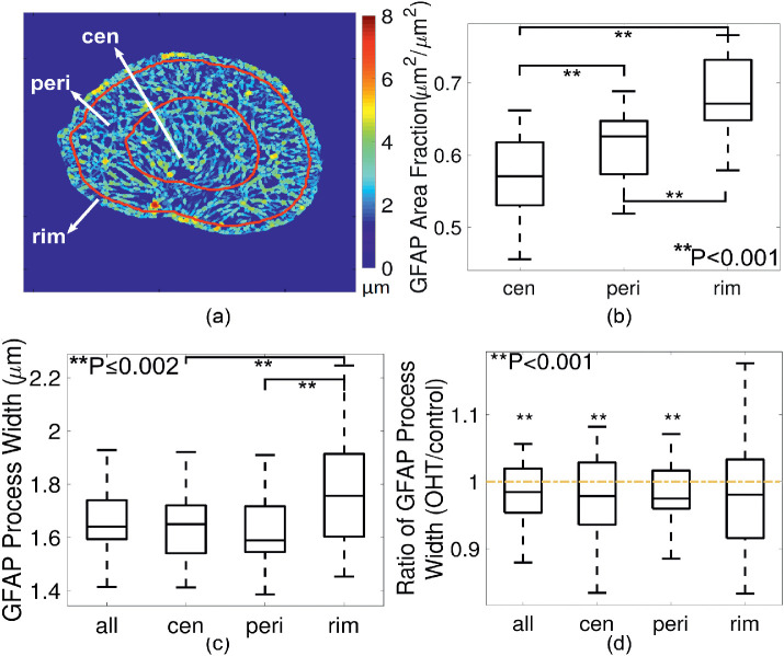
Figures 3a and 3b were misprinted in the published version of the article. Figure 3a should have included distinct boundaries and labels for the central, peripheral and rim region overlaid onto the color map of the measured process width. The labels “(a)” and “(b)” in Figure 3 were also missing in the published article. The correct version of Figure 3 is rendered in this erratum. The article has been corrected online.
Figure 3.
Radial variations in GFAP process width, showing (a) a color map of measured process width and regional divisions. Central zone (cen), peripheral (peri), and rim zone were divided as zones within 0% to 50%, 50% to 90%, and 90% to 100% of radii from fitted center of the ON to the ON boundary. (b) In R1, the GFAP area fraction was lowest in the central zone compared with that in the peripheral and rim zones. (c) Processes were thicker in the rim zone compared with central and peripheral zones (P ≤ 0.002 for both pairwise comparisons from linear mixed model and after Bonferroni adjustment). (d) Processes became significantly thinner overall and in central and peripheral zones after three-day OHT, but not in the rim zone (P = 0.08 for the rim region and ∗∗P < 0.001 for others).


