Operating microscopes are integrally linked to the evolution of microsurgery in ophthalmology. The first binocular surgical microscope used by Perritt in 1945 has given way to sophisticated technological contraptions with integrated aberrometry, optical coherence tomography (OCT) and overlays to guide the surgical steps.[1] Three-dimensional (3D) visualization with heads up display is the latest entrant in this ever-evolving field and may herald a paradigm shift in the way we operate.[2,3,4]
Three-dimensional viewing systems may be active or passive, mainly differing with respect to the generation and projection of the 3D effect.[3] Active 3D systems include the head-mounted systems (HMS) and involve the alternate high-speed projection of consecutive images onto the right and left eyes, with the image in the fellow eye being actively suppressed by the use of electronic glasses. Passive systems involve the horizontal mixing of two images and subsequent separation using polarized glasses to create a 3D effect, which may be viewed on a heads-up display system. The commercially available heads-up display systems with 3D visualization include the TrueVision 3D Visualization System integrated with Leica M822 and Leica M844 ophthalmic microscopes, NGENUITY 3D Visualization System (Alcon Laboratories) and ARTEVO 800 (Carl Zeiss Meditec). The video output is displayed on a 4K ultra-high-definition 55-inch OLED screen which is placed at a distance of 3-4 feet from the surgeon in the NGENUTIY and ARTEVO systems and viewed using polarized glasses [Table 1].
Table 1.
Comparison of technical specifications of two commercially available 3D visualization systems with heads up display
| Features | ARTEVO 800 (Zeiss) | NGENUITY (Alcon) |
|---|---|---|
| Camera | 2 Integrated 4 K Cameras | 1 HDR Camera |
| Latency | <50 ms | 80 milliseconds |
| Cataract Assistance Function and Intraoperative OCT | Integrated iOCT, Callisto Eye and Z Align overlay | Not integrated, based on microscope |
| Fundus Viewing System | RESIGHT 700 with E inverter | Based on microscope |
| Digital and Hybrid Function | Hybrid mode- seamless transition between Heads up Display or microscope binoculars with integrated switch | Not possible to switch between Heads Up Display and microscope binoculars without removing the camera |
| Ergonomics | No change in stack height of microscope- integrated digital optics | Attachment mounted on microscope head- increase in stack height of microscope |
Over the past decade, 3D visualization systems have been used in various vitreoretinal procedures and definitive benefits have been demonstrated in terms of enhanced ergonomics, better magnification and depth perception and the ability to use lower illumination levels.[2] In contrast, the role of these systems in anterior segment surgeries with significantly shorter duration and a limited plane of surgery is tenuous at present.[4,5,6,7,8]
Applications in Anterior Segment Surgeries
The applications of the 3D Visualization Systems with Heads up Display in phacoemulsification, keratoplasty, ocular surface surgeries, glaucoma as well as strabismus surgeries are being explored, with promising initial outcomes.[4,5,6,7,8,9]
The use of the heads-up display in cataract surgery may be traced back to 2010 when Weinstock et al. described their experience of 3D Heads up Display compared with conventional operating microscopes during phacoemulsification. They observed a higher rate of unplanned vitrectomy in cases operated with a standard microscope, highlighting the benefits of greater magnification and better depth perception in enhancing the safety of the surgery.[4] There is no significant increase in the surgical duration and optimal visual and anatomical outcomes have been reported after phacoemulsification. A large series of 2320 eyes comparing phacoemulsification performed using a traditional microscope or 3D heads up visualization reported similar efficacy and safety with both methods.[5] We have performed over 50 phacoemulsification surgeries with ARTEVO 800 system and observed a short learning curve of initial 3-5 cases to get accustomed to the changed dynamics and positioning. There was no difficulty encountered during the surgery and we did not observe any complication in the 50 cases. We did not observe any significant time-lag; however, there was the frequent need to adjust the fine focus for optimal visualization especially during the steps of capsulorhexis, impaling the nucleus at adequate depth while chopping and polishing the posterior capsule.
The viewing monitor displays the surgical video in a 16:9 format and there is adequate space in the sides to project overlay of the phacoemulsification machine settings or intraoperative OCT (iOCT) images. Microscope-integrated iOCT provides a real-time insight into the surgical dynamics and tissue manipulations; however, the overlay projected in the surgeon oculars in traditional microscopes interferes with the surgical field. Surgeons often prefer to use the integrated heads-up display to view the iOCT which involves pausing the surgery and looking away from the surgical field in traditional microscopes. Similarly, modification or verification of the phacoemulsification settings requires the surgeon to glance at the phacoemulsification machine. A heads-up surgery combines the advantage of projecting both the surgical video and iOCT images or machine overlay on the same screen without an overlap of the two and provides an optimized surgical experience. We found simultaneous iOCT display extremely useful in both corneal and cataract surgeries performed using a 3D heads up display. In addition, the overlay for toric IOL alignment is also projected on the 3D heads-up display which guides in precisely aligning the IOL along the intended target axis. The reference overlay for toric IOL alignment consists of three parallel lines, with the central line corresponding to the target axis. The three parallel lines appear closer together when viewed on a 3D heads up display likely due to stretching of the overlay to align with the display screen, which may allow for enhanced precision of toric IOL alignment.
The role of heads up display in corneal surgeries is still being explored with isolated reports of endothelial keratoplasty. Mohamed et al. performed a non-descemet stripping automated endothelial keratoplasty (nDSAEK) with a 3D Heads Up Display and observed the usefulness of the enhanced magnification and depth perception during graft insertion.[7] A frequent adjustment of focus was required to accurately detect the graft depth in the anterior chamber, highlighting the limitation of the system with frequent change in surgical planes. The surgeon did not experience any eye strain or back discomfort, and optimal outcomes could be achieved. Galvis et al. performed Descemet membrane endothelial keratoplasty (DMEK) using the 3D heads up display and observed a significant advantage in determining the correct orientation of the DMEK scroll.[8] They did not observe any appreciable time lag during endothelial keratoplasty.
3D Heads Up Display Systems in the COVID-19 Era
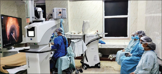
The COVID-19 pandemic has marked a global shift in all aspects of our lives, with a significant impact on the way we practise medicine. As we adjust to a new normal, the conventional operating and teaching methods may need to be modified in order to minimize person-to-person contact and maintain the social distancing norms. 3D Systems with Heads-Up Display have the potential to be excellent teaching tools in the future, with the surgical assistant and students able to view the steps of surgery while maintaining adequate inter-personal distance [Fig. 1]. The 3D visualization enhances the understanding of surgical dynamics by the residents and fellows and promotes interaction with the assisting staff as well. Two-dimensional display systems such as video-output monitors and Callisto heads up display are commonly used for resident training and teaching which provide a limited understanding of the surgical steps. In contrast, 3D heads up display provide an invaluable insight into the surgical process from the operating surgeon's viewpoint and is a more interactive and comprehensive teaching tool.
Figure 1.

3D Systems with Heads-Up Display as an excellent teaching tools, with the surgical assistant and students able to view the steps of surgery while maintaining adequate inter-personal distance
Aerosol generation during phacoemulsification has also been a topic of debate in the COVID-19 era placing the surgeon and assistants at a potential risk of infection.[10] Safety face-shields and goggles have been advocated for operating surgeons as additional protective measures; however, they hamper visualization through the oculars of traditional operating microscopes. Heads-Up Display decreases the proximity between the surgeon and patient without adversely affecting the surgical precision. Additional goggles or face shields may not be mandatory over the polarized glasses as adequate distance is maintained from the surgical field.
Limitations
One of the major benefits of 3D Heads up display system is related to the enhanced ergonomics which allows the surgeon to sit-back on the operating chair during surgery. A reduction in the back strain is well-established; however, the system is associated with unique ergonomic challenges related to the positioning of screen. Most phacoemulsification surgeons operate temporally, with a frequent shifting of position of the surgeon, microscope and display screen required with the change of patient's eye to be operated. It is not feasible to have the viewing screen in an exactly straight-ahead position relative to the operating surgeon, as the viewing system of the operating microscope will impede visualization of the screen. The neck tilt required to compensate for this angulation induces an additional ergonomic strain which may be evident in the long-term and requires further evaluation. Present-day conventional microscopes allow the surgeon to tilt and adjust the oculars in order to assume a comfortable posture while operating without inducing any additional neck strain, and the tilt associated with heads up display may be a step backward rather than forward. A steady head position is maintained throughout surgery in the conventional system unlike a heads-up display.
Moreover, the angulation between the screen and the surgeon is acceptable with left eye surgeries as the microscope arm is towards the right-hand side. Right eye surgeries are relatively more cumbersome with difficulties in optimally adjusting the screen, surgeon position and microscope arm while minimizing the tilt between the surgeon and screen. The impact of the tilt on depth perception and image quality has not been adequately analysed. In addition, there is a learning curve to adjust to instrument handling with respect to either eye.
The assisting surgeon is conventionally seated perpendicular to the operating surgeon. The significant angulation between the assistant and the viewing screen may result in additional back and neck strain and the image quality may be suboptimal. The operating surgeon has the advantage of proprioception as the surgeon's hands rest on the patient's head, providing tactile clues during surgery. The assisting surgeon does not have this advantage, and even simple tasks such as putting a viscoelastic on the ocular surface or cutting the sutures may be extremely tedious while viewing a heads-up display screen. The learning curve for an assistant may in fact be more steep as compared with the operating surgeon. Further studies are required to evaluate if there is any significant difference in adapting to the 3D visualization systems between the experienced and novice surgeons.
Maintenance of an optimal viewing distance and adequate centration are prerequisites for heads-up display systems. We have observed a significant deterioration in image quality and depth perception with even slight decentration of the field of view. Phacoemulsification, unlike posterior segment surgeries, is often performed under topical anaesthesia. Eye movements and drifting of the head sideways are more frequently encountered during anterior segment surgeries and require frequent re-centration of the microscope. Frequent adjustment of fine focussing is also required during surgical steps performed at different depths. In our experience, 3D Heads Up Display System is slightly less tolerant to decentration and defocussing as compared with traditional operating microscopes.
The scrub nurses involved in anterior segment surgeries handle the surgical instruments, adjust the phacoemulsification settings, change the phacoemulsification and irrigation-aspiration handpieces as required and load the intraocular lens (IOL) in the cartridge and injector. Conventionally, the lights in the OR are switched off during surgery to provide optimal resolution and enhanced visualization. Wearing polarized glasses in a dark room may interfere with optimal functioning of the assisting nurses, especially while handling the IOL. In addition, lights in the OR may need to be switched on during IOL loading. It is not feasible for the nursing staff to remove the polarized glasses in between the surgery owing to sterility concerns, and they often do not wear the glasses throughout surgery to avoid any inadvertent delays in instrument handling from their end. This leads to a decreased involvement of the surgical staff as they are less likely to anticipate the surgical steps and requirement for instruments.
A time-lag of up to 80-90 milliseconds has been described with the Ngenuity and TrueVision systems, which is more pronounced in anterior segment surgeries owing to the faster surgical steps. An inadvertent capsulorhexis extension or posterior capsular rupture may be observed due to the lag, especially by surgeons in the initial learning curve. With increasing surgeon experience, the time lag is barely appreciable and does not adversely affect the surgical procedure and outcomes. The ARTEVO 800 has a time lag of less than 50 milliseconds, which is below the detection threshold. We did not appreciate a significant time-lag with the ARTEVO 800 system.
The prohibitive cost of these systems may be an impediment especially for standalone practitioners, as an additional investment may not be feasible in view of no surgical outcome related advantages. The cost-benefit ratio is favourable in teaching institutes keeping in view the advantages of a heads-up display system as a training and teaching tool.
Conclusion
Advancements in ophthalmology are intricately linked to technological developments in all aspects, right from advanced diagnostic modalities, innovative surgical instruments to enhanced operating microscopes. 3D visualization systems with heads up display are associated with enhanced ergonomics and surgeon comfort, better visualization and increased depth of imaging without adversely impacting the surgical duration or outcomes in both anterior and posterior segment surgeries. They have the potential to emerge as an excellent teaching and training tool for residents and fellows. In addition, 3D visualization with heads-up display may be an integral part of the new normal in the post-COVID-19 era with its unique ability to allow for adequate distancing from the patient as well as staff without affecting the surgical precision or resident training. Continual developments in this field may help to overcome the limitations associated with present-day 3D visualization systems and provide a seamless surgical experience for the surgical team as well as the patient.
About the author

Jeevan S Titiyal
Prof. Jeewan S Titiyal heads the Cornea, Cataract and Refractive Surgery Services at RP Centre, AIIMS, New Delhi. He is the Chairman of the National Eye Bank.
He has been conferred with Padma Shri for his outstanding contribution in the field of Medicine (Ophthalmology) by the President of India in 2014. He has also been awarded the AAO Achievement (2009), AAO Senior Achievement (2016), and the APAO Achievement (2015) awards and is the APACRS Certified Educator (2015). He has been honored with over 30 orations and keynote addresses. He has been the 'Teacher of Teachers' and has performed over 100 live surgical demonstrations and has delivered over 1000 lectures and talks. He is the first Indian to perform live surgery in ASCRS, USA. He has been regularly conducting instruction courses at AAO, ASCRS, ESCRS, APAO, APACRS and AIOS. He has two patents to his credit for his innovations.
Prof Titiyal has to his credit more than 250 indexed publications with over 3365 citations. He has co-authored four textbooks in his field of expertise and has written over 52 book chapters. He has completed over 30 funded international and national research projects.
References
- 1.Gorn RA. Ophthalmic microsurgery: Instrumentation, microscopes, technique. Arch Ophthalmol. 1987;105:759. [Google Scholar]
- 2.Moura-Coelho N, Henriques J, Nascimento J, Dutra-Medeiros M. Three-dimensional display systems in ophthalmic surgery – A review. Eur Ophth Rev. 2019;13:31–6. [Google Scholar]
- 3.Martínez-Toldos JJ, Fernández-Martínez C, Navarro-Navarro A. Experience using a 3D head-mounted display system in ophthalmic surgery. Retina. 2017;37:1419–21. doi: 10.1097/IAE.0000000000001664. [DOI] [PubMed] [Google Scholar]
- 4.Weinstock RJ. Boston: 2010. First clinical use of on-screen 3D image guidance templates during small-incision cataract surgery Paper presented at: American Society of Cataract and Refractive Surgery Annual Meeting. [Google Scholar]
- 5.Weinstock RJ, Diakonis VF, Schwartz AJ, Weinstock AJ. Heads-up cataract surgery: Complication rates, surgical duration, and comparison with traditional microscopes. J Refract Surg. 2019;35:318–22. doi: 10.3928/1081597X-20190410-02. [DOI] [PubMed] [Google Scholar]
- 6.Qian Z, Wang H, Fan H, Lin D, Li W. Three-dimensional digital visualization of phacoemulsification and intraocular lens implantation. Indian J Ophthalmol. 2019;67:341–3. doi: 10.4103/ijo.IJO_1012_18. [DOI] [PMC free article] [PubMed] [Google Scholar]
- 7.Mohamed YH, Uematsu M, Inoue D, Kitaoka T. First experience of nDSAEK with heads-up surgery: A case report [published correction appears in Medicine (Baltimore) 2018;97:e12287] Medicine (Baltimore) 2017;96:e6906. doi: 10.1097/MD.0000000000006906. [DOI] [PMC free article] [PubMed] [Google Scholar]
- 8.Galvis V, Berrospi RD, Arias JD, Tello A, Bernal JC. Heads up Descemet membrane endothelial keratoplasty performed using a 3D visualization system. J Surg Case Rep. 2017;2017:rjx231. doi: 10.1093/jscr/rjx231. [DOI] [PMC free article] [PubMed] [Google Scholar]
- 9.Hamasaki I, Shibata K, Shimizu T, Kono R, Morizane Y, Shiraga F, et al. Lights-out surgery for strabismus using a heads-up 3D vision system. Acta Med Okayama. 2019;73:229–33. doi: 10.18926/AMO/56865. [DOI] [PubMed] [Google Scholar]
- 10.Koshy ZR, Dickie D. Aerosol generation from high speed ophthalmic instrumentation and the risk of contamination from SARS COVID19. Eye (Lond) 2020 doi: 10.1038/s41433-020-1000-3. doi:https://doi.org/10.1038/s41433-020-1000-3. [DOI] [PMC free article] [PubMed] [Google Scholar]


