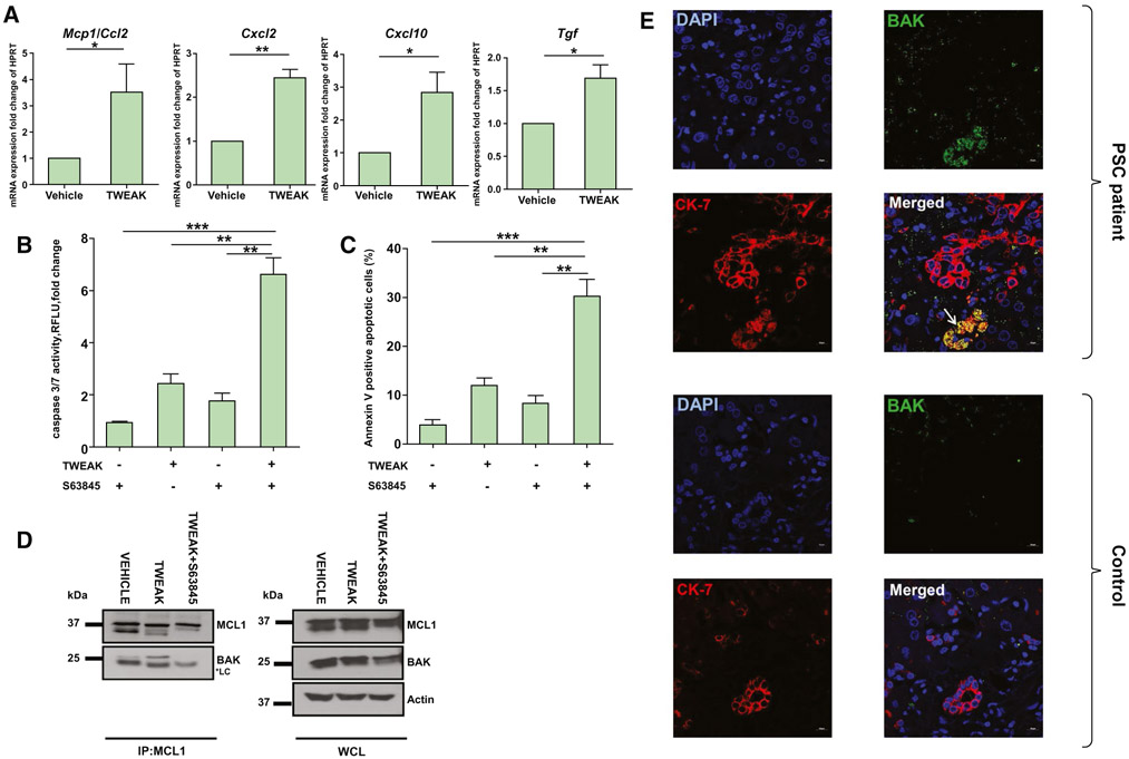
FIG. 3.
DR cells are primed for cell death by increased binding of BAK to MCL1. (A) 603B cholangiocytes were incubated with TWEAK (200 ng/mL) for 48 hours, and expression of proinflammatory genes was analyzed by quantitative real-time PCR (n = 3). (B,C) 603B cholangiocytes were incubated with TWEAK (200 ng/mL) for 48 hours, then S63845 (10 μM) for an additional 24 hours; and apoptosis was assessed by (B) caspase 3/7 activity and (C) annexin V positivity (n = 3). (D) 603B cholangiocytes were incubated with TWEAK (200 ng/mL) for 48 hours or TWEAK (200 ng/mL) for 48 hours followed by S63845 (10 μM) for an additional 24 hours. MCL1 was immunoprecipitated, and association with BAK was assessed by immunoblot. Actin was used as loading control. (E) Representative images of CK-7 (red) and activated BAK (green) coimmunofluorescence (yellow) on stage 3 PSC (upper panel) and healthy (control) human liver specimens (lower panel). White arrow points to location of BAK+CK7+ cells. 40x lens, 1.5 magnification. Scale bars, 10 μm. Data are expressed as mean ± SEM. *P < 0.05, **P < 0.01, ***P < 0.005. Abbreviations: DAPI, 4′,6-diamidino-2-phenylindole; HPRT, hypoxanthine-guanine phosphoribosyltransferase; IP, immunoprecipitation; Mcp1, monocyte chemoattractant protein 1; RFLU, relative fluorescence units; WCL, whole-cell lysate.

