eLife logoLink to eLife
. 2020 Dec 17;9:e63430. doi: 10.7554/eLife.63430

Post-acute COVID-19 associated with evidence of bystander T-cell activation and a recurring antibiotic-resistant bacterial pneumonia

Michaela Gregorova 1,, Daniel Morse 1,, Tarcisio Brignoli 1,, Joseph Steventon 1, Fergus Hamilton 2, Mahableshwar Albur 2, David Arnold 2, Matthew Thomas 2, Alice Halliday 1, Holly Baum 1, Christopher Rice 1, Matthew B Avison 1, Andrew D Davidson 1, Marianna Santopaolo 1, Elizabeth Oliver 1, Anu Goenka 1, Adam Finn 1, Linda Wooldridge 3, Borko Amulic 1, Rosemary J Boyton 4,5, Daniel M Altmann 4, David K Butler 4, Claire McMurray 6, Joanna Stockton 6, Sam Nicholls 6, Charles Cooper 6, Nicholas Loman 6, Michael J Cox 5, Laura Rivino 1,‡,, Ruth C Massey 1,‡,
Editors: Anurag Agrawal7, Mone Zaidi8
PMCID: PMC7775105  PMID: 33331820

Abstract

Here, we describe the case of a COVID-19 patient who developed recurring ventilator-associated pneumonia caused by Pseudomonas aeruginosa that acquired increasing levels of antimicrobial resistance (AMR) in response to treatment. Metagenomic analysis revealed the AMR genotype, while immunological analysis revealed massive and escalating levels of T-cell activation. These were both SARS-CoV-2 and P. aeruginosa specific, and bystander activated, which may have contributed to this patient’s persistent symptoms and radiological changes.

Research organism: Human

Introduction

The COVID-19 pandemic has brought with it the largest ever cohort of patients requiring mechanical ventilation. Complications associated with such severe viral infections are many-fold, and include increased susceptibility to secondary bacterial infections (Zhou et al., 2020; Langford et al., 2020), as well as post-acute COVID-19, where patients experience symptoms extending beyond 3 weeks from the onset of their first COVID-19 symptoms (Greenhalgh et al., 2020). The first report of secondary infections in COVID-19 patients was from Wuhan in March 2020, where 15% of hospitalized patients developed secondary infections, and of those who did not survive their SARS-CoV-2 infection, 50% had a secondary bacterial infection (Zhou et al., 2020). Since then many COVID-19 studies reporting secondary infections have been published, with a recent meta-analysis of 24 independent studies that included 3338 patients from five countries reporting that 14.3% of hospitalized COVID-19 patients developed a secondary bacterial infection, which is associated with significant morbidity, mortality and the financial costs associated with prolonged hospitilisation (Langford et al., 2020). The incidence of post-acute COVID-19 varies depending on the group of patients considered, with approximately 10% of patients who have tested positive for SARS-CoV-2 virus remaining unwell beyond 3 weeks (Greenhalgh et al., 2020). However, this can be as high as 74% when hospitalised patients are considered, where symptoms include breathlessness and excessive fatigue, with abnormal radiological features reported in 12% of this cohort (Arnold et al., 2020).

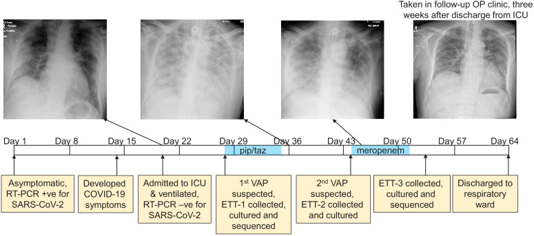
The DISCOVER study (DIagnostic and Severity markers of COVID-19 to Enable Rapid triage, REC: 20/YH/0121) was established in March 2020 to collect and analyse longitudinal samples from COVID-19 patients. One study participant, an otherwise healthy male between 45 and 55 years of age presented to hospital with Type-1 respiratory failure (hypoxaemia), 20 days after he tested positive for SARS-CoV-2 by RT-PCR. At the time of testing he was asymptomatic (tested as a house-hold contact of a health-care worker), and this represents Day one on the time-line presented in Figure 1. He became symptomatic for COVID-19 13 days after this, which is within the incubation period for SARS-CoV-2 as defined by the CDC of 14 days (COVID-19 (Coronavirus Disease), 2020), and his health declined over the following week. Upon admission to hospital a chest X-ray was taken, and he was admitted to the ICU where he was mechanically ventilated (Figure 1). An RT-PCR test on an endotracheal sample collected at this time did not detect SARS-CoV-2 suggesting that he had cleared the viral infection.

Figure 1. The development of a recurring ventilator-associated pneumonia (VAP) by a COVID-19 patient.

A clinical time-line is presented from the point at which the patient tested positive for SARS-CoV-2 (day 1), through to his discharge from the ICU (day 64). Noteworthy clinical features are indicated in the yellow boxes below the time-line. The three x-rays taken during the patient’s time in the ICU are presented, as is a later follow-up x-ray taken in an out-patient (OP) clinic 3 weeks after discharge. The time during which the antibiotics piperacillin and tazobactam (pip/taz) and meropenem were administered to the patient to treat the VAP are indicated in blue. The points at which endotracheal tube aspirates (ETT) were collected and the subsequent analysis of these also are indicated and described.

Figure 1.

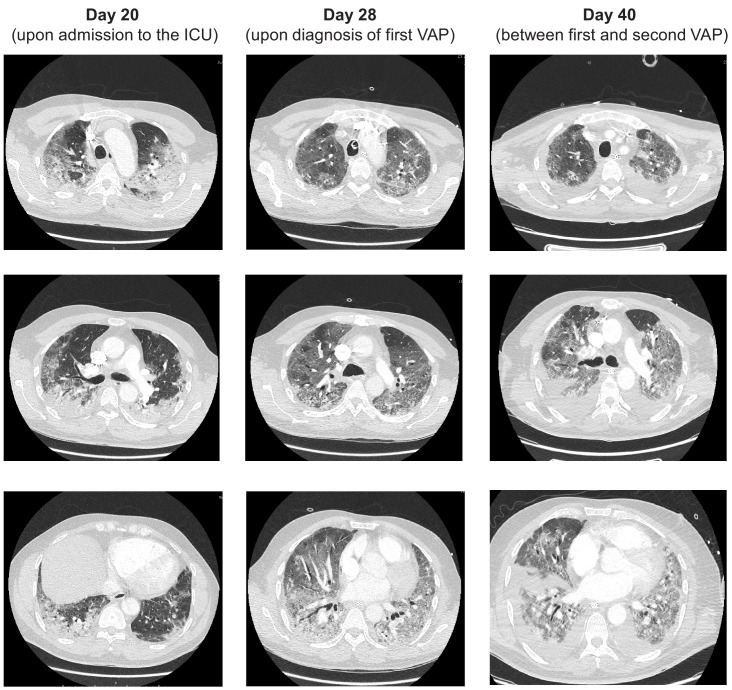
Figure 1—figure supplement 1. CT scans of the patient at three time points during his stay in the ICU, Days 20, 28 and 40.

Figure 1—figure supplement 1.

Features associated with COVID-19 including ground glass opacities are evident throughout. Three separate axial slices from each time point are provided.

Once admitted to our intensive care unit he was assessed, as all our patients are daily, for signs of additional infection through a process led by a consultant medical microbiologist along with senior pharmacist. At these meetings, the clinical status of the patient, radiological features, oxygen requirements, inflammatory indices (WCC, CRP, and procalcitonin) and any culture results are discussed. We had no concerns at the time of admission of this patient to the ICU that he had any infection other than that of SARS-CoV-2. Computerised tomography (CT) scans of the patient were collected through his time in the ICU and the classic features of COVID-19, including ground-glass opacities, were evident throughout (Figure 3—figure supplement 1).

After a week in the ICU he was diagnosed with ventilator-associated pneumonia (VAP) based on the clinical, radiological, biochemical, and microbiological parameters described above. An antibiotic susceptible Pseudomonas aeruginosa strain was cultured from an aspirate collected from his endotracheal tube (ETT-1, Figure 1), (minimum inhibitory concentration (MIC) meropenem 0.5 mg/l; piperacillin-tazobactam (pip/taz) 8 mg/l). He was prescribed a seven-day course of pip/taz (4.5 g every 6 hr), and clinically recovered from this bacterial infection. Eight days after finishing this first course of antibiotics his VAP recurred (diagnosed as described above), and a pip/taz resistant P. aeruginosa was cultured from ETT-2 (minimum inhibitory concentration (MIC) >16 mg/l). He was prescribed a seven-day course of meropenem (1 g every 8 hr) and showed signs of clinical improvement. There were no further concerns about a bacterial infection during his ICU stay (i.e. he did not meet any of the clinical diagnostic criteria described above), although a third P. aeruginosa was cultured from a sample (ETT-3) collected two days after he completed his course of meropenem that was resistant to both pip/taz and meropenem (MICs > 16 mg/l and >8 mg/l respectively). His health continued to improve, and he was discharged to the respiratory ward a week later. The patient attended a follow-up clinic 3 weeks after hospital discharge where he reported on-going symptoms such as breathlessness and myalgia, and there was a significant drop in his oxygen saturations after mild exertion to 84%. Eight months following his positive test he is still reporting breathlessness and fatigue, common symptoms of the newly defined ‘Long COVID’ condition.

To understand the cellular and molecular dynamics of this post-acute COVID-19 case from the perspective of both the pathogen and patient’s immune response, longitudinal respiratory and blood samples were collected and analysed with a view to identifying early diagnostic biomarkers of infection onset and potential opportunities for immunotherapeutic intervention.

Results

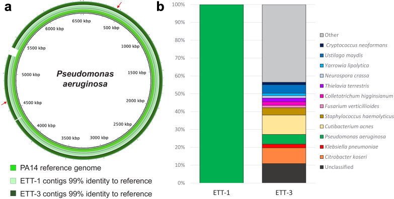
Metagenomics was used to characterise in depth the composition and genomic features of the bacteria present in this patient’s lower respiratory tract from when he first developed VAP (ETT-1, Figure 1) to when he had recovered from his second VAP (ETT-3, Figure 1). ETT-2 collected during his second bout of VAP was unfortunately not available for sequencing. We extracted the entire genetic material from 500 μl of ETTs 1 and 3 with no bacterial enrichment or human DNA depletion steps, and to ensure data for the bacterial component of the samples was generated, these were sequenced on a PromethION (Oxford Nanopore technology). The bacterial DNA from ETT-1 was entirely that of P. aeruginosa corresponding to multi-locus sequence type ST253 (Jolley et al., 2018), a world-wide clone frequently associated with AMR epidemics (Treepong et al., 2018; Figure 2). There was a greater diversity of bacterial DNA in ETT-3, as the patient recovered from his SARS-CoV-2 and P. aeruginosa infections (Figure 2b), and we were able to construct a whole genome of the P. aeruginosa strain from within this, which again corresponded to multi-locus sequence type ST253. P. aeruginosa is a ubiquitous environmental bacterium, and while it can be found as a commensal in the respiratory tract of some individuals, this is typically only when they have some underlying chronic suppurative lung disease such as bronchiectasis. Prior to his COVID-19 diagnosis this relatively young patient was healthy with no prior medical issues, which suggests the P. aeruginosa infections were hospital or ICU acquired.

Figure 2. Direct metagenomic analysis of respiratory samples from a COVID-19 patient who developed a recurring VAP.

Figure 2.

(a) The sequence data were aligned to the genome of a P. aeruginosa reference strain PA14 (inner ring). The P. aeruginosa genomes from both ETT-1 (middle ring) and ETT-3 (outer ring) had 99% identify to the PA14 reference genome and greater than 99% to each other. The position of the AMR conferring SNPs in the P. aeruginosa from ETT-3 are indicated by red arrows. (b) Assembled contigs were taxonomically classified using Kraken two with % of total contigs assigned to each species shown for ETT-1 and ETT-3. Species with only one contig (0.68% of total contigs) were grouped as ‘other’.

Resistance to beta-lactam antibiotics by P. aeruginosa can be multifactorial and includes the acquisition of single nucleotide polymorphisms (SNPs) in efflux and porin genes that affect the passage of the antibiotic into and out of the bacterial cell (Blair et al., 2015). Analysis of the sequence data from ETT-3 generated a whole genome for P. aeruginosa, again corresponding to ST253, that aligned with >99% identify to the genome from ETT-1. There were however two noteworthy SNPs in this later sample that explain the increased AMR of this isolate. The multi-drug efflux pump, MexAB, is transcriptionally regulated by the translated product of the mexR gene, MexR. We found a SNP in mexR that converts a key amino-acid (Arg91-Gln) in the DNA-binding domain of the MexR protein, that would likely result in the de-repression of this efflux system (Saito et al., 2003). A second SNP was found that introduced a premature stop codon (Tyr120-stop) in the gene encoding the outer membrane protein OprD, a protein with a well-established role in the entry of meropenem into the bacterial cell (Quale et al., 2006). The two P. aeruginosa strains could represent a parent strain that acquired resistance upon exposure to antibiotics, that wasn’t fully cleared and so re-emerged; or a new infection or colonisation event with a closely related AMR strain circulating within the ICU. Unfortunately, we are unable to tell which of these scenario is more likely from these data.

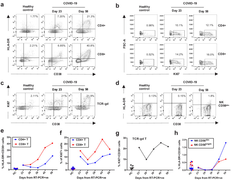
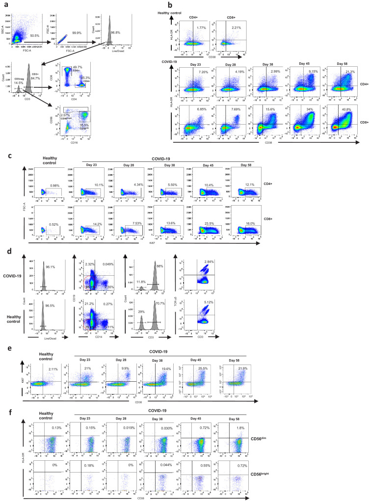
Having recovered from both a viral and a recurring bacterial infection, we also sought to analyse the kinetics of the patient’s innate and adaptive immune response across his time in the ICU (at days 23, 28, 38, 45, and 58 from testing positive for SARS-CoV2, Figure 1), as previous work has demonstrated striking immunological features associated with COVID-19 patients (Mathew et al., 2020). Robust activation of a broad range of immune cell subsets was revealed by flow cytometry, when compared to healthy controls. What was particularly striking was the level of activation and proliferation of CD4+, CD8+, and TCR-γδ T-cells, that appeared to wane between days 23–28 but were subsequently boosted after day 28, concomitant with the onset of the secondary bacterial infection (Figure 3a–c and Figure 3e–g, gating strategies in Figure 3—figure supplement 1). Similar levels of activation of T-cells has been reported for subsets of COVID-19 patients in other studies (Mathew et al., 2020). From day 28 a robust and steady increase in the activation and proliferation of conventional and TCR-γδ-T-cells was evident with approximately 20% and 40% of total CD4+ and CD8+ T-cells co-expressing the activation markers HLA-DR and CD38, respectively by day 58. A similar steady increase in activation levels, albeit at lower magnitudes, was observed for Natural Killer (NK) CD56dim and CD56bright cells (Figure 3d and h, gating strategies in Figure 3—figure supplement 1). Similar perturbations in the frequency and activation phenotypes of monocytes, blood monocyte-derived macrophages as well as neutrophils could be detected from day 28 onwards (Figure 3—figure supplement 2a–e). A robust IgG response to both SARS-CoV-2 and P. aeruginosa antigen was also detected in this patient (Figure 3—figure supplement 3).

Figure 3. Kinetics of the adaptive and innate immune response in a COVID-19 patient during ICU treatment.

(a–d) Representative flow cytometry plots showing the expression of activation and proliferation markers on CD4+ and CD8+ T cells (top and bottom panels, respectively in a, b), TCR-γδ T cells (c) and NK cells (d: plots shown for NK CD56dim cells) in a healthy control and the COVID-19 patient (at days 23 and 58). (e-f) Immune activation and proliferation levels are summarized for healthy controls (HC, n = 7) and five longitudinal time points of the COVID-19 patient and are assessed as co-expression of HLA-DR and CD38 (e) and expression of Ki67 (f) in CD4+ and CD8+ T cells, co-expression of CD38 and Ki67 by TCR-γδ T cells (g), and co-expression of HLA-DR and CD38 in NK CD56dim cells (h). Gating strategies for each population are included in Figure 3—figure supplement 1. All data is obtained by flow cytometry and samples were acquired on a Becton Dickinson LSR Fortessa X-20.

Figure 3.

Figure 3—figure supplement 1. The gating strategies and flow cytometry plots are shown for each immune population.

Figure 3—figure supplement 1.

(a) T and NK cell populations; (b–c) T cell activation and proliferation markers; (d) TCR-γδ T cells; (e) TCR-γδ activation and proliferation markers; (f) NK CD56dim and CD56bright cell activation markers.

Figure 3—figure supplement 2. Kinetics of the innate immune response in the COVID-19 patient.

Figure 3—figure supplement 2.

(a) Percentages of CD14+ CD16- classical monocytes (cMo), CD14+ CD16+ intermediate monocytes (iMo) and CD14- CD16+ non classical monocyes (ncMo). Percentages are calculated in live, CD3-, CD66b-, HLA-DR+ cells in the COVID-19 patient and in healthy controls (n = 6). (b) Percentages of blood monocyte-derived M1 (CD14+ CD80+ CD68+) and M2 (CD14+ CD163+ CD68+) macrophages are shown for the COVID-19 patient and healthy controls. (c–e) Neutrophil degranulation measured as MFI of CD11b (secretory vesicles), CD66b (secondary granules) and CD63 (primary granules) is shown for the patient and healthy controls (n = 4). Neutrophils were immunostained in whole blood, followed by erythrocyte lysis with ACK buffer and fixation with 4% PFA prior for analysis by flow cytometry. Blood monocytes and monocyte-derived macrophages were stained from PBMCs isolated by Ficoll gradient as described in the methods section.

Figure 3—figure supplement 3. Antibody response towards Pseudomonas aeruginosa and SARS-CoV-2.

Figure 3—figure supplement 3.

(a) Serum IgG response to Pseudomonas aeruginosa (PA) protein outer membrane porin F (OprF) was assessed in healthy controls (n = 2), in the COVID-19 patient (n = 1) and in patients with diagnosis of Cystic Fibrosis and PA infection in the lung (PA patient, n = 2). Data are presented as mean ± SD. (b) Plasma IgG responses to SARS-CoV-2 Spike and N were assessed in the COVID-19 patient at five timepoints. For reference we also show levels from serum of healthy control donors (HC; n = 5), healthy pre-pandemic control donors (PP; n = 6) and a pooled serum (PS; n = 3) control per plate (collected from 3 RT-PCR-confirmed convalescent donors). The optical density (OD) at 492 nm was corrected for background (620 nm) and for the blank values. Data are presented as mean ± SD.

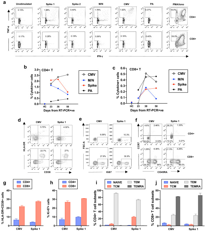
Given the scale of the cellular response of this patient, we investigated whether the large expansions of CD4+ and CD8+ T-cells were due to a T-cell response targeting SARS-CoV-2, the secondary bacterial infection or to bystander T-cell activation (Sandalova et al., 2010; Rivino et al., 2015). We included the analysis of bystander activation, which is T-cell receptor-independent and cytokine-mediated, as it is known to occur during other types of acute viral infection (Sandalova et al., 2010; Rivino et al., 2015). The role of this type of bystander activated T-cells in the recovery of patients remains largely unclear, as these cells can participate in protective immunity towards the virus but can also contribute to tissue damage (Sandalova et al., 2010; Rivino et al., 2015; Kim and Shin, 2019; Maini et al., 2000). We performed a brief stimulation of the patient’s peripheral blood mononuclear cells (PBMCs), with or without overlapping peptides spanning the sequences of SARS-CoV-2 proteins (spike, membrane (M) and nucleoprotein (N)); of an immunodominant P. aeruginosa antigen (OprF); as well as an immunodominant human Cytomegalovirus protein (HCMV pp65) as an indication of non-T-cell receptor (TCR) driven bystander activation. This was followed by intracellular cytokine staining to detect production of IFN-γ and TNF-α by the specific T cells. Following the encounter with specific peptides we observed a robust CD4+ T-cell response to SARS-CoV-2 which decreased from day 23 to day 58, and a more modest SARS-CoV-2-specific CD8+ T-cell response. This is in line with published work, suggesting higher magnitudes of SARS-CoV-2-specific CD4+ versus CD8+ T-cells in severe COVID-19 patients (Peng et al., 2020), as well as consistent detection of virus-specific CD4+ T cells in recovered patients (Grifoni et al., 2020; Figure 4a–c and Figure 4—figure supplement 1a–b). CD4+ and CD8+ T-cell responses targeting P. aeruginosa peptides could also be detected, and increased over time concomitant with the onset of the recurring bacterial infection (Figure 4b–c).

Figure 4. Frequency and phenotype of antigen-specific T cells.

(a) Representative flow cytometry plots showing IFN-γ and TNF-α production by CD4+ and CD8+ T cells from the COVID-19 patient (day 23) as assessed by intracellular cytokine staining after a brief stimulation of PBMCs with HCMV pp65 (CMV), SARS-CoV-2 membrane, nucleoprotein (M/N respectively), spike 1, spike 2, and Pseudomonas aeruginosa OprF (PA) peptides. PMA/ionomycin and unstimulated conditions represent positive and negative controls. (b-c) Summarized are T-cell responses shown as % of IFN-γ and/or TNF-α+ cells in CD4+ or CD8+ T cells after subtraction of the negative controls in the COVID-19 patient at three longitudinal time points and in an age-matched healthy control (spike and M/N). (d-f) Representative flow cytometry plots showing T-cell expression of activation, proliferation and memory markers in CMV-specific and spike1-specific T cells in the COVID-19 patient (day 38), as assessed after a brief stimulation of PBMCs with CMV or spike 1 peptides followed by staining with antibodies targeting the relevant markers. Plots are gated on cytokine-producing cells (TNF-α and/or IFN-γ) in CD4+ or CD8+ T cells (top and bottom panels, respectively). (g-h) T-cell activation and proliferation assessed as co-expression of HLA-DR and CD38, and expression of Ki67 are shown for CD4+ and CD8+ CMV and spike 1-specific T cells (g and h, respectively). (i-j) Percentage of CMV and spike 1-specific T cells contained within each subset identified by expression of CCR7 and CD45RA is shown for CD4+ and CD8+ T cells (i and j, respectively). Naïve: CCR7+CD45RA+; central memory (TCM): CCR7+CD45RA-; T effector memory (TEM):CCR7-CD45RA- and T effector memory RA re-expressing (TEMRA): CCR7-CD45RA+. Columns represent mean +/- SD of the experimental duplicates. All data is obtained by flow cytometry and samples were acquired on a Becton Dickinson LSR Fortessa X-20.

Figure 4.

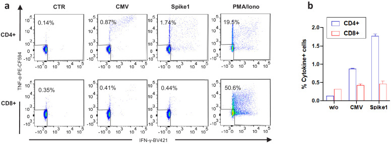
Figure 4—figure supplement 1. Experimental duplicates of intracellular cytokine staining data.

Figure 4—figure supplement 1.

(a) Representative flow cytometry plots showing IFN-γ and TNF-α production by CD4+ and CD8+ T cells from the COVID-19 patient (day 38) as assessed by intracellular cytokine staining after a brief stimulation of PBMCs with or without HCMV pp65 (CMV), SARS-CoV-2 spike 1 and PMA/ionomycin as a positive control. All conditions except for PMA/ionomycin were performed in duplicate wells. (b) Graph showing the average and standard deviation values for the duplicate wells in experiment shown in a. % Cytokine positive cells refers to the % of IFN-γ and/or TNF-α production by CD4+ or CD8+ T cells following stimulation with or without the indicated peptides.

The number of T-cells responding specifically to SARS-CoV-2 and P. aeruginosa represent only a fraction of those activated, which suggests that many of those detected in the patient’s blood may be bystander activated. In support of this we detected an increased magnitude of CMV-specific T-cell responses over time, particularly in the CD8+ compartment (Figure 3b–c). We therefore considered whether CMV-specific T-cells were activated and proliferating in the blood of the COVID-19 patient. To test this we performed a brief stimulation of PBMCs with or without HCMV pp65 peptide (CMV) or SARS-CoV-2 spike 1 one peptide pools (the predominant SARS-CoV-2 target in this patient, within those tested) or with a positive control, and assessed the expression of the activation and proliferation markers HLA-DR, CD38 and Ki67 by the cytokine-producing cells that were responding to the peptide pools. Peptide stimulations and negative controls were performed in duplicate wells (Figure 4—figure supplement 1a–b). Both CMV and spike-1-specific CD8+ T-cells showed increased co-expression of HLA-DR and CD38 and of Ki67, indicating that these cells were activated and proliferating ex vivo, while this was less evident for their CD4+ T-cell counterparts (Figure 4d–e and g–h). This observation is in line with previous reports of bystander T-cell activation for CMV-specific CD8+ T-cell populations (Sandalova et al., 2010; Rivino et al., 2015). We furthermore analysed the phenotype of CMV and spike 1-specific T-cells on the basis of expression of CCR7 and CD45RA, which identifies naïve (CCR7+ CD45RA+), central memory (TCM: CCR7+ CD45RA-), effector memory (TEM: CCR7- CD45RA-) and effector memory re-expressing RA T-cells (TEMRA: CCR7-CD45RA+). CMV and spike 1-specific CD4+ T cells were mainly contained respectively within the TEM and the TEM/TCM subsets, while CMV and spike 1-specific CD8+ T cells were mainly contained respectively within the TEMRA and the TEM subsets. The predominant TEMRA phenotype of SARS-CoV-2 specific CD8+ T-cells in convalescent COVID patients is in line with recent work (Neidleman et al., 2020). The presence of a large proportion of CD8+ TEMRA within the CMV and spike 1-specific CD8+ T cell populations also supports the notion that these cells are stimulated through cytokine receptors rather than through the TCR, as TEMRA cells are believed to arise following antigen withdrawal and cytokine-mediated stimulation (for e.g. IL-15), as TCR ligation downregulates CD45RA expression both in vitro and during responses in vivo (Henson et al., 2012).

In summary, the immune profiles show robust innate and adaptive immune activation in a COVID-19 patient experiencing a secondary bacterial infection, and a detectable T-cell and IgG response targeting both SARS-CoV-2 and P. aeruginosa. Furthermore, we show that CD8+ T-cells targeting viral antigens from SARS-CoV-2 and from the persistent virus HCMV display expression of markers of activation and proliferation and are predominantly TEMRA and TEM cells. Although it is a challenging task to demonstrate that these cells have not encountered their cognate viral antigens in vivo, the TEMRA phenotype of these cells makes it unlikely and suggests that these cells may be proliferating in a bystander way through the action of cytokines present in the inflammatory milieu.

Discussion

In this case report we demonstrate the potential of the application of multidisciplinary technologies to longitudinally-collected patient samples to define the complex dynamics of patient-pathogen-therapy interactions in real-time. Metagenomics directly applied to respiratory samples facilitated the identification of the SNPs responsible for the AMR phenotype of the later P. aeruginosa isolate. However, the most striking feature of this COVID-19 case was the escalating number of circulating activated T-cells more than two months after testing positive for SARS-CoV-2, and 6 weeks after clearing the viral infection. While we could attribute some of this to the recurring bacterial infection, the scale of the activation considered alongside our evidence of increased frequencies of T-cells specific for unrelated antigens, suggests there may be a significant amount of bystander activation.

Studies on large COVID-19 patient cohorts have shown that the period of the peak of T-cell activation in COVID-19 is prolonged compared to other acute viral infections or after vaccination with live attenuated viruses (Mathew et al., 2020), which may be due to the inability of the immune system to downregulate its response. Here, we speculate that due to SARS-CoV2 infection the patient displays a heightened immune system, which was further stimulated by the recurring P. aeruginosa infections, which led to bystander activation of T cells specific for antigens unrelated to either SARS-CoV2 or P. aeruginosa. Bystander T-cell activation could have played a critical role in the severity of illness and the longer-term complications associated with the development of post-acute COVID-19 experienced by this patient (Grifoni et al., 2020). Given the recent appreciation for the role of corticosteroids in reducing the risk of death following SARS-CoV-2 infection by 20% (Sterne et al., 2020), this case suggests that targeting its use to patients with immunological responses as described here, may also improve their long-term recovery.

Materials and methods

Patient recruitment

The patient was enrolled onto the DISCOVER study (Diagnostic and Severity markers of COVID-19 to Enable Rapid triage study), a single centre prospective study recruiting consecutive patients admitted with COVID-19, from 30.03.2020 until present (Ethics approval via South Yorkshire REC: 20/YH/0121, CRN approval no: 45469). Blood/serum samples from pre-pandemic healthy controls and asymptomatic healthy controls were obtained under the Bristol Biobank (NHS Research Ethics Committee approval ref 14/WA/1253).

Clinical microbiology

All ETT samples were processed in the Severn Infection Services laboratory as per standard operating procedures. Antibiotic susceptibility testing was performed by either disk diffusion (according to EUCAST version nine guidance EUCAST, 2019) or on a VITEK two machine (BioMeriux, France).

SARS-CoV-2 test

SARS-CoV-2 test was performed by an in-house RT-PCR at the regional South West Public Health England Regional Virology laboratory, utilising a PHE approved assay at the time of testing.

DNA extractions

DNA from the endotrachaeal aspirates were extracted using the CTAB/Phenol:Chloroform:Isoamyl alcohol and bead-beating approach of Griffiths et al., 2000 with modifications of DeAngelis et al., 2009. Phase lock gel tubes (ThermoFisher) and linear polyacrylamide (Sigma) were included to increase nucleic acid yields and total DNA was resuspended in 50 µl of DNase/RNase free water before storage at −20°C.

DNA sequencing

Samples were prepared for sequencing using SQK-LSK109 kit (Oxford Nanopore) with 1 μg DNA starting input as per manufacture’s protocol. Briefly 1 μg of DNA was end repaired and a tailed using NEBNext Ultra II module E7546 (3.5 μl End Repair Buffer, 2 ul FFPE repair mix, 3.5 μl Ultra II end-prep reaction buffer and 3 μl of Ultra II end-prep enzyme mix to 1 μg DNA in a total of reaction volume of 60 μl). This was incubated at 20°C for 5 min followed by 65°C for 5 min. Clean-up was performed using AMPure XP beads (Beckman Coulter) in a 1X ratio. Adaptors were ligated by adding 5 μl adaptor mix (Oxford Nanopore) 25 μl ligation buffer (Oxford Nanopore) and 10 μl Quick T4 ligase (NEB Module MO202). Following a 20 min incubation at room temperature the adaptor ligated DNA was cleaned using AMPure beads in a 0.8 X ratio and washed using Long Fragment Buffer (Oxford Nanopore) before eluting in 25 μl of elution buffer (Oxford Nanopore). Final quantification by fluorometry (Qubit) was performed and 300 ng DNA prepared for sequencing according to the manufacturer’s instructions (Oxford Nanopore). Sequencing was performed on a PromethION R9.4.1 flow cell (FLO-PRO002) and run for 48 hr using live basecalling, files were outputted in Fast5 and Fastq format. Human-filtered sequencing data for this study have been deposited in the European Nucleotide Archive (ENA) at EMBL-EBI under accession PRJEB40239.

Bioinformatics

Bioinformatics was orchestrated using reticulatus (https://github.com/SamStudio8/reticulatus/ Köster and Rahmann, 2012) with configuration: Flye (v2.6) metagenomic assembly (Kolmogorov et al., 2019), Racon polishing (v1.4.9 + GPU, two rounds) (Vaser et al., 2017) and Medaka polishing (v0.8.0 + GPU, one round). Contigs were assigned to a taxon by Kraken 2 (Wood et al., 2019). SNP differences between the P. aeruginosa contigs for ETT-1 and ETT-3 was determined using Mauve (Darling et al., 2004). Contigs were loaded into BRIG (v0.95) (Alikhan et al., 2011) as concentric rings and compared against the PA14 or JCSC1435 reference genomes using blastn (ncbi-blast 2.10.1+) (Alikhan et al., 2011). MLST types were identified by mapping contigs against P. aeruginosa or S. haemolyticus according to the schemes held in the pubmlst databases (Arnold et al., 2020).

PBMC isolation

Blood samples were collected from the COVID-19 patient in EDTA vacutainer tubes and PBMCs isolated from peripheral blood by Ficoll gradient purification and cryopreserved. Healthy donor PBMCs were obtained from Bristol Biobank (REC: 14/WA/1253).

Synthetic peptides

15-mer peptides overlapping by 10 amino acids and spanning the sequences of SARS-CoV-2 spike (Accession Number: NC_045512.2, Protein ID: YP_009724390.1) and HCMV pp65 (AD169 strain) were purchased from Mimotopes (Australia). The purity of the peptides was >80% (Spike) or >70% (pp65) and peptides were dissolved as described previously (Rivino et al., 2015). SARS-CoV-2 M and N Peptivator peptide pools were purchased from Miltenyi. An OprF (PA1777) peptide library comprising 20-mer peptides overlapping by 10 amino acids was synthesized by GL Biochem Ltd., Shanghai, China (Quigley et al., 2015).

Flow cytometry staining and PBMC stimulation

PBMCs were thawed and either stained ex vivo or stimulated in AIMV 2% FCS with or without peptide pools from SARS-CoV2 spike, M, N, HCMV pp65 (all 1 μg/ml), OrpF PA (10 μg/ml) or with PMA/iono (PMA 10 ng/ml, Iono 100 ng/ml, Sigma Aldrich) for 5 hr at 37°C in the presence of brefeldin A (BD, 5 μg/ml). To assess degranulation, CD107a FITC antibody was added to the cells at the beginning of the stimulation. Cells were stained with a viability dye Zombie Aqua (Biolegend) for 10 min at room temperature and with antibodies targeting surface markers (20 min 4°C, diluted in PBS (HyClone) 1% BSA (Sigma Aldrich)). Cells were fixed for 45 min/overnight in eBioscience Foxp3/Transcription factor fixation/permeabilization buffer (Invitrogen) and intracellular staining was performed using eBioscience Foxp3/Transcription factor permeabilization buffer (Invitrogen) for Ki67 or intracellular cytokines (30 min on ice). Cells were acquired on a BD Fortessa X20 and data analysed using FlowJo software v10.7. A complete list of antibodies is included in Supplementary file 1.

Antibody quantification

a. Pseudomonas Aeruginosa: The PA antigen OprF was diluted in coating buffer, added to the wells of MaxiSorp flat-bottom 96-Well immunoplates (Thermo Fisher Scientific, USA) and incubated for 2 hr at 37°C. Immunoplates were blocked with 1% BSA/PBS for 1 hr at 37°C. Sera from each sample were diluted and added to the immunoplates in triplicate and incubated at 4°C overnight. Biotin mouse anti-human IgG (BD Pharmingen, USA) was added to the plate and incubated for 1 hr at RT. Streptavidin-HRP (R and D Systems, UK) was added and incubated for 30 mins at RT. ELISAs were developed using TMB substrate. Results were recorded using a Multiskan GO Microplate Spectrophotometer (Thermo Scientific, USA) and absorbance values (OD 450) were converted to ELISA units. OprF antigen was produced using the recombinant vector pSUMO-OprF as described previously (Quigley et al., 2015). b. ELISA methods were based on the protocols for measuring SARS-CoV-2 seroconversion (Amanat et al., 2020; Stadlbauer et al., 2020) with some modifications. A full-length trimeric stabilized version of the SARS-CoV-2 Spike protein was expressed in insect cells using the MultiBac baculovirus expression system and affinity purified, whereas the SARS-CoV-2 N protein was expressed in, and purified from, E. coli. High-binding plates (MaxiSorp, NUNC) were coated with either 10 mg/ml Spike or 20 mg/ml N protein in PBS and left overnight at 4°C followed by blocking for 1 hr (3% BSA/0.1% Tween/PBS). Dilution buffer (1% BSA/0.1% Tween/PBS) containing patient plasma samples collected at different time points, or serum controls, were added to wells either in duplicate (for patient, pre-pandemic (PP) or pooled serum standard (PS)) or in single wells (healthy controls, HC) at a final dilution of 1 in 450 (adjusting for dilution of plasma if obtained via PBMC collection) and left for 2 hr at RT. Goat anti-human IgG-HRP (Southern Biotech) was added at 1 in 25,000 in dilution buffer for 1 hr, followed by development with OPD solution (Sigma) as per the manufacturer’s instructions. Development was quenched after 30 min using 3M HCl and absorbance measured at 492 and 620 nm using a BMG FLUOstar Omega Spectrophotometer. Plots show mean and standard deviation where samples were run in duplicate.

Control samples: Serum from n = 6 adult pre-pandemic donors recruited in 2008 for a vaccine trial were obtained from the Bristol Biobank (NHS REC 14/WA/1253) and were used as known-negative controls on the same plate as the COVID19 case samples, in duplicate. In addition, we have included the OD values from the healthy control donor samples which were used as controls for the T cell assays; these donors were collected during the pandemic but reporting no exposure or symptoms prior to sampling (also obtained via the Biobank as explained in the manuscript methods section). These samples were run in single wells on different plates to the COVID19 case samples; however, all plates included a pooled serum (PS) standard control, in duplicate, to assess for inter-plate variability. The PS average ODs across six plates are therefore shown. The PS, HC and PP samples used in this experiment were heat inactivated at 56°C for 30 min, while the patient plasma were not (after testing negative for SARS-CoV-2). Despite the difference in sample type and handling, we are confident these do not impact on antibody levels or ELISA signal from our own in-house comparisons (data not shown) as well as data from others (Amanat et al., 2020).

Acknowledgements

We would like to thank Kapil Gupta and Imre Berger for kindly providing us the spike protein, Natalie Di Bartolo and Ashley Toye for kindly providing us the N protein, both used for the SARS-CoV-2 serology work. The authors wish to acknowledge the assistance of Dr. Andrew Herman and Helen Rice and the University of Bristol Faculty of Biomedical Sciences Flow Cytometry Facility. We would also like to thank Keith Jolley and the Bristol University UNCOVER team for helpful discussions during the execution of this work and preparation of the manuscript. This work was supported by donations to Southmead Hospital Charity (Registered Charity Number: 1055900), by the Wellcome Trust (reference number: 212258/Z/18/Z) and by the Elizabeth Blackwell Institute, University of Bristol, with funding from the University’s alumni and friends. DKB is supported by a Cystic Fibrosis Trust PhD studentship (CF Trust SRC 015). RJB and DMA are supported by UKRI (MR/S019553/1 and MR/R02622X/1)

Funding Statement

The funders had no role in study design, data collection and interpretation, or the decision to submit the work for publication.

Contributor Information

Laura Rivino, Email: laura.rivino@bristol.ac.uk.

Ruth C Massey, Email: ruth.massey@bristol.ac.uk.

Anurag Agrawal, CSIR Institute of Genomics and Integrative Biology, India.

Mone Zaidi, Icahn School of Medicine at Mount Sinai, United States.

Funding Information

This paper was supported by the following grants:

  • Southmead Hospital Charity to Fergus Hamilton.

  • Wellcome Trust 212258/Z/18/Z to Ruth C Massey.

  • Elizabeth Blackwell Institute to Laura Rivino.

  • UK Research and Innovation MR/S019553/1 to Rosemary J Boyton.

  • UK Research and Innovation MR/R02622X/1 to Daniel M Altmann.

  • Cystic Fibrosis Trust CF Trust SRC 015 to David K Butler.

Additional information

Competing interests

No competing interests declared.

Author contributions

Conceptualization, Resources, Data curation, Formal analysis, Supervision, Funding acquisition, Validation, Investigation, Visualization, Methodology, Writing - original draft, Project administration, Writing - review and editing.

Data curation, Formal analysis, Investigation.

Data curation, Formal analysis, Investigation.

Data curation, Formal analysis, Investigation, Methodology.

Conceptualization, Resources, Methodology, Writing - review and editing.

Conceptualization, Resources, Writing - review and editing.

Conceptualization, Resources, Writing - review and editing.

Conceptualization, Resources, Writing - review and editing.

Conceptualization, Resources, Formal analysis, Investigation, Methodology.

Formal analysis, Investigation, Methodology.

Formal analysis, Investigation, Methodology.

Formal analysis, Investigation, Methodology, Writing - review and editing.

Formal analysis, Investigation, Writing - review and editing.

Formal analysis, Investigation, Writing - review and editing.

Formal analysis, Investigation, Methodology.

Resources, Investigation, Methodology, Writing - review and editing.

Resources, Supervision, Writing - review and editing.

Resources, Supervision, Funding acquisition, Writing - review and editing.

Resources, Formal analysis, Supervision, Funding acquisition, Writing - review and editing.

Resources, Formal analysis, Supervision, Funding acquisition, Writing - review and editing.

Resources, Formal analysis, Supervision, Funding acquisition, Writing - review and editing.

Resources, Formal analysis, Supervision, Funding acquisition, Investigation, Writing - review and editing.

Formal analysis, Investigation, Methodology.

Investigation, Methodology.

Data curation, Formal analysis, Investigation, Methodology.

Data curation, Formal analysis, Visualization.

Conceptualization, Resources, Formal analysis, Supervision, Visualization.

Conceptualization, Resources, Supervision, Funding acquisition, Project administration, Writing - review and editing.

Conceptualization, Resources, Data curation, Formal analysis, Supervision, Funding acquisition, Investigation, Methodology, Writing - original draft, Project administration, Writing - review and editing.

Conceptualization, Resources, Data curation, Formal analysis, Supervision, Funding acquisition, Validation, Investigation, Visualization, Methodology, Writing - original draft, Project administration, Writing - review and editing.

Ethics

Human subjects: The patient was enrolled onto the DISCOVER study (Diagnostic and Severity markers of COVID-19 to Enable Rapid triage study), a single centre prospective study recruiting consecutive patients admitted with COVID-19, from 30.03.2020 until present (Ethics approval via South Yorkshire REC: 20/YH/0121, CRN approval no: 45469). Blood/serum samples from pre-pandemic healthy controls and asymptomatic healthy controls were obtained under the Bristol Biobank (NHS Research Ethics Committee approval ref 14/WA/1253).

Additional files

Supplementary file 1. Details of the antibodies used for the flow cytometry experiments.
elife-63430-supp1.pdf (51.3KB, pdf)
Transparent reporting form

Data availability

Human-filtered sequencing data for this study have been deposited in the European Nucleotide Archive (ENA) at EMBL-EBI under accession PRJEB40239.

The following dataset was generated:

Gregorova M. 2020. Metagenomic analysis of respiratory samples from a COVID-19 ICU patient. European Nucleotide Archive. PRJEB40239

References

  1. Alikhan NF, Petty NK, Ben Zakour NL, Beatson SA. BLAST ring image generator (BRIG): simple prokaryote genome comparisons. BMC Genomics. 2011;12:402. doi: 10.1186/1471-2164-12-402. [DOI] [PMC free article] [PubMed] [Google Scholar]
  2. Amanat F, Stadlbauer D, Strohmeier S, Nguyen THO, Chromikova V, McMahon M, Jiang K, Arunkumar GA, Jurczyszak D, Polanco J, Bermudez-Gonzalez M, Kleiner G, Aydillo T, Miorin L, Fierer DS, Lugo LA, Kojic EM, Stoever J, Liu STH, Cunningham-Rundles C, Felgner PL, Moran T, García-Sastre A, Caplivski D, Cheng AC, Kedzierska K, Vapalahti O, Hepojoki JM, Simon V, Krammer F. A serological assay to detect SARS-CoV-2 seroconversion in humans. Nature Medicine. 2020;26:1033–1036. doi: 10.1038/s41591-020-0913-5. [DOI] [PMC free article] [PubMed] [Google Scholar]
  3. Arnold DT, Hamilton FW, Milne A, Morley AJ, Viner J, Attwood M, Noel A, Gunning S, Hatrick J, Hamilton S, Elvers KT, Hyams C, Bibby A, Moran E, Adamali HI, Dodd JW, Maskell NA, Barratt SL. Patient outcomes after hospitalisation with COVID-19 and implications for follow-up: results from a prospective UK cohort. medRxiv. 2020 doi: 10.1101/2020.08.12.20173526. [DOI] [PMC free article] [PubMed]
  4. Blair JM, Webber MA, Baylay AJ, Ogbolu DO, Piddock LJ. Molecular mechanisms of antibiotic resistance. Nature Reviews Microbiology. 2015;13:42–51. doi: 10.1038/nrmicro3380. [DOI] [PubMed] [Google Scholar]
  5. COVID-19 (Coronavirus Disease) Interim clinical guidance for management of patients with confirmed coronavirus disease (COVID-19) [November 4, 2020];2020 https://www.cdc.gov/coronavirus/2019-ncov/hcp/clinical-guidance-management-patients.html
  6. Darling AC, Mau B, Blattner FR, Perna NT. Mauve: multiple alignment of conserved genomic sequence with rearrangements. Genome Research. 2004;14:1394–1403. doi: 10.1101/gr.2289704. [DOI] [PMC free article] [PubMed] [Google Scholar]
  7. DeAngelis KM, Brodie EL, DeSantis TZ, Andersen GL, Lindow SE, Firestone MK. Selective progressive response of soil microbial community to wild oat roots. The ISME Journal. 2009;3:168–178. doi: 10.1038/ismej.2008.103. [DOI] [PubMed] [Google Scholar]
  8. EUCAST . The European Committee on Antimicrobial Susceptibility Testing. EUCAST; 2019. https://eucast.org/ast_of_bacteria/previous_versions_of_documents/?debug=1 [Google Scholar]
  9. Greenhalgh T, Knight M, A’Court C, Buxton M, Husain L. Management of post-acute covid-19 in primary care. BMJ. 2020;13:m3026. doi: 10.1136/bmj.m3026. [DOI] [PubMed] [Google Scholar]
  10. Griffiths RI, Whiteley AS, O'Donnell AG, Bailey MJ. Rapid method for coextraction of DNA and RNA from natural environments for analysis of ribosomal DNA- and rRNA-based microbial community composition. Applied and Environmental Microbiology. 2000;66:5488–5491. doi: 10.1128/AEM.66.12.5488-5491.2000. [DOI] [PMC free article] [PubMed] [Google Scholar]
  11. Grifoni A, Weiskopf D, Ramirez SI, Mateus J, Dan JM, Moderbacher CR, Rawlings SA, Sutherland A, Premkumar L, Jadi RS, Marrama D, de Silva AM, Frazier A, Carlin AF, Greenbaum JA, Peters B, Krammer F, Smith DM, Crotty S, Sette A. Targets of T cell responses to SARS-CoV-2 coronavirus in humans with COVID-19 disease and unexposed individuals. Cell. 2020;181:1489–1501. doi: 10.1016/j.cell.2020.05.015. [DOI] [PMC free article] [PubMed] [Google Scholar]
  12. Henson SM, Riddell NE, Akbar AN. Properties of end-stage human T cells defined by CD45RA re-expression. Current Opinion in Immunology. 2012;24:476–481. doi: 10.1016/j.coi.2012.04.001. [DOI] [PubMed] [Google Scholar]
  13. Jolley KA, Bray JE, Maiden MCJ. Open-access bacterial population genomics: bigsdb software, the PubMLST.org website and their applications. Wellcome Open Research. 2018;3:124. doi: 10.12688/wellcomeopenres.14826.1. [DOI] [PMC free article] [PubMed] [Google Scholar]
  14. Kim T-S, Shin E-C. The activation of bystander CD8+ T cells and their roles in viral infection. Experimental & Molecular Medicine. 2019;51:1–9. doi: 10.1038/s12276-019-0316-1. [DOI] [PMC free article] [PubMed] [Google Scholar]
  15. Kolmogorov M, Yuan J, Lin Y, Pevzner PA. Assembly of long, error-prone reads using repeat graphs. Nature Biotechnology. 2019;37:540–546. doi: 10.1038/s41587-019-0072-8. [DOI] [PubMed] [Google Scholar]
  16. Köster J, Rahmann S. Snakemake--a scalable bioinformatics workflow engine. Bioinformatics. 2012;28:2520–2522. doi: 10.1093/bioinformatics/bts480. [DOI] [PubMed] [Google Scholar]
  17. Langford BJ, So M, Raybardhan S, Leung V, Westwood D, MacFadden DR, Soucy J-PR, Daneman N. Bacterial co-infection and secondary infection in patients with COVID-19: a living rapid review and meta-analysis. Clinical Microbiology and Infection. 2020;26:1622–1629. doi: 10.1016/j.cmi.2020.07.016. [DOI] [PMC free article] [PubMed] [Google Scholar]
  18. Maini MK, Boni C, Lee CK, Larrubia JR, Reignat S, Ogg GS, King AS, Herberg J, Gilson R, Alisa A, Williams R, Vergani D, Naoumov NV, Ferrari C, Bertoletti A. The role of Virus-Specific Cd8+ cells in liver damage and viral control during persistent hepatitis B virus infection. Journal of Experimental Medicine. 2000;191:1269–1280. doi: 10.1084/jem.191.8.1269. [DOI] [PMC free article] [PubMed] [Google Scholar]
  19. Mathew D, Giles JR, Baxter AE. Deep immune profiling of COVID-19 patients reveals distinct immunotypes with therapeutic implications. Science. 2020;6508:eabc8511. doi: 10.1126/science.abc8511. [DOI] [PMC free article] [PubMed] [Google Scholar]
  20. Neidleman J, Luo X, Frouard J, Xie G, Gill G, Stein ES, McGregor M, Ma T, George AF, Kosters A, Greene WC, Vasquez J, Ghosn E, Lee S, Roan NR. SARS-CoV-2-Specific T cells exhibit phenotypic features of helper function, lack of terminal differentiation, and high proliferation potential. Cell Reports Medicine. 2020;1:100081. doi: 10.1016/j.xcrm.2020.100081. [DOI] [PMC free article] [PubMed] [Google Scholar]
  21. Peng Y, Mentzer AJ, Liu G. Broad and strong memory CD4+ and CD8+ T cells induced by SARS-CoV-2 in UK convalescent COVID-19 patients. bioRxiv. 2020 doi: 10.1101/2020.06.05.134551. [DOI] [PMC free article] [PubMed]
  22. Quale J, Bratu S, Gupta J, Landman D. Interplay of efflux system, ampC, and oprD expression in carbapenem resistance of Pseudomonas aeruginosa clinical isolates. Antimicrobial Agents and Chemotherapy. 2006;50:1633–1641. doi: 10.1128/AAC.50.5.1633-1641.2006. [DOI] [PMC free article] [PubMed] [Google Scholar]
  23. Quigley KJ, Reynolds CJ, Goudet A, Raynsford EJ, Sergeant R, Quigley A, Worgall S, Bilton D, Wilson R, Loebinger MR, Maillere B, Altmann DM, Boyton RJ. Chronic infection by Mucoid Pseudomonas aeruginosa Associated with Dysregulation in T-Cell Immunity to Outer Membrane Porin F. American Journal of Respiratory and Critical Care Medicine. 2015;191:1250–1264. doi: 10.1164/rccm.201411-1995OC. [DOI] [PMC free article] [PubMed] [Google Scholar]
  24. Rivino L, Kumaran EA, Thein TL, Too CT, Gan VC, Hanson BJ, Wilder-Smith A, Bertoletti A, Gascoigne NR, Lye DC, Leo YS, Akbar AN, Kemeny DM, MacAry PA. Virus-specific T lymphocytes home to the skin during natural dengue infection. Science Translational Medicine. 2015;7:278ra35. doi: 10.1126/scitranslmed.aaa0526. [DOI] [PubMed] [Google Scholar]
  25. Saito K, Akama H, Yoshihara E, Nakae T. Mutations affecting DNA-binding activity of the MexR repressor of mexR-mexA-mexB-oprM operon expression. Journal of Bacteriology. 2003;185:6195–6198. doi: 10.1128/JB.185.20.6195-6198.2003. [DOI] [PMC free article] [PubMed] [Google Scholar]
  26. Sandalova E, Laccabue D, Boni C, Tan AT, Fink K, Ooi EE, Chua R, Shafaeddin Schreve B, Ferrari C, Bertoletti A. Contribution of herpesvirus specific CD8 T cells to anti-viral T cell response in humans. PLOS Pathogens. 2010;6:e1001051. doi: 10.1371/journal.ppat.1001051. [DOI] [PMC free article] [PubMed] [Google Scholar]
  27. Stadlbauer D, Amanat F, Chromikova V. Sars‐cov‐2 seroconversion in humans: a detailed protocol for a serological assay, antigen production, and test setup. Current Protocols. 2020;57:e100. doi: 10.1002/cpmc.100. [DOI] [PMC free article] [PubMed] [Google Scholar]
  28. Sterne JAC, Murthy S, Diaz JV, Slutsky AS, Villar J, Angus DC, Annane D, Azevedo LCP, Berwanger O, Cavalcanti AB, Dequin PF, Du B, Emberson J, Fisher D, Giraudeau B, Gordon AC, Granholm A, Green C, Haynes R, Heming N, Higgins JPT, Horby P, Jüni P, Landray MJ, Le Gouge A, Leclerc M, Lim WS, Machado FR, McArthur C, Meziani F, Møller MH, Perner A, Petersen MW, Savovic J, Tomazini B, Veiga VC, Webb S, Marshall JC, WHO Rapid Evidence Appraisal for COVID-19 Therapies (REACT) Working Group Association between administration of systemic corticosteroids and mortality among critically ill patients with COVID-19: a Meta-analysis. Jama. 2020;324:1330–1341. doi: 10.1001/jama.2020.17023. [DOI] [PMC free article] [PubMed] [Google Scholar]
  29. Treepong P, Kos VN, Guyeux C, Blanc DS, Bertrand X, Valot B, Hocquet D. Global emergence of the widespread Pseudomonas aeruginosa ST235 clone. Clinical Microbiology and Infection. 2018;24:258–266. doi: 10.1016/j.cmi.2017.06.018. [DOI] [PubMed] [Google Scholar]
  30. Vaser R, Sović I, Nagarajan N, Šikić M. Fast and accurate de novo genome assembly from long uncorrected reads. Genome Research. 2017;27:737–746. doi: 10.1101/gr.214270.116. [DOI] [PMC free article] [PubMed] [Google Scholar]
  31. Wood DE, Lu J, Langmead B. Improved metagenomic analysis with Kraken 2. Genome Biology. 2019;20:257. doi: 10.1186/s13059-019-1891-0. [DOI] [PMC free article] [PubMed] [Google Scholar]
  32. Zhou F, Yu T, Du R, Fan G, Liu Y, Liu Z, Xiang J, Wang Y, Song B, Gu X, Guan L, Wei Y, Li H, Wu X, Xu J, Tu S, Zhang Y, Chen H, Cao B. Clinical course and risk factors for mortality of adult inpatients with COVID-19 in Wuhan, China: a retrospective cohort study. The Lancet. 2020;395:1054–1062. doi: 10.1016/S0140-6736(20)30566-3. [DOI] [PMC free article] [PubMed] [Google Scholar]

Decision letter

Editor: Anurag Agrawal1

In the interests of transparency, eLife publishes the most substantive revision requests and the accompanying author responses.

Acceptance summary:

The article demonstrates the capacity of multidisciplinary technologies to provide illustrative insights into hitherto hidden aspects of the evolution of immune responses during a complicated SARS-CoV2 infection and recovery. In particular, the prominent bystander T cell activation in hospital and escalating number of circulating activated T-cells more than two months after testing positive for SARS-CoV-2, and six weeks after clearing the viral infection are notable. It is unusual to get this level of detail, albeit for a single patient, and hopefully this would lead to further thinking and research about how to approach and treat patients with long COVID.

Decision letter after peer review:

[Editors’ note: the authors submitted for reconsideration following the decision after peer review. What follows is the decision letter after the first round of review.]

Thank you for choosing to send your work, "Post-acute COVID-19 associated with evidence of bystander T-cell activation and a recurring AMR bacterial pneumonia", for consideration at eLife. Your submission has been assessed by a Senior Editor in consultation with a member of the Board of Reviewing Editors. Although the work is of interest, we regret to inform you that the findings at this stage are too preliminary for further consideration at eLife.

Specifically, the major conclusion, namely bystander T cell activation in severe COVID19, even though plausible, is not justified from the data presented in the manuscript. Even with the short form assessment, the manuscript would require significant amount of additional data from multiple patients of different severity and co-infection patterns to make any claims on the finding of bystander activation.

Reviewer #1:

Gregarova et al., present a clinical case of COVID-19 and ventilator acquired P. aeruginosa co-infection with persistent T cell activation, mostly of a bystander nature. While the Pseudomonasaeruginosa strain was initially found to be antibiotic sensitive and treated accordingly, within eight days the bacterial infection re-emerged. Whole genome sequencing revealed a mixed infection of P. aeruginosa associated with AMR infections and a coagulase negative S. haemolyticus strain. The authors profiled activation status of monocytes and T cells during the course of the hospitalization of the patient. By profiling antigen specific responses, they identified that a large proportion of activated T cells that neither recognized SARS-CoV2 nor P. aeruginosa antigens. Responses to HCMV were evident in the patient. Such bystander T cell responses are known in other viral infections also, although the relevance of these is not well understood.

1) This study uses elegant molecular tools to quantify T cell responses in the patient. The finding that there is an increase in activated bystander T cells during the course of the disease may be important. The authors make an important point about the relevance of this finding to potential use of corticosteroids in treatment of severe COVID19. However, this study does not have the power to inform whether ventilator acquired co-infections are the drivers of this bystander activated state in COVID19 patients. A larger study with defined groups of SARS-CoV2 with and without VAP infection, and VAP infections without SARS-CoV2 infection would be required to evaluate the relevance of the bystander T cell response in COVID19 disease severity/outcome.

Reviewer #2:

The manuscript by Gregorova et al., investigates a COVID-19 patient with an antibiotic-resistant secondary bacterial infection by Pseudomonas aeruginosa evident with bystander T cell activation. The case study demonstrates the potential application of multidisciplinary technologies to longitudinally collected patient samples to define the complex dynamics of patient-pathogen-therapy interactions in real-time. Though the importance and novelty of the timely research, the lack of appropriate sample size in the key experiment limits the overall significance of this work.

1) This is a practical matter, but a significant one. How confident are the authors to claim any statement from a single patient case study? Some of the immune cell response data look dramatic. However, with only one data point, no biological replicates and no possible statistics is not very convincing.

2) It's unclear how the authors chose and prepared the controls, the "pre-pandemic healthy controls" and "asymptomatic healthy controls" are not well explained. And this part of immune assay is not clearly documented while this is critical, like are they undergone the same treatment, and what are the vital statistics?

3) It's not clear how the presence of P. aeruginosa is associated with the disease severity or pneumonia. One of such confusing statement is "There were no further concerns about a bacterial infection during his ICU stay although a third P. aeruginosa was cultured from a sample (ETT- 3) collected two days after he completed his course of meropenem, that was resistant to both pip/taz and meropenem (MICs >16 mg/l and >8mg/l respectively)" in the Introduction. Does this mean in ETT-3, this bacterium is still detected and resistant to antibiotics? But why it becomes not a concern? If it's because the patient is in his remission stage, it makes people wonder if the earlier concern from the bacterial infection during ETT-1 and ETT-2 is a side effect from the severe SARS-CoV2 symptoms instead of any causation or association effects.

4) How's the level of P. aeruginosa after the patient is discharged from hospital and even before the development of SARS-CoV-2? Although it's too late to ask such a question about the level before SARS-CoV-2, is it possible that P. aeruginosa is an existing bacterium in this patient other than from a secondary bacterial infection after SARS-CoV-2?

5) What is the logic of performing sequencing on ETT-1 and ETT-3 but not ETT-2? ETT-2 genome sequencing might help to show some evidence of evolution of the mutation points.

6) During metagenomics, did the authors see other microbes? It's surprising if there are only two bacterial species detected in those samples using metagenomic sequencing. Or is it because these two strains are the most dominant ones that detected? If so, it would be useful to show the whole microbial community profile from this sequencing.

7) Since there are two strains that are fully sequenced, although Staphylococcus haemolyticus is "not recognized as lung pathogens" as stated by the authors, if this particular strain is resistant to methicillin, would it also be a potential target strain with AMR deteriorating the disease in this scenario? Did the authors also detect it from ETT-3? Would it be necessary for the authors to carry out a similar assay as Figure 3E and f using the S. haemolyticus antigen to show if it has an effect?

8) It's very interesting observation that CD4+, CD8+ and TCR-gd T-cells appear to wane between days 23-28 and boosted after day 28, is there an antibiotic effect here as pip/taz was administrated ~day 28? How to explain a decrease around day 45 on Figure 3B? Was that also an antibiotic effect by the starting of meropenem? It could be further discussed in the paper. And authors should explain a little more in the text about the difference between Figure 3A and 3B.

Reviewer #3:

Dr Gregorova and colleagues present an interesting case report entitled "Post-acute COVID-19 associated with evidence of bystander T-cell activation and a recurring antibiotic-resistant bacterial pneumonia." The report details a patient that presented with respiratory failure 20 days after testing positive for COVID-19 on screening following exposure to a family member. The patient's course was complicated by two episodes of VAP with metagenomics serially revealing complete genomes of P. aeruginosa encoding antibiotic resistance. The fundamental thrust of the paper is the inference that both the initial respiratory failure and the subsequent VAP episodes result directly or indirectly from immunopathology driven by bystander T cell activation, which the investigators assess by determining CD4 and CD8 cell cytokine expression following PBMCs exposure to viral and bacterial peptides.

The report is easy to read and proposes an explanation for the patient's respiratory failure that is congruent with known immune responses to SARS-CoV-2. Indeed, as the authors note, this may help explain the observed efficacy of dexamethasone in patients with respiratory insufficiency. However, there are limitations that reduce enthusiasm.

1) The paper broadly suggests/implies that bystander activation of T cells is a proximate cause of the VAP episodes without any direct evidence to support this notion. It is well established in the literature that post-viral leukocyte dysfunction (most often lymphocytes and monocyte/macrophage lineage cells) enhance susceptibility to secondary bacterial infections. In this case, the authors demonstrate variably elevated numbers of T cells but do not demonstrate failures of P. aeruginosa sensing/responding/clearance by any cell population.

2) The essential arguments for bystander activation are the stimulation experiments in Figure 3E-F. These data are widely variable and are difficult to interpret in the absence of data from the times that VAP was first suspected. The only shown dates are from ICU admission and two dates each after several days of VAP therapy.

3) In a single case report, it is always challenging to confidently know what is driving an observed immune response. This is particularly so in a patient who was well at the time of viral diagnosis but presents 20 days later. Certainly, the authors' inference that the respiratory failure represents COVID-19 immunopathology is plausible, even likely. However, since the interval between diagnosis and respiratory failure is longer than the mean interval reported in recent large series and since longer intervals provide more opportunities for patients to acquire new pathogens/be exposed to new insults, more robust evidence than a single case would typically be expected to make such a claim.

4) The metagenomics data are impressively generated, but don't drive new understanding. That an intubated patient might acquire two related strains of P. aeruginosa during a single episode of critical illness is unsurprising. As above, there seems to be an underlying implication that the T cell activation drives the SNP differences between the isolates, but that is not further demonstrated. Metagenomic assessment at the time of ICU admission might have been supportive of the authors' assessment that there was no active viral infection (SARS-CoV-2 or otherwise) driving the acute decompensation that led to intubation.

[Editors’ note: further revisions were suggested prior to acceptance, as described below.]

Thank you for your note appealing the decision on your article "Post-acute COVID-19 associated with evidence of bystander T-cell activation and a recurring AMR bacterial pneumonia" for reconsideration by eLife.

We would like to assure you that the single case did not underscore rejection. We do consider case reports if they change the practice of medicine, bring to the fore important clinical questions and/or trigger new patient-centered research. Your report fell into the first category. With that said, there were significant concerns that I have listed below in consultation with the Reviewing Editor who handled your paper, and we would be happy to send it out for re-review, without a guarantee of acceptance, once we receive a point-to-point response to the critique in the reviews that were sent to you previously.

Specifically, we would consider a rebuttal that focuses on the comments of reviewer 3 and broadly answers the summarised concerns listed below. This may be done through additional clinical data and experimental data that the authors find feasible.

The case study's value is in suggesting that bystander T cell activation in SARS-CoV2 infection may have led to a severe COVID-19 presentation. This supports the benefit of corticosteroids in COVID-19. The message is indeed plausible and important. The authors need to tell us how much it adds to what we know (see Mathew et al., 2020).

The essence of the case report seems to be that a patient with asymptomatic SARS CoV2 infection developed respiratory symptoms suggestive of COVID19 at 2 weeks post-infection, requiring hospitalisation. After more than 96 hrs of hospitalisation the patient required ICU admission and was SARS CoV2 negative, but resistant P. Aeurginosa positive on ETT aspirate. At this point bystander T cell activation was noted. The authors link the bystander activation to the SARS-CoV2 infection and disease progression, which is plausible but uncertain due to questions: namely (a) whether an additional infection may have occurred in 2 weeks, (b) whether bystander activation measured at 3 weeks is confidently determined and relatable to original infection, and (c) whether the hospital-acquired multi drug resistant pneumonia is related to the immune dysfunction.

The specific concerns that need to be considered and addressed are:

a) Is this severe COVID-19 or a new insult given that 15 days passed from PCR positivity for onset of symptoms? A clear comparison of this to reported intervals between onset of infection and clinical deterioration would help make the case stronger. If this is very unusual, then alternative explanations, such as a new infection, become more likely than severe COVID-19. Clinical data would be helpful, including any repeat PCR at admission.

b) P. aeruginosa infection is probably hospital-acquired and multiple strains are quite possible. The link to bystander cell activation only arises if it led to ineffectual sensing/clearance. Can this be supported by any experimental evidence beyond clinical deterioration?

c) The essential arguments for bystander activation are illustrated in Figure 3E-F – that were found not to be particularly convincing. At the very least, please provide full data and experimental replicates.

eLife. 2020 Dec 17;9:e63430. doi: 10.7554/eLife.63430.sa2

Author response


[Editors’ note: The authors appealed the original decision. What follows is the authors’ response to the first round of review.]

Reviewer #1:

Gregarova et al., present a clinical case of COVID-19 and ventilator acquired P. aeruginosa co-infection with persistent T cell activation, mostly of a bystander nature. While the Pseudomonas aeruginosa strain was initially found to be antibiotic sensitive and treated accordingly, within eight days the bacterial infection re-emerged. Whole genome sequencing revealed a mixed infection of P. aeruginosa associated with AMR infections and a coagulase negative S. haemolyticus strain. The authors profiled activation status of monocytes and T cells during the course of the hospitalization of the patient. By profiling antigen specific responses, they identified that a large proportion of activated T cells that neither recognized SARS-CoV2 nor P. aeruginosa antigens. Responses to HCMV were evident in the patient. Such bystander T cell responses are known in other viral infections also, although the relevance of these is not well understood.

1) This study uses elegant molecular tools to quantify T cell responses in the patient. The finding that there is an increase in activated bystander T cells during the course of the disease may be important. The authors make an important point about the relevance of this finding to potential use of corticosteroids in treatment of severe COVID19. However, this study does not have the power to inform whether ventilator acquired co-infections are the drivers of this bystander activated state in COVID19 patients. A larger study with defined groups of SARS-CoV2 with and without VAP infection, and VAP infections without SARS-CoV2 infection would be required to evaluate the relevance of the bystander T cell response in COVID19 disease severity/outcome.

We are in absolute agreement with this reviewer on this matter. As an entirely descriptive case study of a single patient, and we make no statements within the manuscript as to there being a causal link between the VAP and the bystander activation observed. However, if the reviewer feels this has been inadvertently inferred anywhere within the manuscript, we would be more than happy to address this.

As discussed above, the editor has confirmed that case studies are considered suitable for publication in eLife if they “change the practice of medicine, bring to the fore important clinical questions and/or trigger new patient-centred research”. With this in mind we hope the reviewer will find themselves in agreement with us that our study satisfies this remit in that, just as they suggest, a study of a wider population of patients is warranted based on these findings.

Reviewer #2:

The manuscript by Gregorova et al., investigates a COVID-19 patient with an antibiotic-resistant secondary bacterial infection by Pseudomonas aeruginosa evident with bystander T cell activation. The case study demonstrates the potential application of multidisciplinary technologies to longitudinally collected patient samples to define the complex dynamics of patient-pathogen-therapy interactions in real-time. Though the importance and novelty of the timely research, the lack of appropriate sample size in the key experiment limits the overall significance of this work.

This is an entirely descriptive case study of a single patient. As discussed above, the editor has confirmed that these types of studies are considered suitable for publication in eLife if they “change the practice of medicine, bring to the fore important clinical questions and/or trigger new patient-centred research”. With this in mind we hope the reviewer will find themselves in agreement with us that our study satisfies this remit in that it is an interesting study that raises important clinical questions and warrants further investigation in a wider cohort of patients. We will address the specific issue raised with regards to replicates below.

1) This is a practical matter, but a significant one. How confident are the authors to claim any statement from a single patient case study? Some of the immune cell response data look dramatic. However, with only one data point, no biological replicates and no possible statistics is not very convincing.

We are in complete agreement with the reviewers that no statement can be made with any confidence based on the findings from a single patient, which is why we were careful not to make any. We have re-read the manuscript to ensure this remains the case, however if the reviewer can point us to any, we may have inadvertently written we will happily temper them.

The issue with regards to biological replicates in a case study of a severely ill patient is complex. Biological replicates for larger immunological studies would typically include samples from multiple patients, which is beyond the remit of a case study as we were limited by the numbers of samples we could collect for research purposes. However, with what we were able to obtain, our immunophenotyping was performed on PBMCs from five longitudinal time points (Figure 3E-F) while intracellular cytokine staining was performed on 3 time points (Figure 4B-C) due to limiting cell numbers. For this revised manuscript we now provide technical replicates for the experiments described in the original manuscript consistent (Figure 4—figure supplement 1B). We also provide new data (in duplicate) strengthening the case for the bystander activation we have observed for this patient (Figure 4D-J). We hope this is sufficient to alleviate the reviewers concerns on this matter.

2) It's unclear how the authors chose and prepared the controls, the "pre-pandemic healthy controls" and "asymptomatic healthy controls" are not well explained. And this part of immune assay is not clearly documented while this is critical, like are they undergone the same treatment, and what are the vital statistics?

We thank the reviewer for pointing out this omission in the original manuscript. Details on the pre-pandemic and asymptomatic healthy controls and a more detailed description of the methodology for the serology assay have been included (Materials and methods section). These are copied below for your reference:

“ELISA methods were based on the protocols for measuring SARS-CoV-2 seroconversion outlined in Amanat et al., (2020) and Stadlbauer et al., (2020) with modifications. […] Despite the difference in sample type and handling, we are confident these do not impact on antibody levels or ELISA signal from our own in-house comparisons (data not shown) as well as data from others (Amanat et al., 2020).”

3) It's not clear how the presence of P. aeruginosa is associated with the disease severity or pneumonia. One of such confusing statement is "There were no further concerns about a bacterial infection during his ICU stay although a third P. aeruginosa was cultured from a sample (ETT- 3) collected two days after he completed his course of meropenem, that was resistant to both pip/taz and meropenem (MICs >16 mg/l and >8mg/l respectively)" in the Introduction. Does this mean in ETT-3, this bacterium is still detected and resistant to antibiotics? But why it becomes not a concern? If it's because the patient is in his remission stage, it makes people wonder if the earlier concern from the bacterial infection during ETT-1 and ETT-2 is a side effect from the severe SARS-CoV2 symptoms instead of any causation or association effects.

The diagnosis of VAP in intubated patients is complex. In our intensive care unit, there is a daily infection review led by the Consultant Medical Microbiologist along with a senior Pharmacist. At these meetings, the clinical progress of all patients, inflammatory indices (WCC, CRP, and procalcitonin) and the relevance of concurrently cultured isolates is discussed. For this patient, the initial samples ETT-1 and ETT-2 coincided with an increasing oxygen requirement, fever, and the patient becoming more clinically unwell, and therefore a clinical diagnosis of likely VAP was made and patient was treated with antibiotics for same. At the time of ETT-3, the patient was clinically well and did not meet any clinical diagnostic criteria for VAP, so this was not deemed significant. We explain this in greater detail in the revised manuscript (Introduction).

4) How's the level of P. aeruginosa after the patient is discharged from hospital and even before the development of SARS-CoV-2? Although it's too late to ask such a question about the level before SARS-CoV-2, is it possible that P. aeruginosa is an existing bacterium in this patient other than from a secondary bacterial infection after SARS-CoV-2?

Measuring levels of bacterial load in VAP is extremely difficult and not routinely done in clinical practice. P. aeruginosa is a ubiquitous environmental bacteria, and while it can be found as a commensal in the respiratory tract of some individuals, this is typically only when they have some underlying chronic suppurative lung disease such as bronchiectasis. Prior to his COVID-19 diagnosis this relatively young patient was healthy with no prior medical issues, which makes us think it is more likely the positive cultures and metagenomic data represent hospital or ICU acquired colonisation and subsequent infection of this patient. We have made a comment to explain this in the manuscript (Results section).

5) What is the logic of performing sequencing on ETT-1 and ETT-3 but not ETT-2? ETT-2 genome sequencing might help to show some evidence of evolution of the mutation points.

There is no logic to this, just an unfortunate situation that arose due to the pressures the diagnostic lab was under during the first wave of the pandemic. ETT-2 was not made available for research and likely disposed of in error, and this has been explaining in the revised manuscript (Results section).

6) During metagenomics, did the authors see other microbes? It's surprising if there are only two bacterial species detected in those samples using metagenomic sequencing. Or is it because these two strains are the most dominant ones that detected? If so, it would be useful to show the whole microbial community profile from this sequencing.

In the original manuscript we only reported the whole genomes we were able sequence from the clinical samples. However, as correctly pointed out by the reviewer, the omission of information on what other organisms were present is unhelpful. In the revised manuscript we now described these data (Figure 2B).

7) Since there are two strains that are fully sequenced, although Staphylococcus haemolyticus is "not recognized as lung pathogens" as stated by the authors, if this particular strain is resistant to methicillin, would it also be a potential target strain with AMR deteriorating the disease in this scenario?

Having revisited our metagenomic data we found a labelling error such that the S. haemolyticus genome we originally reported as being part of ETT-1 was in fact part of ETT-3. Given the relatively healthy status of the patient at the time ETT-3 was collected and the likelihood that it was a commensal as opposed to an infecting organism, we have removed specific mention of this from the revised manuscript. Instead, we report its presence as part of the microbial make-up of ETT-3 in Figure 2B.

Did the authors also detect it from ETT-3? Would it be necessary for the authors to carry out a similar assay as Figure 3E and f using the S. haemolyticus antigen to show if it has an effect?

As mentioned above, when we revisited our metagenomic data we found a labelling error such that the S. haemolyticus genome we originally reported as being part of ETT-1 was in fact part of ETT-3. In the revised manuscript we now include greater details on the microbial composition of both ETT-1 and ETT-3, where the only bacterial DNA found in ETT-1 was that of the infecting P. aerunginosa strains; whereas ETT-3, where the patient had recovered from his VAP there was greater microbial diversity, including the S. haemolyticus strain. As such we don’t believe it to be necessary to perform ICS experiment with a S. haemolyticus antigen.

8) It's very interesting observation that CD4+, CD8+ and TCR-gd T-cells appear to wane between days 23-28 and boosted after day 28, is there an antibiotic effect here as pip/taz was administrated ~day 28? How to explain a decrease around day 45 on Figure 3B? Was that also an antibiotic effect by the starting of meropenem? It could be further discussed in the paper. And authors should explain a little more in the text about the difference between Figure 3A and 3B.

The initial waning of T-cell activation and proliferation shown in Figure 3E-F could suggest a declining response to a viral infection, which would be in line with another case report that looked at T cell kinetics longitudinally in a COVID-19 patient and shows that T-cell activation during SARS-CoV-2 infection declines from around 20 days post-infection (Theravajan et al., 2020). While there is some evidence that antibiotics can interact with aspects of host immunity, the available data suggests the activity is typically suppressive. With this in mind, we feel the increase in activity that coincided with the administration of pip/taz is more likely due to the response of the immune system to the VAP. Regarding the decrease following meropenem treatment, it is possible it plays a roll in the observed decrease in proliferation of some of the cell types we studied (i.e. the CD8+ and gd T-cells) but as this was not observed for the other cells type studied, we feel it is too speculative to bring into this descriptive study.

Reviewer #3:

Dr Gregorova and colleagues present an interesting case report entitled "Post-acute COVID-19 associated with evidence of bystander T-cell activation and a recurring antibiotic-resistant bacterial pneumonia." The report details a patient that presented with respiratory failure 20 days after testing positive for COVID-19 on screening following exposure to a family member. The patient's course was complicated by two episodes of VAP with metagenomics serially revealing complete genomes of P. aeruginosa encoding antibiotic resistance. The fundamental thrust of the paper is the inference that both the initial respiratory failure and the subsequent VAP episodes result directly or indirectly from immunopathology driven by bystander T cell activation, which the investigators assess by determining CD4 and CD8 cell cytokine expression following PBMCs exposure to viral and bacterial peptides.

The report is easy to read and proposes an explanation for the patient's respiratory failure that is congruent with known immune responses to SARS-CoV-2. Indeed, as the authors note, this may help explain the observed efficacy of dexamethasone in patients with respiratory insufficiency. However, there are limitations that reduce enthusiasm.

1) The paper broadly suggests/implies that bystander activation of T cells is a proximate cause of the VAP episodes without any direct evidence to support this notion. It is well established in the literature that post-viral leukocyte dysfunction (most often lymphocytes and monocyte/macrophage lineage cells) enhance susceptibility to secondary bacterial infections. In this case, the authors demonstrate variably elevated numbers of T cells but do not demonstrate failures of P. aeruginosa sensing/responding/clearance by any cell population.

As this is a case study with a single patient, we were very careful not to make any statements or infer any specific thoughts we had on what has happened with this patient. It’s always possible that such inferences slip in inadvertently, and if the reviewer can direct us to where these are to be found we will modify/temper that language used.

It is interesting that the reviewer felt we were inferring the bystander activation was a proximate cause of the VAP, as we believe the opposite is more likely to be true. With or without prior viral infection, the mechanical ventilation of patients leaves them highly susceptible, such that between 10 and 30% will typically succumb to a bacterial VAP, and this patient has fallen into this category. If we were pushed to make a statement on what we believe might be going on (given the limitation of a single patient) it’s that the onslaught of an invasive bacterial infection immediately after the viral infection (and the heightened and prolonged immune response this triggered) is responsible for the observed bystander activation. This activation may be responsible for the ongoing inflammatory associated problems that patient is experiencing, but again, we believe we have been very careful not to be explicit about this in the manuscript, give its limitation as a case study.

It is worth noting here that although we detect both an antibody and T-cell response specific to P. aerunginosa, we make no comments or assertion as to their role in the clearance of the VAPs, as in both cases the patient was treated with appropriate antibiotics, which will have had a dominant affect on clearing these infections.

2) The essential arguments for bystander activation are the stimulation experiments in Figure 3E-F. These data are widely variable and are difficult to interpret in the absence of data from the times that VAP was first suspected. The only shown dates are from ICU admission and two dates each after several days of VAP therapy.

Unfortunately, we were unable to perform T-cell stimulation and ICS on samples from all time points from this patient, due to the limited blood samples that we could ethically collect from a severly ill patient. As explained above, we have repeated the T-cell stimulation with CMV and SARS-CoV-2 spike peptides on the last vial of PBMCs made available to us, and we now also include data (in duplicate) on the T cell response to both CMV and SARS-CoV-2 (Figure 4—figure supplement 1). We were also able to measure the expression of activating and proliferating markers on CMV-specific T cells and show that CMV-specific CD8+T cells are activated and proliferating in line with other studies on bystander activation, showing increased expression of activation and proliferation markers by CMV-specific CD8+ cells when compared to the CMV-specific CD4+ cells. We also found that the CD8+ CMV-specific T cells were predominantly TEMRA and TEM cells while CD4+ CMV-specific T cells were predominantly TEM cells (Figure 4F,I), which is in line with previous reports on this phenomenon.

3) In a single case report, it is always challenging to confidently know what is driving an observed immune response. This is particularly so in a patient who was well at the time of viral diagnosis but presents 20 days later. Certainly, the authors' inference that the respiratory failure represents COVID-19 immunopathology is plausible, even likely. However, since the interval between diagnosis and respiratory failure is longer than the mean interval reported in recent large series and since longer intervals provide more opportunities for patients to acquire new pathogens/be exposed to new insults, more robust evidence than a single case would typically be expected to make such a claim.

As discussed above, we were careful not to make any claims in relation to what is driving the observed immune response, given the limitation of the study. However, if the reviewer can point us to where these are inferred we will be more than happy to address/temper these.

We are however confident that the SARs-CoV-2 infection was responsible for this patient’s respiratory failure. There was a gap of 13 days from this patient testing positive to the development of symptoms, and a further week before his symptoms became such that he needed hospitalisation. The CDC, based on the findings of several studies, advise that the incubation period for COVID-19 can be as long as 14 days, which is a day longer than that experienced by this patient. (https://www.cdc.gov/coronavirus/2019-ncov/hcp/clinical-guidance-management-patients.html).

Also, although the patient tested negative for SARs-CoV-2 upon admission, none of his cultures up until ETT-1 grew anything to suggest a secondary infection. He also he had no other clinical features prior to the collection of ETT-1 to suggest any other clinical bases for his symptoms. The patient also had three sets CT scans take; upon admission to the ICU, when the first VAP was diagnosed and between his first and second VAP. The ground glass opacities typical of COVID-19 were evident throughout. These have now been included as supplementary data (Figure 1—figure supplement 1), as they are supportive of the fact that the patient required hospitalisation as a result of COVID-19.

4) The metagenomics data are impressively generated, but don't drive new understanding. That an intubated patient might acquire two related strains of P. aeruginosa during a single episode of critical illness is unsurprising. As above, there seems to be an underlying implication that the T cell activation drives the SNP differences between the isolates, but that is not further demonstrated. Metagenomic assessment at the time of ICU admission might have been supportive of the authors' assessment that there was no active viral infection (SARS-CoV-2 or otherwise) driving the acute decompensation that led to intubation.

As a study of a single patient, it’s difficult to say that what is presented drives any new understanding, but instead demonstrates the potential for what could be achieved/learned if applied to a wider cohort of patients. We include the metagenomics here as an illustration of what can be achieved above and beyond routine culture-based approaches, including the detection of AMR conferring mutations.

We are again interested to see that the reviewer has concluded that we felt the T-cell activity was driving the SNPs in the strains. This isn’t the case at all, and if the section in the manuscript that led the reviewer to think this could be highlighted to us, we will address the language we used there. Instead, we believe it is far more likely that the antibiotics used to treat the VAPs led to the selection of the AMR strains. Whether it is one strain that acquires the mutations to become resistant, or sequential infections with a different strain of P. aerunginosa with differing AMR profiles is impossible to say, but we discuss both of these scenarios in greater detail in the revised manuscript (Results section).

[Editors’ note: what follows is the authors’ response to the second round of review.]

The case study's value is in suggesting that bystander T cell activation in SARS-CoV2 infection may have led to a severe COVID-19 presentation. This supports the benefit of corticosteroids in COVID-19. The message is indeed plausible and important. The authors need to tell us how much it adds to what we know (see Mathew et al., 2020).

The Mathew et al., 2020 paper describes distinct immunophenotypes that associate with disease in 125 hospitalized COVID-19 patients at a single timepoint. There is information within that paper describing some COVID-19 patients that acquired secondary infections (e.g. Figure S2E) but little information regarding the nature of the secondary infection, and only partial immunophenotyping for these patients is reported.

Our current case report, which was submitted prior to publication of the Mathew et al., study, provides a detailed and in-depth longitudinal analysis of the kinetics of immune activation of conventional T cells, gd T cells and other immune cells during a secondary bacterial infection post COVID-19, albeit in a single patient. We also show evidence of bystander activation of T-cells, which was not examined in the Mathew et al., paper. In the revised manuscript we now discuss the parallels between our work and what was reported in the Mathew et al., paper (Results section and Discussion section).

The essence of the case report seems to be that a patient with asymptomatic SARS CoV2 infection developed respiratory symptoms suggestive of COVID19 at 2 weeks post-infection, requiring hospitalisation. After more than 96 hrs of hospitalisation the patient required ICU admission and was SARS CoV2 negative, but resistant P. Aeurginosa positive on ETT aspirate. At this point bystander T cell activation was noted. The authors link the bystander activation to the SARS-CoV2 infection and disease progression, which is plausible but uncertain due to questions: namely (a) whether an additional infection may have occurred in 2 weeks, (b) whether bystander activation measured at 3 weeks is confidently determined and relatable to original infection, and (c) whether the hospital-acquired multi drug resistant pneumonia is related to the immune dysfunction.

a) Is this severe COVID-19 or a new insult given that 15 days passed from PCR positivity for onset of symptoms? A clear comparison of this to reported intervals between onset of infection and clinical deterioration would help make the case stronger. If this is very unusual, then alternative explanations, such as a new infection, become more likely than severe COVID-19. Clinical data would be helpful, including any repeat PCR at admission.

There were 13 days between the patient testing positive for SARS-CoV-2 and developing symptoms. Once admitted to our intensive care unit he was assessed, as all our patients are daily, for signs of additional infection through a process led by a consultant medical microbiologist along with senior pharmacist. At these meetings, the clinical status of the patient, inflammatory indices (WCC, CRP, and procalcitonin) and any culture results are discussed. We had no concerns at the time of admission of this patient to the ICU that he had any infection other than that of SARS-CoV-2. We agree that the time between him testing positive by RT-PCR and develop symptoms is atypical, however the CDC, based on the findings of several studies, advise that the incubation period for COVID-19 can be as long as 14 days, which is a day longer than that experienced by this patient. (https://www.cdc.gov/coronavirus/2019-ncov/hcp/clinical-guidance-management-patients.html). We have discussed this in greater details within the revised manuscript (Introduction)

With regards to additional clinical data, he was tested for SARS-CoV-2 upon admission to hospital as well as upon admission to the ICU the following day, both tests were negative. Until ETT-1, none of his cultures grew anything to suggest a secondary infection. The patient had three sets of CT scans take; upon admission to the ICU, when the first VAP was diagnosed and between his first and second VAP. The ground glass opacities typical of COVID-19 were evident throughout. These have now been included as supplementary data (Figure 1—figure supplement 1).

b) P. aeruginosa infection is probably hospital-acquired and multiple strains are quite possible. The link to bystander cell activation only arises if it led to ineffectual sensing/clearance. Can this be supported by any experimental evidence beyond clinical deterioration?

If we have understood this question correctly, we are being asked if we believe there to be a link between the clearance of the recurring VAPs and the observed bystander activation; and whether we have any additional experimental evidence to support this? While we provide additional data (described below) in support of the observed bystander activation, we have been careful not to make any causal links between what we have observed for this patient, except to propose that this massive level of immune activation may be contributing to his ongoing inflammatory-associated symptoms. With regards to clearance of the VAPs, having been treated with antibiotics, the effect of the immune system is likely to be minimal, so we believe it unlikely that the bystander activation contributed positively or negatively to this. We have ensured within the revised manuscript that this is made clearer

c) The essential arguments for bystander activation are illustrated in Figure 3E-F – that were found not to be particularly convincing. At the very least, please provide full data and experimental replicates.

As there are strict ethical constraints on how many blood samples can be collected from severely ill patients, we endeavoured throughout this study to make the best use of what we were able to obtain, which was what was presented in the original manuscript. However, we have since managed to obtain one final vial of PBMCs from this patient collected during his time in the ICU at day 38 post RT-PCR+ test. To address the concern raised here, we have used these cells to provide replicate data that shows the presence of CMV-specific CD4+ and CD8+T cells within the sample. We also show new data that demonstrates (in duplicate) that these CMV-specific CD4+ and CD8+T cells were activated and proliferating, based on their expression of activation and proliferation markers, which supports that they are bystander activated (Sandalova et al., 2014; Rivino et al., 2015) (Figure 4D-E,G-H and Figure 4—figure supplement 4A-B). The expression of activation and proliferation markers by CMV-specific CD8+T cells during an infection with an unrelated pathogen is in line with other studies on bystander activation, showing increased expression of activation and proliferation markers by CMV-specific CD8+ cells when compared to the CMV-specific CD4+ cells. We also found that the CD8+ CMV-specific T cells were predominantly TEMRA and TEM cells while CD4+ CMV-specific T cells were predominantly TEM cells (Figure 4F,I), which is in line with previous reports on this phenomenon.

Associated Data

    This section collects any data citations, data availability statements, or supplementary materials included in this article.

    Data Citations

    1. Gregorova M. 2020. Metagenomic analysis of respiratory samples from a COVID-19 ICU patient. European Nucleotide Archive. PRJEB40239

    Supplementary Materials

    Supplementary file 1. Details of the antibodies used for the flow cytometry experiments.
    elife-63430-supp1.pdf (51.3KB, pdf)
    Transparent reporting form

    Data Availability Statement

    Human-filtered sequencing data for this study have been deposited in the European Nucleotide Archive (ENA) at EMBL-EBI under accession PRJEB40239.

    The following dataset was generated:

    Gregorova M. 2020. Metagenomic analysis of respiratory samples from a COVID-19 ICU patient. European Nucleotide Archive. PRJEB40239


    Articles from eLife are provided here courtesy of eLife Sciences Publications, Ltd

    RESOURCES