Coronavirus disease 2019 (COVID-19) is a disease that has become a pandemic in the world with very high transmission rates. COVID-19 is caused by coronavirus which initially infects animals (bats, camels, birds, and anteater). This virus is transmitted by animals to humans, then transmitted from human to human. Coronavirus that infects humans causes acute respiratory distress syndrome (ARDS). 1 2
COVID-19 infection begins with the invasion of severe acute respiratory syndrome coronavirus 2 (SARS-CoV-2) in host cells. SARS-CoV-2 has a life cycle in host cells to be able to replicate so that viral load will increase and cause symptoms of the disease. The life cycle of SARS-CoV-2 in host cells can be divided into attachment, endocytosis, membrane fusion phases, biosynthesis, and maturation. The presence of SARS-CoV-2 in the host body will trigger a series of immune responses that involve complex intersection signaling. 1 2 3 4
Transmission of Disease
SARS-CoV-2 is transmitted through saliva by droplet, airborne, and aerosol transmission. Droplets are formed when COVID-19 sufferers talk, cough, or sneeze causing saliva to splash around (± 1 m). SARS-CoV-2 in saliva can last 29 days. SARS-CoV-2 droplet transmission can occur directly or indirectly. 5 6 Direct droplet transmission occurs when healthy people are splashed with oral fluid when in close contact with an infected patient. Indirect droplet transmission occurs when healthy people touch the patient or the surface of objects or objects around the infected patient. Droplet infectious fluid will evaporate into a lighter fluid and spread through the air (airborne) up to 10 m from the initial location of the droplet. This if inhaled, healthy people become infected. Aerosol transmission is an airborne transmission that occurs indoors and SARS-CoV-2 can last for 3 days in a closed room. Aerosol transmission causes SARS-CoV-2 to infect large numbers of people at one time and in a fast time. 5 6 7 8
Clinical Manifestations
Symptoms of SARS-CoV-2 infection include an upper respiratory tract infection (URTI) (mild–severe), ARDS, sepsis, and septic shock. 7 Complaint of the oral cavity in COVID-19 patients in the form of mouth and throat pain due to tonsillitis, epiglottitis, or pharyngodynia. SARS-CoV-2 infection also causes inflammation of the nasopharynx region. 9 10 Complaints of dry mouth and reduced taste sensation occur at a chronic stage. This condition occurs because a high SARS-CoV-2 viral load causes damage to the salivary glands. 11 These complaints can be one indicator of patients suspected of being infected with SARS CoV-2. 9 10 11
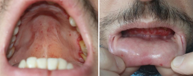
In COVID-19 patients, lesions were found in the skin and oral cavity. Skin lesions are exanthem (47%), pseudo-chilblain (erythematous vesicles or erythematous pustules) (19%), urticaria (19%) vesicular eruption (9%), and necrotic (6%). 12 Enanthem is the term exanthem in the oral mucosa. 12 13 Exanthem is an erythematous rash that develops together with fever or together with a host of other symptoms. Exanthema lesions have morphological variations, including erythematous macules, erythematous papules, erythematous maculopapular, erythematous maculopapular accompanied by petechiae, erythematous vesicles, pustules with erythematous, and urticaria 12 13 14 15 ( Fig. 1 Table 1 ).
Fig. 1.

Enanthem lesions on palatal and labial mucosa accompanied by desquamation of gingival patients positive for COVID-19. 12
Table 1. Differential diagnosis of COVID-19.
| COVID-19 7 12 | Hand, foot, and mouth diseases 15 16 | Measles 15 16 |
|---|---|---|
| Abbreviation: COVID-19, coronavirus disease 2019. | ||
|
|
|
Case Management
To reduce pain in the oral cavity and inactivate coronavirus, an antiseptic mouthwash medication containing 0.2% iodine povidone is given. The ability of iodine povidone has been proven in the case of SARS-CoV and Middle East respiratory syndrome coronavirus (MERS CoV). 8 9 17 18 Hydrogen peroxide 1% can be used as an alternative mouthwash, although no specific mechanism is known for deactivating coronavirus. 8 9 Mouthwash containing chlorhexidine is not effective in COVID-19 cases. 9 Anti-inflammatory mouthwash can be used to reduce pain in the oral cavity,19-23 but the authors have not found a case report journal of this drug used in COVID-19 patients.
Table 2. Anti-inflammatory mouthwash.
Footnotes
Disclosure History and understanding of clinical characteristics in the initial screening of patients with complaints of the intraoral are the starting points for COVID-19 identification.
References
- 1.Melo Neto CLM, Bannwart LC, de Melo Moreno AL, Goiato MC. SARS-CoV-2 and dentistry-review. Eur J Dent 2020;14(suppl S1):S130–S139 doi:10.1055/s-0040-1716438 [DOI] [PMC free article] [PubMed]
- 2.Li X, Geng M, Peng Y, Meng L, Lu S. Molecular immune pathogenesis and diagnosis of COVID-19. J Pharm Anal. 2020;10(02):102–108. doi: 10.1016/j.jpha.2020.03.001. [DOI] [PMC free article] [PubMed] [Google Scholar]
- 3.Mason R J. Pathogenesis of COVID-19 from a cell biology perspective. Eur Respir J. 2020;55(04):9–11. doi: 10.1183/13993003.00607-2020. [DOI] [PMC free article] [PubMed] [Google Scholar]
- 4.
- 5.Morawska L, Cao J. Airborne transmission of SARS-CoV-2: the world should face the reality. Environ Int. 2020;139:105730. doi: 10.1016/j.envint.2020.105730. [DOI] [PMC free article] [PubMed] [Google Scholar]
- 6.Sabino-Silva R, Jardim A CG, Siqueira W L. Coronavirus COVID-19 impacts to dentistry and potential salivary diagnosis. Clin Oral Investig. 2020;24(04):1619–1621. doi: 10.1007/s00784-020-03248-x. [DOI] [PMC free article] [PubMed] [Google Scholar]
- 7.
- 8.Ather A, Patel B, Ruparel N B, Diogenes A, Hargreaves K M.Coronavirus disease 19 (COVID-19): Implications for clinical dental care J Endod 2020. May4605584–595. 10.1016/j.joen.2020.03.008 [DOI] [PMC free article] [PubMed] [Google Scholar]
- 9.Meng L, Hua F, Bian Z. Coronavirus disease 2019 (COVID-19): emerging and future challenges for dental and oral medicine. J Dent Res. 2020;99(05):481–487. doi: 10.1177/0022034520914246. [DOI] [PMC free article] [PubMed] [Google Scholar]
- 10.Lovato A, de Filippis C.Clinical presentation of COVID-19: a systematic review focusing on upper airway symptoms Ear Nose Throat J 2020. Nov9909569–576. 10.1177/0145561320920762 [DOI] [PubMed] [Google Scholar]
- 11.
- 12.Galván Casas C, Català A, Carretero Hernández G et al. Classification of the cutaneous manifestations of COVID-19: a rapid prospective nationwide consensus study in Spain with 375 cases. Br J Dermatol. 2020;183(01):71–77. doi: 10.1111/bjd.19163. [DOI] [PMC free article] [PubMed] [Google Scholar]
- 13.Recalcati S. Cutaneous manifestations in COVID-19: a first perspective. J Eur Acad Dermatol Venereol. 2020;34(05):e212–e213. doi: 10.1111/jdv.16387. [DOI] [PubMed] [Google Scholar]
- 14.Drago F, Rampini E, Rebora A. Atypical exanthems: morphology and laboratory investigations may lead to an aetiological diagnosis in about 70% of cases. Br J Dermatol. 2002;147(02):255–260. doi: 10.1046/j.1365-2133.2002.04826.x. [DOI] [PubMed] [Google Scholar]
- 15.Drago F, Ciccarese G, Gasparini G et al. Contemporary infectious exanthems: an update. Future Microbiol. 2017;12(02):171–193. doi: 10.2217/fmb-2016-0147. [DOI] [PubMed] [Google Scholar]
- 16.Kadambari S, Segal S, Acute viral exanthems. Medicine 2017;45(12):788-793 doi:10.1016/j.mpmed.2017.09.011
- 17.Eggers M, Koburger-Janssen T, Eickmann M, Zorn J. In vitro bactericidal and virucidal efficacy of povidone-iodine gargle/mouthwash against respiratory and oral tract pathogens. Infect Dis Ther. 2018;7(02):249–259. doi: 10.1007/s40121-018-0200-7. [DOI] [PMC free article] [PubMed] [Google Scholar]
- 18.Eggers M. Infectious disease management and control with povidone iodine. Infect Dis Ther. 2019;8(04):581–593. doi: 10.1007/s40121-019-00260-x. [DOI] [PMC free article] [PubMed] [Google Scholar]
- 19.Farah B, Visintini S. 2018. Benzydamine for Acute Sore Throat: A Review of Clinical Effectiveness and Guidelines. [PubMed] [Google Scholar]
- 20.Altenburg A, El-Haj N, Micheli C, Puttkammer M, Abdel-Naser M B, Zouboulis C C. The treatment of chronic recurrent oral aphthous ulcers. Dtsch Arztebl Int. 2014;111(40):665–673. doi: 10.3238/arztebl.2014.0665. [DOI] [PMC free article] [PubMed] [Google Scholar]
- 21.Pignataro L, Marchisio P, Ibba T, Torretta S. Topically administered hyaluronic acid in the upper airway: a narrative review. Int J Immunopathol Pharmacol. 2018;32:2.058738418766739E15. doi: 10.1177/2058738418766739. [DOI] [PMC free article] [PubMed] [Google Scholar]
- 22.Dalessandri D, Zotti F, Laffranchi L et al. Treatment of recurrent aphthous stomatitis (RAS; aphthae; canker sores) with a barrier forming mouth rinse or topical gel formulation containing hyaluronic acid: a retrospective clinical study. BMC Oral Health. 2019;19(01):153. doi: 10.1186/s12903-019-0850-1. [DOI] [PMC free article] [PubMed] [Google Scholar]
- 23.Mehdipour M, Taghavi Z enooz, A, Sohrabi A, Gholizadeh N, Bahramian A, Jamali Z. A comparison of the effect of triamcinolone ointment and mouthwash with or without zinc on the healing process of aphthous stomatitis lesions. J Dent Res Dent Clin Dent Prospect. 2016;10(02):87–91. doi: 10.15171/joddd.2016.014. [DOI] [PMC free article] [PubMed] [Google Scholar]


