Figure 3.
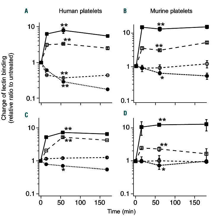
Changes in lectin bindings to platelets following treatment of α2,3,6,8- or α2,3-neuraminidase in vitro. Citrated (A, C) human or (B, D) wild-type (WT) murine platelets were collected and treated with (A, B) 10 mU/ml α2,3,6,8-neuraminidase or (C, D) α2,3-neuraminidase at the equivalent activity level at 37˚C for 15, 60, and 180 minutes (min) before being analyzed for lectin binding. The glycan content was detected by flow cytometry using Fluorescein isothiocyanate (FITC)- conjugated PNA (), FITCECL (), FITC-SNA() and biotinconjugated MAL-ΙΙ combined with FITC-streptavidin (). The mean fluorescence intensity was quantified for the entire cell population and normalized to untreated sample (0 min). Data are shown as mean ± standard deviation, n=4. Results at 60 min are compared to those of untreated sample by unpaired t-test. *P<0.05; **P<0.01.

