A 60-year-old woman is brought to the emergency department following an out-of-hospital cardiac arrest with non-shockable rhythm. Return of spontaneous circulation was achieved in the field with cardio-pulmonary resuscitation and one dose of epinephrine, but she remains comatose. She is intubated, fluid resuscitated, and admitted to the intensive care unit (ICU) where she is cooled to a temperature of 33 °C. Continuous multiparameter physiological monitoring is initiated, including 5-lead electrocardiography (cECG), pulse oximetry (SpO2), core and peripheral thermometry, invasive arterial and central venous blood pressure monitoring, and electroencephalography (cEEG).
Over the next few days, her care team works to optimise the chance of a meaningful recovery. Many questions arise: Is she at risk of a second arrest? Are infection and sepsis lurking? Will she develop organ-threatening hypotension? In the event that she does, would she benefit from additional fluid resuscitation? Most importantly, the team must inform her family as to the likelihood of survival with a good functional outcome.
After more than half a century of innovation in critical care, the modern intensivist has access to a wide array of sophisticated diagnostic tools with which to answer these and other questions. Yet the mainstay of clinical evaluation—and that which in part defines the ICU itself—is the continuous monitoring of physiological waveforms. These waveforms, also called telemetry data, are usually displayed locally at the bedside, but can now be monitored remotely as well, in the context of eICU or tele-ICU practices. These signs are deemed “vital” with good reason; even basic values like heart rate, blood pressure, and oxygen saturation tell us a lot about a patient’s condition.
But beneath these simple numbers lies more information still. Complex measures, such as heart rate variability (HRV) [1], pulse pressure variation [2], and pulse contour [3], describe cardiopulmonary and autonomic functioning, in unexpectedly rich detail. What is more, these signals are collected throughout a patient’s ICU stay, providing not only a snapshot of the current physiological state, but also medium- and long-term trends. These time series telemetry data are a key constituent of the rich ICU data ecosystem that also includes data from EHR systems, imaging, pharmacy records, and other data types, each of which provides important context and complementary detail.
Unlike these latter data types, telemetry data are ubiquitous; most are collected for the vast majority of patients, simply by virtue of their being admitted to the ICU. As such, they are largely without bias. By contrast, lab values, X-rays, and medication records, exist only if someone decided to order a test or administer a drug, and therefore encode patterns of care [4]. Instead, where telemetry is universally applied the resulting data are largely robust to practice variation, and may implicitly mitigate bias associated with sex, gender, and ethnicity [5].
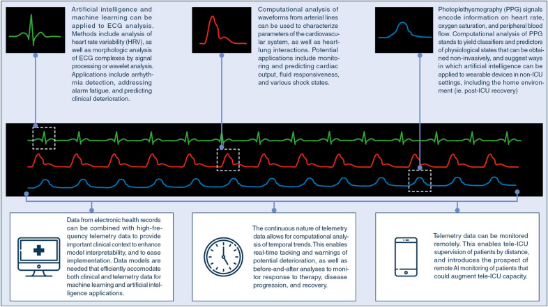
Advances in artificial intelligence are beginning to unlock the potential of telemetry data to directly inform diagnosis and personalized treatment [6] (Fig. 1). We can see hints of this in considering the case above. Is a second arrest on the horizon? Retrospective studies have shown how machine learning might be used to make such predictions based on changes in continuous ECG signals [7]. Will she develop sepsis? Similar studies have suggested that machine learning analysis of HRV may serve as a harbinger of sepsis, even days in advance of its clinical onset [8]. Will hypotension arise as a result? Machine learning deployed across high-frequency vital signs data may well prove capable of providing some measure of advanced warning [9], and has been shown to reduce the duration of hypotension during non-cardiac surgery [10]. In the area of neuroprognostication, deep neural networks have been used with cEEG signals to accurately classify patients post-cardiac arrest, identifying those with little to no chance of a meaningful neurological recovery [11].
Fig. 1.
Data sources and potential applications for artificial intelligence (AI) and telemetry data in the intensive care unit
Promising results have been achieved in artificial intelligence (AI) research using telemetry data, but much computational, translational, and clinical research remains to bring these tools to the bedside [12]. Experts are needed with multi-disciplinary training, as familiar with critical illness as they are with machine learning and signal processing. Expert teams are needed that bring together engineers, computer scientists, and clinicians. These research teams need broad access to data, vast computing resources, a framework for implementation provided by an incentivised healthcare environment, and a clear regulatory environment [13]. The ESICM and SCCM have convened a Joint Task Force to meet this need by establishing multinational collaborations, and setting shared priorities for research and implementation.
The main currency in this space are the data themselves, and here there is a paradox to be reconciled; nearly all patients in the ICU have telemetry data streaming across their bedside monitors in real time, yet for the most part, these data are purged at the end of their stay, and lost to further analysis. Software and hardware systems are needed that can capture and store these data, and make them available for research [14]. Ideally such tools should be open source, and supported by data standards and ethics protocols that maximize sharing and collaboration.
These efforts have begun in earnest. The MIMIC dataset, hosted by MIT, is now widely known and broadly used as a public access repository of clinical and waveform data. MIMIC is part of a larger collection called Physionet, which hosts other waveform datasets. More large datasets are coming online, such as the eICU Collaborative Research Database (eICU-CRD) and the first freely available European ICU database, AmsterdamUMCdb [15]. The coronavirus disease 2019 (COVID-19) pandemic has significantly accelerated efforts at producing rich multi-center, multi-domain, interoperable datasets to enhance rapid understanding of this generational challenge. Work is ongoing to add telemetry data to existing clinical datasets, and to develop data standards and open source tools that will help other stakeholders to do the same.
Despite the considerable potential of pairing AI and telemetry data, challenges remain before clinical tools can be successfully deployed [16]. Telemetry data contain frequent artefacts, and though AI systems may be capable of identifying noise, a preponderance of artifact could create a drag on algorithm performance. Telemetry data must be accompanied by clinical data to provide context, and current EHR-based systems are not without shortcomings [17, 18]. Where AI systems can predict clinical deterioration, there are no guarantees that the decline can be prevented. Even when an algorithm performs well in silico, or in an initial prospective cohort, it may not necessarily yield durable benefits when evaluated in other settings [19]. Clinical implementation remains an important barrier, and it is as yet unclear what sort of evidence will be needed to recommend widespread adoption of a new AI system.
What, then, should the critical care clinician know about AI and telemetry data in the ICU? Increased literacy in machine learning and AI will serve clinicians well as studies using these methods make their way into clinical journals. Cautious optimism is warranted; while there is tremendous promise in the use of high-frequency waveforms to diagnose physiologic states and predict clinical sequelae, it is also likely that as datasets proliferate, so too will the number of classification and prediction models being generated. Not all of these will be of equal reliability and generalizability, and the majority of these may in fact never see clinical deployment.
Returning to our case, AI analysis of cEEG data at 12 h allowed the team to rapidly determine that a good 6-month functional outcome might still be achieved. Machine learning analysis of telemetry data empowered the team to detect sepsis in its earliest stages, and determine the optimal timing for antibiotics and additional fluids. The patient improved, was discharged to the ward, and eventually back home, where a sensor-enabled smart watch allowed her doctors to monitor key parameters of her recovery in real time, including heart rate, step count, and sleep quality. While this vignette describes a future state of practice, it is one that may well be close at hand. Cross-disciplinary, collaborative efforts supported by open source tools and a view to solving clinical problems stands to breathe new life into the vital signs that have for decades informed practice in the ICU.
Author contribution
All authors contributed to the conception, writing, and revision of the manuscript.
Funding
DMM is supported by a Southeastern Ontario Academic Medical Organization (SEAMO) Clinician Scientist award.
Availability of data and material
Not applicable.
Code availability
Not applicable.
Compliance with ethical standards
Conflicts of interest
The authors have no conflicts or competing interests to report.
Footnotes
Publisher's Note
Springer Nature remains neutral with regard to jurisdictional claims in published maps and institutional affiliations.
References
- 1.Buchman TG, Stein PK, Goldstein B. Heart rate variability in critical illness and critical care. Curr Opin Crit Care. 2002;8:311–315. doi: 10.1097/00075198-200208000-00007. [DOI] [PubMed] [Google Scholar]
- 2.Magder S. The meaning of blood pressure. Crit Care. 2018;22:257. doi: 10.1186/s13054-018-2171-1. [DOI] [PMC free article] [PubMed] [Google Scholar]
- 3.Tusman G, Bohm SH, Suarez-Sipmann F. Advanced uses of pulse oximetry for monitoring mechanically ventilated patients. Anesth Analg. 2017;124:62–71. doi: 10.1213/ANE.0000000000001283. [DOI] [PubMed] [Google Scholar]
- 4.Sharafoddini A, Dubin JA, Maslove DM, Lee J. A new insight into missing data in intensive care unit patient profiles: observational study. JMIR Med Inform. 2019;7:e11605. doi: 10.2196/11605. [DOI] [PMC free article] [PubMed] [Google Scholar]
- 5.Desai AN. Artificial intelligence: promise, pitfalls, and perspective. JAMA. 2020;323:2448. doi: 10.1001/jama.2020.8737. [DOI] [PubMed] [Google Scholar]
- 6.Rush B. Applying machine learning to continuously monitored physiological data. J Clin Monit Comput. 2019;33:887–893. doi: 10.1007/s10877-018-0219-z. [DOI] [PMC free article] [PubMed] [Google Scholar]
- 7.Do DH, Kuo A, Lee ES, et al. Usefulness of trends in continuous electrocardiographic telemetry monitoring to predict in-hospital cardiac arrest. Am J Cardiol. 2019;124:1149–1158. doi: 10.1016/j.amjcard.2019.06.032. [DOI] [PMC free article] [PubMed] [Google Scholar]
- 8.Bravi A, Green G, Longtin A, Seely AJE. Monitoring and identification of sepsis development through a composite measure of heart rate variability. PLoS ONE. 2012;7:e45666. doi: 10.1371/journal.pone.0045666. [DOI] [PMC free article] [PubMed] [Google Scholar]
- 9.Chan B, Chen B, Sedghi A, et al. Generalizable deep temporal models for predicting episodes of sudden hypotension in critically ill patients: a personalized approach. Sci Rep. 2020 doi: 10.1038/s41598-020-67952-0. [DOI] [PMC free article] [PubMed] [Google Scholar]
- 10.Wijnberge M, Geerts BF, Hol L, et al. Effect of a machine learning-derived early warning system for intraoperative hypotension vs standard care on depth and duration of intraoperative hypotension during elective noncardiac surgery. JAMA. 2020 doi: 10.1001/jama.2020.0592. [DOI] [PMC free article] [PubMed] [Google Scholar]
- 11.Tjepkema-Cloostermans MC, da Silva LC, Ruijter BJ, et al. Outcome prediction in postanoxic coma with deep learning. Crit Care Med. 2019 doi: 10.1097/CCM.0000000000003854. [DOI] [PubMed] [Google Scholar]
- 12.Fleuren LM, Thoral P, Shillan D, et al. Machine learning in intensive care medicine: ready for take-off? Intensive Care Med. 2020;46:1486–1488. doi: 10.1007/s00134-020-06045-y. [DOI] [PubMed] [Google Scholar]
- 13.Matheny M, Israni ST, Ahmed M, Med DWNA (2019) Artificial intelligence in health care: The hope, the hype, the promise, the perilperil. NAM Special Publication
- 14.Burykin A, Peck T, Buchman TG. Using “off-the-shelf” tools for terabyte-scale waveform recording in intensive care: computer system design, database description and lessons learned. Comput Methods Programs Biomed. 2011;103:151–160. doi: 10.1016/j.cmpb.2010.10.004. [DOI] [PubMed] [Google Scholar]
- 15.Elbers PWG, Hoogendoorn M Amsterdam Medical Data Science. In: httpsamsterdammedicaldatascience.nl. https://amsterdammedicaldatascience.nl/. Accessed 5 Aug 2020
- 16.Michard F, Teboul J-L. Predictive analytics: beyond the buzz. Ann Intensive Care. 2019 doi: 10.1186/s13613-019-0524-9. [DOI] [PMC free article] [PubMed] [Google Scholar]
- 17.Maslove DM, Dubin JA, Shrivats A, Lee J. Errors, omissions, and outliers in hourly vital signs measurements in intensive care. Crit Care Med. 2016 doi: 10.1097/CCM.0000000000001862. [DOI] [PubMed] [Google Scholar]
- 18.O’Halloran H, Kwong K, Veldhoen RA, Maslove DM (2020) Characterizing the patients, hospitals, and data quality of the eICU collaborative research database. Crit Care Med (in press) [DOI] [PubMed]
- 19.Maheshwari K, Shimada T, Yang D, et al. Hypotension prediction index for prevention of hypotension during moderate- to high-risk noncardiac surgery. Anesthesiology. 2020 doi: 10.1097/ALN.0000000000003557. [DOI] [PubMed] [Google Scholar]
Associated Data
This section collects any data citations, data availability statements, or supplementary materials included in this article.
Data Availability Statement
Not applicable.
Not applicable.


