Description
Vulvar pruritus is a common symptom that often markedly impairs the affected women’s quality of life (QoL). Causes of vulvar pruritus are vast and may be environmental, inflammatory, infectious or neoplastic. Often, several causes may coexist. Diagnosis may be challenging due to the unique anatomy and inherent properties of the genital and perianal skin.1 Contact irritant dermatitis is a common cause of vulvar pruritus, followed by lichen sclerosus (LS), fungal infection and psoriasis.2 3 We describe four cases of women who presented at our department due to persistent vulvar pruritus and burning.
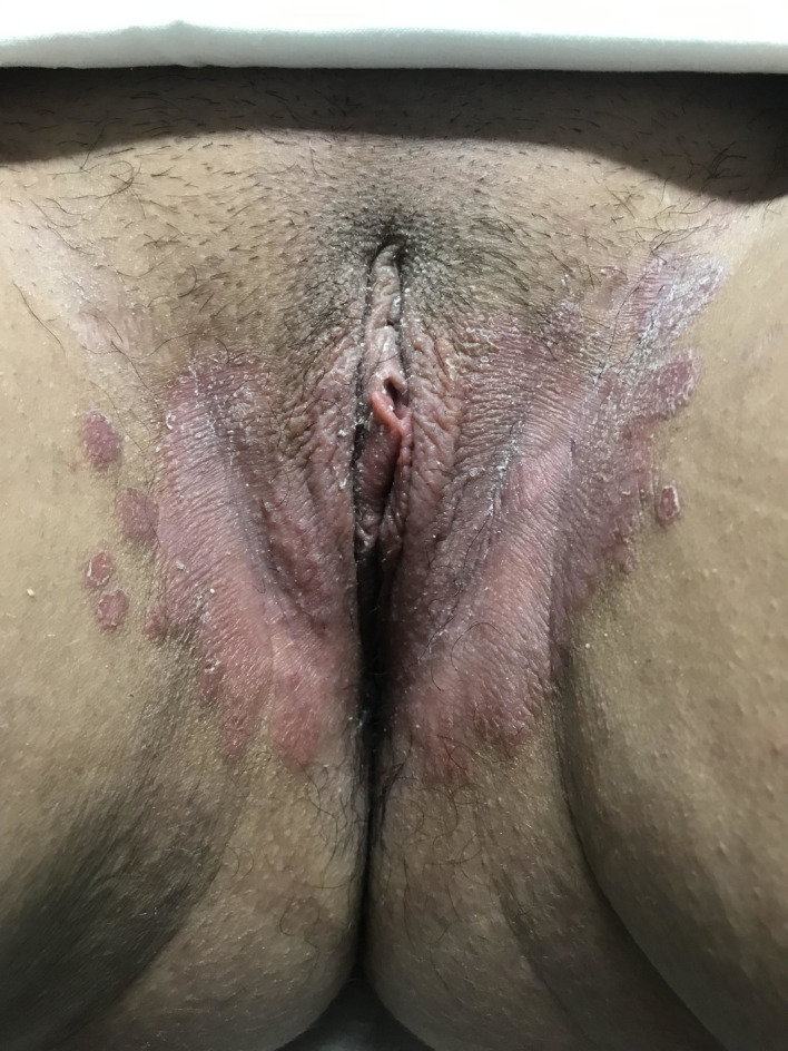
Figure 1 shows a 32-year-old pregnant woman with a red scaling plaque and satellite lesions. The typical discharge and swelling of acute candidiasis were absent. The symptoms and lesions resolved after treatment with topical antifungal. All these aspects were consistent with a fungal infection.
Figure 1.

Fungal infection.
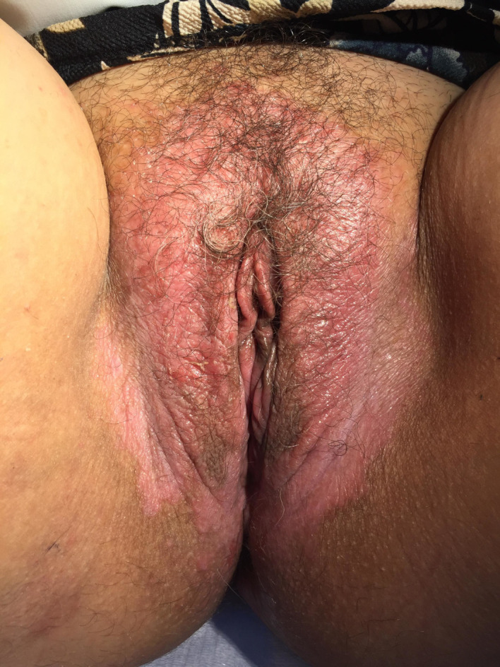
Figure 2 displays a severe erythema of the vulvar skin with evidence of surface disruption with some scaling in dry areas and excoriations. The onset of symptoms in this 48-year-old woman was related to over cleaning and use of sanitary pads due to abundant menses. There was a complete resolution of symptoms avoiding the identified triggering factors. Biopsy was not performed but clinical and physical examination was consistent with contact irritant dermatitis.
Figure 2.

Contact irritant dermatitis.
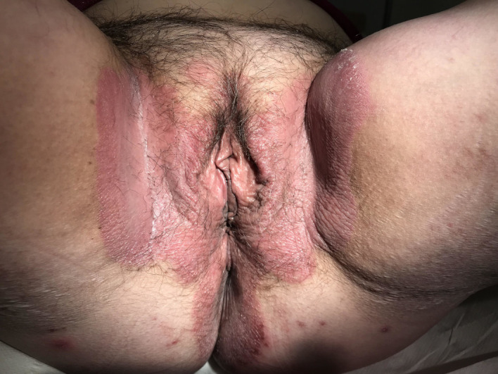
Figure 3 belongs to a 42-year-old woman with a sudden onset of vulvar erythema and pruritus. Physical examination revealed a symmetrical well-demarcated and smooth pink/red plaques, with absent or minimal scaling, involving the perineal and inguinal regions. A biopsy confirmed the diagnosis of inverse psoriasis. This entity is often misdiagnosed as intertriginous fungal or bacterial infections. Genital involvement is prevalent in patients presenting with extragenital psoriasis, markedly affecting QoL.4 Clinicians should take this into account in order to optimise global care.
Figure 3.

Inverse psoriasis.
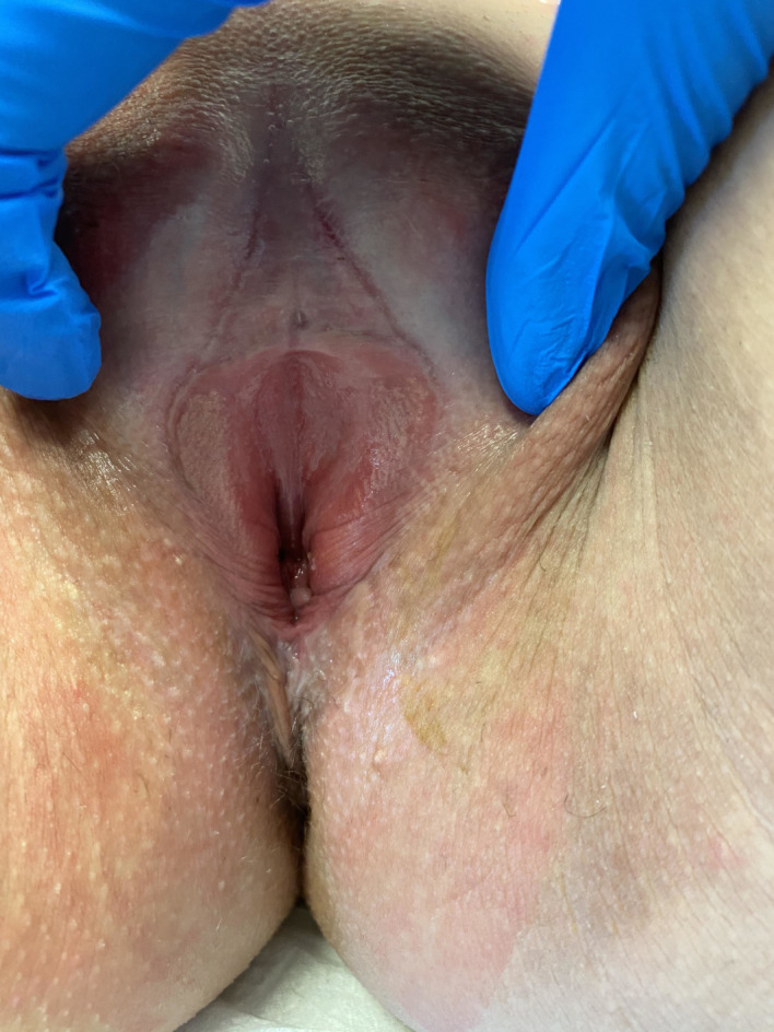
Figure 4 reveals a distortion of the normal vulvar architecture without distinction between the labia majora and minora and a white hyperkeratotic plaque with fissures at the posterior fourchette. This 31-year-old woman presented with intense pruritus and dyspareunia despite numerous topical treatments. A biopsy was made and confirmed the diagnosis of LS. Although LS can occur in all age groups, its prevalence has two peaks (prepubertal and postmenopausal) with most cases diagnosed in postmenopausal women. Usually, the vulvar architecture remains intact early in the course of the disease. The lesions most frequently affect the labia minora although the whitening may extend over the perineum and around the anus in a keyhole fashion. As the disease progresses, the distinction between the labia majora and minora is lost and the clitoris becomes buried under the fused prepuce. Vaginal involvement has not been considered a feature of LS, but few cases have been reported in the literature. Not all cases of adult-onset vulvar LS require a confirmatory biopsy.5
Figure 4.

Lichen sclerosus.
Learning points.
Vulvar pruritus may be challenging for healthcare providers from both a diagnostic and therapeutic point of view.
Vulvar skin fungal infections, contact irritant dermatitis, psoriasis and differentiated vulvar intraepithelial neoplasia can all present with similar physical findings.
Clinical history and physical signs are essential, but may not be sufficient to make the correct diagnosis. Biopsy should be performed if clinical doubt persists, if initial treatment fails or if there is concern about possible underlying neoplasia.
Footnotes
Contributors: CE, ES, APC and FC examined and treated the patient. CE wrote the manuscript and all the authors have read and approved it for publication.
Funding: The authors have not declared a specific grant for this research from any funding agency in the public, commercial or not-for-profit sectors.
Competing interests: None declared.
Patient consent for publication: Obtained.
Provenance and peer review: Not commissioned; externally peer reviewed.
References
- 1.Stockdale CK, Boardman L. Diagnosis and treatment of vulvar dermatoses. Obstet Gynecol 2018;131:371–86. 10.1097/AOG.0000000000002460 [DOI] [PubMed] [Google Scholar]
- 2.Fischer GO. The commonest causes of symptomatic vulvar disease: a dermatologist's perspective. Australas J Dermatol 1996;37:12–18. 10.1111/j.1440-0960.1996.tb00988.x [DOI] [PubMed] [Google Scholar]
- 3.ACOG practice Bulletin No. 93: diagnosis and management of vulvar skin disorders. Obstet Gynecol 2008;111:1243–54. 10.1097/AOG.0b013e31817578ba [DOI] [PubMed] [Google Scholar]
- 4.Larsabal M, Ly S, Sbidian E, et al. GENIPSO: a French prospective study assessing instantaneous prevalence, clinical features and impact on quality of life of genital psoriasis among patients consulting for psoriasis. Br J Dermatol 2019;180:647–56. 10.1111/bjd.17147 [DOI] [PubMed] [Google Scholar]
- 5.Kirtschig G, Becker K, Günthert A, et al. Evidence-Based (S3) guideline on (anogenital) lichen sclerosus. J Eur Acad Dermatol Venereol 2015;29:e1–43. 10.1111/jdv.13136 [DOI] [PubMed] [Google Scholar]


