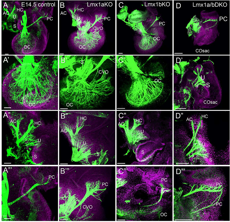
Fig. 2. Auditory and vestibular inner ear innervation abnormalities of Lmx1a/b DKO embryos at e14.5.
Anti-acetylated α-Tubulin immunostaining (green) in the inner ear of indicated genotypes. Some samples were co-stained with Hoechst nuclear stain (lilac) to visualize tissue.
Panels A-D show innervation in the entire inner ear at low magnification; panels A’-D’ show auditory inner ear innervation at higher magnification; panels A”- D” and A”’- D”’ show vestibular inner ear innervation at higher magnification.
(A-D’) Organ of Cori (OC) innervation was clearly detectable in control, Lmx1a KO and Lmx1b KO embryos (A-C, A’-C’) but no cochlear innervation was found in Lmx1a/b DKO embryos, in which a cochlear sac (COSac) formed (D, D’). Consistent with previously described partial transformation of the OC into an irregular vestibular epithelium (NICHOLS et al., 2008), innervation to an abnormal cochlear-vestibular organ (CVO) was detected in Lmx1a KO mice (B, B’).
(A”-D”) In Lmx1a/b DKO mice, branches innervating anterior crista (AC) and horizontal crista (HC) appeared abnormally thin compared to control, Lmx1a KO and Lmx1b KO ears. Some innervation to utricle (U) and saccule (S) was present in Lmx1a KO, Lmx1b KO and Lmx1a/b DKO ears.
(A”’-D”’) Innervation of posterior crista (PC) appeared extended in Lmx1a KO and Lmx1a/b DKO embryos, consisting of multiple small branches in Lmx1a KO and multiple large branches in Lmx1a/b DKO mutants.
Scale bar: 100 μm for all images.

