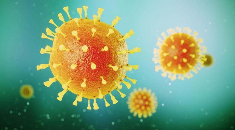
Severe acute respiratory syndrome coronavirus 2 (SARS-CoV-2) is present in the neurons of the olfactory mucosa in some individuals who died with COVID-19, according to new research published in Nature Neuroscience. The finding suggests that the virus could invade the CNS via the olfactory nerve and possibly other cranial nervses.
Although COVID-19 is predominantly a respiratory illness, evidence for an effect of SARS-CoV-2 infection on the brain is accumulating. In previous studies, SARS-CoV-2 was detected in the CNS of individuals with COVID-19, but the details of the relationship between the virus and the nervous system are still far from clear. The aim of the new study was to investigate one possible route of SARS-CoV-2 entry to the brain — the olfactory system — and to perform a detailed analysis of the regional and cellular distribution of the virus within the brain.

sixam / Alamy Stock Photo
“As a loss of smell is one of the most prominent clinical features of SARS-CoV-2 infection, we focused on the presumed olfactory entry route,” explains Frank Heppner, who led the study with Helena Radbruch. “We did this by starting to screen for SARS-CoV-2 at the sensory neurons in the olfactory mucosa, and tracing their neuroanatomical path into the brain.”
The researchers analysed post-mortem tissue from 33 individuals with COVID-19, including samples from the olfactory mucosa, olfactory bulb, uvula, trigeminal ganglion and medulla oblongata. They used real-time quantitative PCR (rt-qPCR), and in-situ hybridization to detect SARS-CoV-2 RNA, as well as immunohistochemistry and electron microscopy to detect the protein.
The results of the rt-qPCR revealed that viral RNA was most common in the olfactory mucosa, where it was detected in 20 of 30 participants. The next most common locations were the uvula and the medulla oblongata, both of which were SARS-CoV-2 RNA-positive in six participants. Electron microscopy analysis of tissue from one individual with a high viral RNA load revealed intact viral particles in the olfactory mucosa.
The olfactory mucosa includes the olfactory epithelium, connective tissue and olfactory neurons. To establish whether the virus had entered neurons, the researchers performed immunohistochemistry for the neuronal markers TuJ1, NF-200 and OMP on olfactory mucosa samples from three of the participants with COVID-19 (the only three from whom appropriate tissue samples had been collected) and two controls without COVID-19. They also probed the same samples with a SARS-CoV spike protein antibody, which is expected to recognize the spike protein on SARS-CoV-2.
Spike immunoreactivity was detected in TuJ1-positive, NF-200-positive and OMP-positive cells in participants with COVID-19, indicating that the virus is present in olfactory neurons. No spike immunoreactivity was detected in these cell types in the controls.
“These observations provide unequivocal evidence for SARS-CoV-2 infection of neurons in the olfactory mucosa, which, in context, supports the notion that SARS-CoV-2 is able to use the olfactory mucosa as a port of entry into the brain,” explains Radbruch.
The researchers then looked for signs of a SARS-CoV-2-mediated inflammatory response in the CNS by probing for markers of activated microglia and macrophages in the medulla oblongata. Compared with samples from individuals without SARS-CoV-2 infection, levels of HLA-DR were increased in samples from individuals with the virus, suggesting an ongoing innate immune response.
These new findings add to our understanding of the interactions between SARS-CoV-2 and the brain, but whether viral invasion of the CNS via the olfactory system is responsible for the neurological symptoms observed in patients with COVID-19 is not entirely clear.
These new findings add to our understanding of the interactions between SARS-CoV-2 and the brain
“We are planning a follow-up study to further investigate the immunological responses of the CNS following SARS-CoV-2 infection,” says Heppner. “This will include analysis of the CNS-related long-term effects of COVID-19 and a more detailed correlation of clinical findings with pathological findings, as well as genomics and proteomics approaches.”
References
Original article
- Meinhardt J, et al. Olfactory transmucosal SARS-CoV-2 invasion as a port of central nervous system entry in individuals with COVID-19. Nat. Neurosci. 2020 doi: 10.1038/s41593-020-00758-5. [DOI] [PubMed] [Google Scholar]
Related article
- Pezzini A, Padovani A. Lifting the mask on neurological manifestations of COVID-19. Nat. Rev. Neurol. 2020;16:636–644. doi: 10.1038/s41582-020-0398-3. [DOI] [PMC free article] [PubMed] [Google Scholar]


