Abstract
Purpose
Expanded indications for endoscopic submucosal dissection (ESD) in early gastric cancer (EGC) remain controversial due to the potential risk of undertreatment after adequate lymph node dissection (LND). Regional LND (RLND) is a novel technique used for limited lymphadenectomy to avoid gastrectomy. This study established the safety and effectiveness of RNLD as an additional treatment option after ESD for expanded indications.
Materials and Methods
A total of 69 patients who met the expanded indications for ESD were prospectively enrolled from 2014 to 2017. The tumors were localized using intraoperative esophagogastroduodenoscopy (EGD) before RLND. All patients underwent RLND first, followed by conventional radical gastrectomy with LND. The locations of the preoperative and intraoperative EGD were compared. Pathologic findings of the primary lesion and the RLND status were analyzed.
Results
The concordance rates of tumor location between the preoperative and intraoperative EGD were 79.7%, 76.8%, and 63.8% according to the longitudinal, circumferential, and regional locations, respectively. Of the 4 patients (5.7%) with metastatic LNs, 3 were pathologically classified as beyond the expanded indication for ESD and 1 had a single LN metastasis in the regional lymph node.
Conclusions
RLND is a safe additional option for the treatment of EGC in patients meeting expanded indications after ESD.
Keywords: Stomach neoplasms, Regional lymph node dissection, Minimally invasive surgical procedures, Endoscopic mucosal resection, Endoscopy
INTRODUCTION
The standard treatment for gastric cancer (GC) is gastrectomy with radical lymph node dissection (LND) [1]; however, post-gastrectomy syndromes such as dumping syndrome, nutritional deficiency, or alkaline gastritis can occur [2,3,4]. Endoscopic resection such as endoscopic submucosal dissection (ESD) is performed in limited cases to reduce these complications and improve the quality of life (QOL); however, ESD has the disadvantage of not being able to confirm LN status [5].
The incidence of lymph node (LN) metastasis in early GC (EGC) reportedly ranges from 2% to 20% according to the tumor characteristics [5,6]. Therefore, ESD can be safely implemented in patients with a low risk of LN metastasis. To meet the absolute indications for ESD, a tumor should be less than 2 cm in diameter, only within the mucosal layer without ulceration (UL), and histologically differentiated. The expanded indications for ESD remain a subject of debate due to relatively high incidence of LN metastasis [1], which can reach up to 4% [7,8]. Despite the high survival rate of EGC after gastrectomy, this risk of LN metastasis after ESD remains high [9].
Recently, the safety and efficacy of limited lymphadenectomy, such as sentinel LND, to avoid radical gastrectomy have been related to sentinel LNs (SLNs), which require special materials or instruments such as dyes or radioisotopes for identification [10,11,12]. However, it would be difficult to provide a clear answer about limited LND due to the complex lymphatic flow and skip metastasis of GC [13,14]. Regional LN (RLN) is a novel concept that refers to LNs in a region with potential for lymphatic drainage, based on the location of the tumor, in a wider range than SLN. Because RLND can cause gastric ischemia, its safety should be established.
This study investigated the feasibility and safety of RLND for patients with EGC who met the expanded criteria for ESD.
MATERIALS AND METHODS
Study population and data collection
From September 30, 2014, to April 3, 2017, patients with histologically confirmed clinical stage T1N0M0 gastric adenocarcinoma were enrolled for the validation of safety in the RLND for EGC (REALLY) trial conducted at Seoul St. Mary's Hospital in Seoul, South Korea. The expanded indications for ESD were as follows: a) depth within the mucosal layer, differentiated type, UL(−), and 2–3 cm in diameter; b) depth within the mucosal layer, differentiated type, UL(+), and ≤3 cm in diameter; c) depth within the submucosal layer (≤ 500 µm depth), differentiated type, and ≤3 cm in diameter; d) depth within the mucosal layer, UL(−), ≤2 cm, and undifferentiated type according to the Japanese guidelines [5]. The enrollment criteria were as follows: expanded indication for ESD, non-curative resection after ESD, between 18 and 80 years of age, and Eastern Cooperative Oncology Group score ≤2 (Supplementary Fig. 1). The exclusion criteria were as follows: metachronous or synchronous malignancy, remnant GC, history of prior gastric surgery or adjacent organ surgery around the stomach, absolute indication for ESD, history of chemotherapy or radiation therapy, body mass index < 18.5, and pregnancy or planning for pregnancy. The REALLY trial was approved by the Institutional Review Board (IRB) of Seoul St. Mary's Hospital (KC14EISE0492). Written informed consent was obtained from all patients prior to gastrectomy.
Design
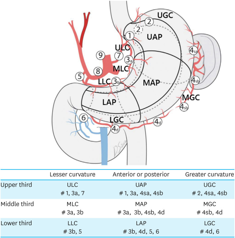
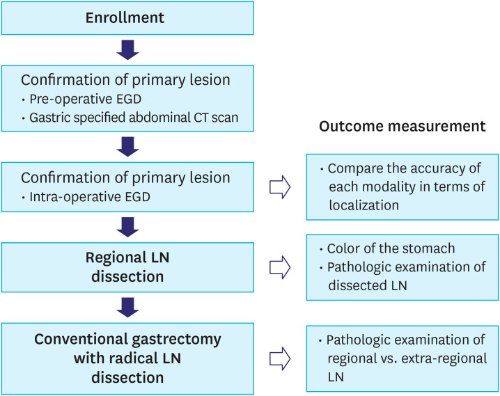
The present study was a single-arm prospective study. The stomach was divided into 9 regions according to the tumor location (Fig. 1). The 9 regions were defined by the anatomical boundaries separating the stomach. A total of 166 patients who met the expanded indication for ESD in pathologic results and underwent curative radical gastrectomy from 2010 to 2016 in the same institution were retrospectively analyzed to identify the RLNs (data not shown). The RLNs were defined based on these data and the anatomical structures. For all patients, the clinical region was confirmed by esophagogastroduodenoscopy (EGD), and the clinical stage was confirmed using endoscopic ultrasound (EUS) and computed tomography (CT) preoperatively. On the day of surgery, intraoperative EGD was performed to confirm the surgical region while directly observing the stomach right after insertion of the laparoscopic camera or laparotomy. Methylene blue or indocyanine green solution was injected around the tumor endoscopically to confirm the intraoperative location. For tumors spanning 2 regions, the location was determined based on the epicenter of the tumor. All preoperative and intraoperative EGD procedures were performed by gastroenterologists or surgeons who had experienced EGD localization in more than 500 cases. The location of the tumor was described according to a predetermined method, and the preoperative location was confirmed by reviewing it by the authors. The station for RLND was decided based on the determined surgical region. After RLND, the color of the stomach was observed for approximately 10 to 30 minutes to confirm that no ischemic changes had occurred. Subsequently, all patients underwent conventional gastrectomy with radical LND according to the guidelines for GC treatment [1]. Although the tumor was EGC, D2 LND was often performed according to the surgeon's preference, especially for younger patients. After gastrectomy, the retrieved RLNs and extra-RLNs were separately sent to pathologists (Fig. 2). The metastatic status of the LNs according to region, concordance between the location of pre- and intraoperative EGD, and short-term postoperative complications were determined to measure the outcomes.
Fig. 1. Definition of 9 regions and station of regional lymph node according to the tumor location.
Fig. 2. Flow chart of the study protocol.
EGD = esophagogastroduodenoscopy; CT = computed tomography; LN = lymph node.
Sample size and statistical analysis
This was a preliminary study to establish the feasibility and safety of RLND prior to a noninferiority study of RLND compared to conventional radical LND. The sample size was calculated as follows: the success rate of conventional radical LND was assumed to be 98%, the limit of 95% confidence interval was set to within 3%, and the alpha value was 0.05. The success rate of conventional radical LND was defined as 5-year disease-free survival after conventional gastrectomy with radical LND performed in EGC. We determined that 84 patients were required; assuming a 10% dropout rate, a total of 92 patients were required [15,16]. The χ2 test, Fisher's exact test, and Student's t-test were used to compare the groups. P<0.05 was considered statistically significant. All statistical analyses were performed using Statistical Package for the Social Sciences software for Windows (ver. 21.0.; SPSS Inc., Chicago, IL, USA).
RESULTS
A total of 69 patients were enrolled in this study. The trial was terminated prematurely with IRB approval because the recruitment period was longer than expected, and some significant LN metastasis findings were observed. The mean patient age was 58.6 years, and 63.8% were male. Fifty-nine patients met the expanded indications of ESD in the preoperative evaluation, and ten patients were enrolled due to non-curative ESD. Twelve patients underwent open surgery, and fifty-seven underwent minimally invasive surgery. The mean operation time was 209.7 min, and no ischemic changes in the stomach after RLND were observed. Five (7.2%) patients had a Clavien-Dindo classification of 3. These results suggest that RLND seems to be feasible and safe in terms of gastric ischemia and short-term postoperative outcomes (Table 1).
Table 1. Clinical characteristics and reasons for gastrectomy and operation details.
| Variable | Value (n=69) | |
|---|---|---|
| Age (yrs) | 58.6±10.6 | |
| Sex | ||
| Male | 44 (63.8) | |
| Female | 25 (36.2) | |
| BMI (kg/m2) | 24.2±3.2 | |
| ECOG | ||
| 0 | 59 (85.5) | |
| 1 | 9 (13.0) | |
| 2 | 1 (1.4) | |
| Tumor size in EGD (cm) | 1.6±0.7 | |
| Reason of gastrectomy (data was duplicated) | ||
| Tumor size >2 cm in preoperative EGD | 12 (17.4) | |
| Ulcer in preoperative EGD | 17 (24.6) | |
| SM invasion in preoperative EUS | 30 (43.5) | |
| Undifferentiated type in preoperative EGD biopsy | 34 (49.3) | |
| SM invasion in specimen from ESD | 2 (2.9) | |
| LVI in specimen from ESD | 8 (11.6) | |
| Approach | ||
| Open | 12 (17.4) | |
| Laparoscopy | 53 (76.8) | |
| Robot-assisted | 4 (5.8) | |
| Resection | ||
| Total gastrectomy | 10 (14.5) | |
| Distal gastrectomy | 59 (85.5) | |
| Extent of LN dissection | ||
| D1+ | 28 (40.6) | |
| D2 | 41 (59.4) | |
| Reconstruction | ||
| Billroth-I | 10 (14.5) | |
| Billroth-II | 48 (69.6) | |
| Roux-en-Y | 11 (15.9) | |
| OP time (min) | 209.7±44.0 | |
| EBL (mL) | 91.0±70.7 | |
| Color change of stomach after regional LN dissection | 0 (0) | |
| Duration to flatus (days) | 3.4±0.6 | |
| SOW (days) | 3.5±0.6 | |
| SD (days) | 5.6±0.7 | |
| Hospital stay (days) | 9.1±6.0 | |
| Complication | ||
| No | 56 (81.2) | |
| CDC I | 1 (1.4) | |
| CDC II | 7 (10.1) | |
| CDC IIIa | 4 (5.8) | |
| CDC IIIb | 1 (1.4) | |
Data are shown as mean±standard deviation or number (%).
BMI = body mass index; ECOG = Eastern Cooperative Oncology Group; EGD = esophagogastroduodenoscopy; EUS = endoscopic ultrasound; ESD = endoscopic submucosal dissection; SM = submucosa; LVI = lymphovascular invasion; LN = lymph node; OP = operation; EBL = estimated blood loss; SOW = sips of water; SD = soft diet; CDC = Clavien-Dindo classification.
Considering the pathologic results, the mean tumor size was 2.0 cm, and the mean length of the proximal margin was 4.4 cm. The mean number of total retrieved LNs, RLNs, and extra-RLNs was 40.6, 12.0, and 28.5, respectively. The histologic type from the preoperative biopsy of one patient changed from a differentiated to an undifferentiated type. Submucosal invasion and lymphovascular invasion (LVI) were observed in 21 (30.4%) and 8 (11.6%) patients, respectively, 4 (5.7%) patients had LN metastasis (Table 2).
Table 2. Pathologic results.
| Variable | Value (n=69) | |
|---|---|---|
| Tumor size in pathology (cm) | 2.0±1.1 | |
| PRM (cm) | 4.4±2.1 | |
| DRM (cm) | 6.8±3.9 | |
| No. of retrieved LNs | 40.6±14.8 | |
| No. of metastatic LNs | 0.1±0.6 | |
| Histologic type from pathology | ||
| Differentiated | 34 (49.3) | |
| Undifferentiated | 35 (50.7) | |
| Lymphovascular invasion | 8 (11.6) | |
| Neural invasion | 1 (1.4) | |
| Depth of invasion in pathology | ||
| Mucosa | 48 (69.6) | |
| SM1 | 7 (10.1) | |
| SM2 | 6 (8.7) | |
| SM3 | 8 (11.6) | |
| N stage | ||
| N0 | 65 (94.2) | |
| N1 | 3 (4.3) | |
| N2 | 1 (1.4) | |
Data are shown as mean±standard deviation or number (%).
PRM = proximal resection margin; DRM = distal resection margin; LN = lymph node; SM = submucosa.
All patients were classified by indication of ESD based on the pathologic results. Twelve patients with a clinically overestimated absolute indication for ESD had no LN metastasis. One (3.6%) patient had LN metastasis within the RLN among twenty-eight patients with expanded indications for ESD. Of the 29 patients with underestimated indications for ESD, 3 (10.3%) patients had LN metastasis, 1 patient (3.4%) had RLN metastasis, whereas the other 2 (6.9%) had extra-RLN metastasis (Table 3).
Table 3. LN meta rate according to endoscopic submucosal dissection indication.
| Variable | Absolute (n=12) | Expanded (n=28) | Beyond (n=29) |
|---|---|---|---|
| LN metastasis | 0 | 1 (3.6) | 3 (10.3) |
| RLN | 0 | 1 (3.6) | 1 (3.4) |
| Extra-RLNs | 0 | 0 | 2 (6.9) |
LN = lymph node; RLN = regional lymph node.
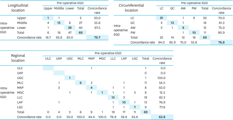
We determined the tumor location at 3 individual points: preoperative EGD, intraoperative EGD, and pathologic findings. Interestingly, according to the methods, the tumor locations were poorly concordant with each other. Therefore, we compared the location of preoperative EGD to that of intraoperative EGD, which is most important for determining the tumor region (Fig. 3). The concordance rates of the longitudinal, circumferential, and regional locations were 79.7%, 76.8%, and 63.8%, respectively; the concordance rate of the middle third of the longitudinal location was only 55.6%, which was significantly lower than that of the lower third (97.5%). In addition, the middle third location, regardless of the circumferential location, was the main reason for the lower concordance rate with statistical significance in the regional location. On the other hand, there was no significant difference according to the circumferential location (Fig. 3).
Fig. 3. Concordance status between pre- and intraoperative EGD.
EGD = esophagogastroduodenoscopy; LC = lesser curvature; GC = greater curvature; AW = anterior wall; PW = posterior wall; ULC = upper lesser curvature; UAP = upper anterior to posterior; UGC = upper greater curvature; MLC = middle lesser curvature; MAP = middle anterior to posterior; MGC = middle greater curvature; LLC = lower lesser curvature; LAP = lower anterior to posterior; LGC = lower greater curvature.
Of the 4 patients with LN metastasis, 1 patient (No. 1) had 4 metastatic LNs beyond the expanded indications of ESD. The region was the lower third and anterior or posterior abdominal wall (LAP), and the RLN stations were 3b, 4d, 5, and 6. However, the number of metastatic LNs was 6, 8a, and 9. Metastatic LNs were observed not only in the RLNs, but also in the extra-RLNs, especially in the D2 group. Another patient (No. 2) had 2 metastatic LNs in the extra-RLNs. The tumor was beyond the expanded indications of ESD. The region was the lower third with lesser curvature, whereas metastatic LNs were observed in the extra-RLNs at stations 1 and 6. The third patient (No. 3) had a tumor that met the expanded indications of ESD. The region was the middle third with greater curvature, and 2 metastatic LNs (4d) within the RLN station (4sb, 4d) were observed. The fourth patient (No. 4) had a tumor that was beyond the indication of ESD. The region was LAP, and 1 metastatic LN (6) was detected within the RLNs (3b, 4d, 5, and 6) (Table 4).
Table 4. Details of No. positive patients.
| No. | No. of metastatic LNs | Regional LNs | Station of metastatic LNs | Tumor size in preoperative EGD | Tumor size in pathology | Depth of invasion | Differentiation | Ulceration | LVI | ESD indication |
|---|---|---|---|---|---|---|---|---|---|---|
| 1 | 4 | LAP (3b, 4d, 5, 6) | 6, 8a, 9 | 2 | 6.2 | SM2 | WD | (+) | (+) | Beyond |
| 2 | 2 | LLC (3b, 5) | 1, 6 | 2 | 2.8 | SM2 | PD | (−) | (+) | Beyond |
| 3 | 2 | MGC (4sb, 4d) | 4d (RLN) | 1.3 | 2 | M | SRC | (−) | (−) | Expanded |
| 4 | 1 | LAP (3b, 4d, 5, 6) | 6 (RLN) | 1.2 | 0.8 | M | MD | (−) | (+) | Beyond |
LN = lymph node; EGD = esophagogastroduodenoscopy; LVI = lymphovascular invasion; ESD = endoscopic submucosal dissection; LAP = lower anterior or posterior; SM = submucosa; M = mucosa; WD = well differentiated; LLC = lower lesser curvature; PD = poorly differentiated; MGC = middle greater curvature; RLN = regional lymph node; SRC = signet ring cell; MD = moderately differentiated.
DISCUSSION
As the survival rate of patients with GC has improved, interest in QOL after gastrectomy has also increased [17,18,19]. Numerous studies have reported limited surgery or endoscopic resection for the treatment of EGC to improve QOL and avoid complications, including post-gastrectomy syndrome [2,5,20,21,22]. The most important disadvantage of ESD is that the LN status cannot be identified pathologically. The sensitivity and specificity of LN status on preoperative CT or EUS are limited [23,24]. Therefore, ESD is limited to patients with a low risk of LN metastasis on preoperative examination. Currently, indications of ESD are classified into absolute, expanded, and beyond according to tumor size, ulcer, depth of invasion, and histologic type [5]. The incidence of LN metastasis is low for absolute indications and high for beyond indications [25,26]; accordingly, ESD and gastrectomy are recommended. However, the feasibility and risk of ESD for expanded indications have been a subject of debate [7,8,27,28]. Therefore, ESD for expanded indications has been performed according to the consensus of each institution. The biggest obstacle to ESD application is that of the various discrepancies, as the results from preoperative examinations, including tumor size or depth of invasion, which are measured by CT or EUS, often differ from the pathologic results. EUS and CT have T-stage accuracies of 41%–67% and 4%–82%, respectively [29,30,31]. The indication of ESD was established based on the pathologic results, which were inferred using the clinical stage despite its low accuracy. A discrepancy in histologic type between the preoperative biopsy sample and the surgical specimen is frequently observed, although the histologic type is one of the most important parameters of ESD application [32,33]. Discrepancies in tumor size, depth of invasion, or histologic type are important reasons for non-curative ESD. At the time of patient enrollment for this study, 59 patients manifested expanded indications of ESD and 10 of non-curative ESD. However, the final pathologic results showed that 28 (40.6%) showed expanded indications, 12 (17.4%) overestimated absolute indications, and 29 (42.0%) underestimated indications for ESD. These results suggest that more accurate preoperative examinations are necessary to avoid undertreatment or overtreatment, and the treatment modality should be decided to avoid undertreatment. It is considered appropriate to attempt RLND when the patient is classified as an expanded indication by pathologic report after ESD.
Another discrepancy could be tumor location. Accurate localization is crucial not only for the determination of proper RLN, but also for performing adequate wedge resection. Generally, the tumor is localized on preoperative EGD or CT before surgery. However, these methods are limited in their ability to accurately locate tumors. Although there are various methods to overcome this inaccuracy, their effectiveness remains controversial [34,35,36]. In this study, we determined tumor location through preoperative EGD, intraoperative EGD, and pathologic reports. Among these 3 methods, intraoperative EGD was the most accurate localization method for several reasons. First, the region from the pathologic report does not affect clinical practice because LND according to the region was performed during surgery. Second, in the case of distal gastrectomy because the stomach was cut, the accuracy of the pathologic location was inevitable. In terms of preoperative EGD, it was difficult to localize accurately because the structures that are the basis for localization were difficult to locate inside the stomach, and the shape and size of the stomach were different in each patient. Another possible cause of location discordance was ESD scar distortion. Gastrectomy is usually performed between 2 and 6 weeks after ESD, and the location of the ESD scar can be moved due to scar distortion. However, regarding intraoperative EGD because tumor localization is performed under direct intra-abdominal observation, the most accurate localization is possible. In this study, the concordance rates of the longitudinal, circumferential, and regional locations of the tumor between preoperative and intraoperative EGD were 79.7%, 76.8%, and 63.8%, respectively. Therefore, tumor localization by intraoperative EGD is mandatory for accurate RLND.
Recently, the concept of SLN has emerged for patients with expanded indications of ESD with an intermediate risk of LN metastasis [10,11,12]. SLN refers to the first possible site of metastasis along the route of lymphatic flow from the tumor and is widely used in breast cancer surgery [37]. In terms of GC, the role of SLN is still controversial because of the heterogeneity of lymphatic flow and the possibility of skip metastasis [13,14]. RLN is a broader concept than SLN, and refers to the entire LN station and surrounding tissue that can be drained from the region according to the tumor location. Therefore, more LNs can be retrieved in RLND than in SLND, and there might be a reduction in false-positive results. A potential problem that may occur during RLND is that 1 or 2 feeding vessels of the stomach can be ligated in each region. Practically, the short gastric artery, left gastroepiploic artery, right gastric artery, right gastroepiploic artery, and left gastric artery should be ligated during dissection of LN stations 4sa, 4sb, 5, 6, and 7, respectively, which could lead to postoperative complications due to gastric ischemia. In this study, the color of the stomach was routinely checked in all patients after RLND, and no ischemic changes were observed. In addition, no perforation or anastomosis leakage occurred after the RLND. Thus, RLND can be considered a treatment option in combination with gastric wedge resection, ESD, or full-thickness endoscopic resection without the risk of ischemia.
This study included 3 of 29 patients who had LN metastasis beyond the expanded indication of ESD according to the pathologic results, of whom 2 had extra-RLN metastasis. For patients with large tumors with submucosal invasion and LVI, RLND was not applicable because metastatic LNs were identified in the extra-RLNs and D2 LN zones. On the other hand, 28 patients met the expanded criteria, and there was no extra-RLN metastasis, and only one had RLN metastasis. Patient 3 was classified as an expanded indication of ESD pathologically because of the small tumor size within the mucosal layer with signet ring cell carcinoma (SRC), and metastatic LNs were identified within the RLNs. SRC has the lowest risk of LN metastasis among the conditions for expanded indications of ESD [38]. Although more evidence is required, patients who have a relatively lower risk of LN metastasis could be a potential option for RLND, which can be used to evaluate RLNs pathologically. These results suggest that RLND may be performed within the absolute or expanded indications of ESD (Table 4).
The limitation of this study is that it was a single-arm trial without a comparison-arm conducted by a single institution. Moreover, patients classified as not only pathologically but also clinically expanded indications of ESD were included. It would be better to include only pathologically expanded indications after ESD to obtain more accurate results. However, it was a preliminary study to establish the feasibility and safety of RLND. The results of this study can be used as a basis for future comparative studies. Second, more analyses in patients with LN metastasis were difficult due to the small number of cases. However, the incidence of LN metastasis is low in patients with EGC, especially for expanded indications of ESD. After verifying the surgical safety of RLND, a large-scale study should be performed to confirm the oncologic safety in the future. Third, the safety of RLND is limited. Since conventional gastrectomy was performed after observing the color change for about 10 to 30 minutes after RLND, the actual post-RLND complication was not directly observed. In the case of local tumor resection, such as a wedge or segmental resection in addition to RLND, there is risk of ischemia, stricture, or kinking of the resected stomach. RLND without radical gastrectomy and observation is needed to confirm the safety of RLND. However, since the ethical problem that RLND is a procedure that has not yet been verified, conventional radical gastrectomy was performed after RLND to overcome the ethical issue. Further studies are needed to confirm the surgical safety of RLND.
In conclusion, RLND would be a safe additional treatment option for expanded indications of ESD, as ESD has the risk of undertreatment in terms of adequate LND, and conventional radical gastrectomy has the risk of complications. RLND seems to be safely performed without the risk of complications, including gastric ischemia. Conventional radical gastrectomy is needed considering extra-RLN metastasis in some cases of expanded indications of ESD. Additionally, intraoperative EGD is recommended to confirm accurate tumor location.
ACKNOWLEDGMENTS
The authors would like to thank nurses Yunjung Song and Hyunkyo Kim for assisting with the research. Language editing services for this manuscript were provided by TextCheck.
Footnotes
- Conceptualization: Y.H.M., C.M.G, P.C.H.
- Data curation: S.H.S., Y.H.M., J.Y.J.
- Formal analysis: S.H.S., Y.H.M.
- Investigation: S.H.S., Y.H.M., P.C.H.
- Methodology: Y.H.M., L.S.H., P.J.M., S.K.Y., J.E.S., C.M.G., P.C.H.
- Project administration: S.H.S., Y.H.M., P.C.H.
- Resources: S.H.S., J.Y.J., L.S.H., P.J.M., S.K.Y., J.E.S., C.M.G., P.C.H.
- Software: S.H.S.
- Supervision: P.C.H.
- Writing - original draft: S.H.S.
- Writing - review & editing: S.H.S., Y.H.M., J.Y.J., L. S.H., P.J.M., S.K.Y., J.E.S., C.M.G., P.C.H.
Conflict of Interest: No potential conflict of interest relevant to this article was reported.
SUPPLEMENTARY MATERIAL
Summary of eligible criteria.
References
- 1.Guideline Committee of the Korean Gastric Cancer Association (KGCA), Development Working Group & Review Panel. Korean practice guideline for gastric cancer 2018: an evidence-based, multi-disciplinary approach. J Gastric Cancer. 2019;19:1–48. doi: 10.5230/jgc.2019.19.e8. [DOI] [PMC free article] [PubMed] [Google Scholar]
- 2.Bolton JS, Conway WC., 2nd Postgastrectomy syndromes. Surg Clin North Am. 2011;91:1105–1122. doi: 10.1016/j.suc.2011.07.001. [DOI] [PubMed] [Google Scholar]
- 3.Carvajal SH, Mulvihill SJ. Postgastrectomy syndromes: dumping and diarrhea. Gastroenterol Clin North Am. 1994;23:261–279. [PubMed] [Google Scholar]
- 4.Eagon JC, Miedema BW, Kelly KA. Postgastrectomy syndromes. Surg Clin North Am. 1992;72:445–465. doi: 10.1016/s0039-6109(16)45689-6. [DOI] [PubMed] [Google Scholar]
- 5.Gotoda T. Endoscopic resection of early gastric cancer: the Japanese perspective. Curr Opin Gastroenterol. 2006;22:561–569. doi: 10.1097/01.mog.0000239873.06243.00. [DOI] [PubMed] [Google Scholar]
- 6.Seo HS, Lee GE, Kang MG, Han KH, Jung ES, Song KY. Mixed histology is a risk factor for lymph node metastasis in early gastric cancer. J Surg Res. 2019;236:271–277. doi: 10.1016/j.jss.2018.11.055. [DOI] [PubMed] [Google Scholar]
- 7.Oh SY, Lee KG, Suh YS, Kim MA, Kong SH, Lee HJ, et al. Lymph node metastasis in mucosal gastric cancer: reappraisal of expanded indication of endoscopic submucosal dissection. Ann Surg. 2017;265:137–142. doi: 10.1097/SLA.0000000000001649. [DOI] [PubMed] [Google Scholar]
- 8.Abdelfatah MM, Barakat M, Othman MO, Grimm IS, Uedo N. The incidence of lymph node metastasis in submucosal early gastric cancer according to the expanded criteria: a systematic review. Surg Endosc. 2019;33:26–32. doi: 10.1007/s00464-018-6451-2. [DOI] [PubMed] [Google Scholar]
- 9.Kim HH, Han SU, Kim MC, Kim W, Lee HJ, Ryu SW, et al. Effect of laparoscopic distal gastrectomy vs open distal gastrectomy on long-term survival among patients with stage I gastric cancer: the KLASS-01 randomized clinical trial. JAMA Oncol. 2019;5:506–513. doi: 10.1001/jamaoncol.2018.6727. [DOI] [PMC free article] [PubMed] [Google Scholar]
- 10.An JY, Min JS, Lee YJ, Jeong SH, Hur H, Han SU, et al. Safety of laparoscopic sentinel basin dissection in patients with gastric cancer: an analysis from the SENORITA prospective multicenter quality control trial. J Gastric Cancer. 2018;18:30–36. doi: 10.5230/jgc.2018.18.e6. [DOI] [PMC free article] [PubMed] [Google Scholar]
- 11.Lee CM, Park S, Park SH, Jung SW, Choe JW, Sul JY, et al. Sentinel node mapping using a fluorescent dye and visible light during laparoscopic gastrectomy for early gastric cancer: result of a prospective study from a single institute. Ann Surg. 2017;265:766–773. doi: 10.1097/SLA.0000000000001739. [DOI] [PubMed] [Google Scholar]
- 12.Bok GH, Kim YJ, Jin SY, Chun CG, Lee TH, Kim HG, et al. Endoscopic submucosal dissection with sentinel node navigation surgery for early gastric cancer. Endoscopy. 2012;44:953–956. doi: 10.1055/s-0032-1310162. [DOI] [PubMed] [Google Scholar]
- 13.Shida A, Mitsumori N, Fujioka S, Takano Y, Fujisaki M, Hashizume R, et al. Sentinel node navigation surgery for early gastric cancer: analysis of factors which affect direction of lymphatic drainage. World J Surg. 2018;42:766–772. doi: 10.1007/s00268-017-4226-x. [DOI] [PubMed] [Google Scholar]
- 14.Choi YY, An JY, Guner A, Kang DR, Cho I, Kwon IG, et al. Skip lymph node metastasis in gastric cancer: is it skipping or skipped? Gastric Cancer. 2016;19:206–215. doi: 10.1007/s10120-015-0472-5. [DOI] [PubMed] [Google Scholar]
- 15.Sano T, Sasako M, Kinoshita T, Maruyama K. Recurrence of early gastric cancer. Follow-up of 1475 patients and review of the Japanese literature. Cancer. 1993;72:3174–3178. doi: 10.1002/1097-0142(19931201)72:11<3174::aid-cncr2820721107>3.0.co;2-h. [DOI] [PubMed] [Google Scholar]
- 16.Sue-Ling HM, Johnston D, Martin IG, Dixon MF, Lansdown MR, McMahon MJ, et al. Gastric cancer: a curable disease in Britain. BMJ. 1993;307:591–596. doi: 10.1136/bmj.307.6904.591. [DOI] [PMC free article] [PubMed] [Google Scholar]
- 17.Hong S, Won YJ, Park YR, Jung KW, Kong HJ, Lee ES, et al. Cancer statistics in Korea: incidence, mortality, survival, and prevalence in 2017. Cancer Res Treat. 2020;52:335–350. doi: 10.4143/crt.2020.206. [DOI] [PMC free article] [PubMed] [Google Scholar]
- 18.Yu W, Park KB, Chung HY, Kwon OK, Lee SS. Chronological changes of quality of life in long-term survivors after gastrectomy for gastric cancer. Cancer Res Treat. 2016;48:1030–1036. doi: 10.4143/crt.2015.398. [DOI] [PMC free article] [PubMed] [Google Scholar]
- 19.Park KB, Yu B, Park JY, Kwon OK, Yu W. Impact of body mass index on quality of life after distal gastrectomy for gastric cancer. Ann Surg Treat Res. 2019;96:250–258. doi: 10.4174/astr.2019.96.5.250. [DOI] [PMC free article] [PubMed] [Google Scholar]
- 20.Hahn KY, Park CH, Lee YK, Chung H, Park JC, Shin SK, et al. Comparative study between endoscopic submucosal dissection and surgery in patients with early gastric cancer. Surg Endosc. 2018;32:73–86. doi: 10.1007/s00464-017-5640-8. [DOI] [PubMed] [Google Scholar]
- 21.Park JC, Lee YK, Kim SY, Roh Y, Hahn KY, Shin SK, et al. Long-term outcomes of endoscopic submucosal dissection in comparison to surgery in undifferentiated-type intramucosal gastric cancer using propensity score analysis. Surg Endosc. 2018;32:2046–2057. doi: 10.1007/s00464-017-5901-6. [DOI] [PubMed] [Google Scholar]
- 22.Kinami S, Funaki H, Fujita H, Nakano Y, Ueda N, Kosaka T. Local resection of the stomach for gastric cancer. Surg Today. 2017;47:651–659. doi: 10.1007/s00595-016-1371-z. [DOI] [PMC free article] [PubMed] [Google Scholar]
- 23.D'Elia F, Zingarelli A, Palli D, Grani M. Hydro-dynamic CT preoperative staging of gastric cancer: correlation with pathological findings. A prospective study of 107 cases. Eur Radiol. 2000;10:1877–1885. doi: 10.1007/s003300000537. [DOI] [PubMed] [Google Scholar]
- 24.Puli SR, Batapati Krishna Reddy J, Bechtold ML, Antillon MR, Ibdah JA. How good is endoscopic ultrasound for TNM staging of gastric cancers? A meta-analysis and systematic review. World J Gastroenterol. 2008;14:4011–4019. doi: 10.3748/wjg.14.4011. [DOI] [PMC free article] [PubMed] [Google Scholar]
- 25.Yamao T, Shirao K, Ono H, Kondo H, Saito D, Yamaguchi H, et al. Risk factors for lymph node metastasis from intramucosal gastric carcinoma. Cancer. 1996;77:602–606. doi: 10.1002/(SICI)1097-0142(19960215)77:4<602::AID-CNCR3>3.0.CO;2-I. [DOI] [PubMed] [Google Scholar]
- 26.Kim SJ, Choi CW, Kang DH, Kim HW, Park SB, Nam HS, et al. Preoperative predictors of beyond endoscopic submucosal dissection indication or lymphovascular invasion in endoscopic resection for early gastric cancer. Surg Endosc. 2018;32:2948–2957. doi: 10.1007/s00464-017-6009-8. [DOI] [PubMed] [Google Scholar]
- 27.Fukunaga S, Nagami Y, Shiba M, Ominami M, Tanigawa T, Yamagami H, et al. Long-term prognosis of expanded-indication differentiated-type early gastric cancer treated with endoscopic submucosal dissection or surgery using propensity score analysis. Gastrointest Endosc. 2017;85:143–152. doi: 10.1016/j.gie.2016.06.049. [DOI] [PubMed] [Google Scholar]
- 28.Hasuike N, Ono H, Boku N, Mizusawa J, Takizawa K, Fukuda H, et al. A non-randomized confirmatory trial of an expanded indication for endoscopic submucosal dissection for intestinal-type gastric cancer (cT1a): the Japan Clinical Oncology Group study (JCOG0607) Gastric Cancer. 2018;21:114–123. doi: 10.1007/s10120-017-0704-y. [DOI] [PubMed] [Google Scholar]
- 29.Razavi SM, Khodadost M, Sohrabi M, Keshavarzi A, Zamani F, Rakhshani N, et al. Accuracy of endoscopic ultrasonography for determination of tumor invasion depth in gastric cancer. Asian Pac J Cancer Prev. 2015;16:3141–3145. doi: 10.7314/apjcp.2015.16.8.3141. [DOI] [PubMed] [Google Scholar]
- 30.Fairweather M, Jajoo K, Sainani N, Bertagnolli MM, Wang J. Accuracy of EUS and CT imaging in preoperative gastric cancer staging. J Surg Oncol. 2015;111:1016–1020. doi: 10.1002/jso.23919. [DOI] [PubMed] [Google Scholar]
- 31.Kim JW, Shin SS, Heo SH, Choi YD, Lim HS, Park YK, et al. Diagnostic performance of 64-section CT using CT gastrography in preoperative T staging of gastric cancer according to 7th edition of AJCC cancer staging manual. Eur Radiol. 2012;22:654–662. doi: 10.1007/s00330-011-2283-3. [DOI] [PubMed] [Google Scholar]
- 32.Takao M, Kakushima N, Takizawa K, Tanaka M, Yamaguchi Y, Matsubayashi H, et al. Discrepancies in histologic diagnoses of early gastric cancer between biopsy and endoscopic mucosal resection specimens. Gastric Cancer. 2012;15:91–96. doi: 10.1007/s10120-011-0075-8. [DOI] [PubMed] [Google Scholar]
- 33.Kim JM, Sohn JH, Cho MY, Kim WH, Chang HK, Jung ES, et al. Pre- and post-ESD discrepancies in clinicopathologic criteria in early gastric cancer: the NECA-Korea ESD for Early Gastric Cancer Prospective Study (N-Keep) Gastric Cancer. 2016;19:1104–1113. doi: 10.1007/s10120-015-0570-4. [DOI] [PMC free article] [PubMed] [Google Scholar]
- 34.Matsuda T, Iwasaki T, Hirata K, Tsugawa D, Sugita Y, Ishida S, et al. Simple and reliable method for tumor localization during totally laparoscopic gastrectomy: intraoperative laparoscopic ultrasonography combined with tattooing. Gastric Cancer. 2017;20:548–552. doi: 10.1007/s10120-016-0635-z. [DOI] [PubMed] [Google Scholar]
- 35.Kim BS, Yook JH, Kim BS, Jung HY. A simplified technique for tumor localization using preoperative endoscopic clipping and radio-opaque markers during totally laparoscopic gastrectomy. Am Surg. 2014;80:1266–1270. [PubMed] [Google Scholar]
- 36.Kim HI, Hyung WJ, Lee CR, Lim JS, An JY, Cheong JH, et al. Intraoperative portable abdominal radiograph for tumor localization: a simple and accurate method for laparoscopic gastrectomy. Surg Endosc. 2011;25:958–963. doi: 10.1007/s00464-010-1288-3. [DOI] [PubMed] [Google Scholar]
- 37.Giuliano AE, Jones RC, Brennan M, Statman R. Sentinel lymphadenectomy in breast cancer. J Clin Oncol. 1997;15:2345–2350. doi: 10.1200/JCO.1997.15.6.2345. [DOI] [PubMed] [Google Scholar]
- 38.Bang CS, Park JM, Baik GH, Park JJ, Joo MK, Jang JY, et al. Therapeutic outcomes of endoscopic resection of early gastric cancer with undifferentiated-type histology: a Korean ESD registry database analysis. Clin Endosc. 2017;50:569–577. doi: 10.5946/ce.2017.017. [DOI] [PMC free article] [PubMed] [Google Scholar]
Associated Data
This section collects any data citations, data availability statements, or supplementary materials included in this article.
Supplementary Materials
Summary of eligible criteria.


