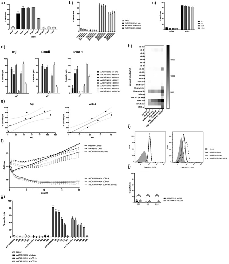
Figure 3.

AdCAR NK-92-mediated tumor cell lysis. AdCAR NK-92 cells were co-incubated with calcein-labeled lymphoma cell lines in the presence or absence of indicated biotinylated antibodies for 2 h. Specific lysis is shown as mean ± SD, n = 3. Using the Raji cell line as target, an optimal bAb concentration of 100 ng/ml was chosen after titration of bCD19 (a). Free biotin added in excess of physiological concentrations showed no impairment of AdCAR-mediated lysis of lymphoma cells (b). Various E:T ratios utilizing AdCAR NK-92 cells as effectors and Raji cells as target were analyzed using a calcein release assay (c). Biotinylated antibodies, already utilized for the flow cytometry screening panel (Table 1), were tested with Raji, Daudi and JeKo-1 cells as target in a CRA (d). Target antigen expression levels were correlated with their respective AdCAR NK-92-mediated tumor cell lysis (e). Kinetics of AdCAR-mediated lysis of Raji cells was assessed using the xCELLigence real-time cell-analysis system at an E:T ratio of 5:1 (f). Cytotoxic effector function of AdCAR NK-92 cells irradiated with 10 Gy at indicated time points was assessed using Raji cells as target (g). The release of cytokines by NK-92 cells was measured using the Bio-Plex Pro human cytokine 17-plex assay and is shown as a heatmap (h). AdCAR NK-92 cells were co-incubated with Raji cells in the presence or absence of bCD19 for 6 h and screened for expression of FasL (CD178) and TRAIL (CD253) via flow cytometry (i). Finally, AdCAR NK-92-mediated lysis of calcein-labeled parental NK-92 cells using bCD56 as adapter molecule (j) was tested for the assessment of possible fratricide.
