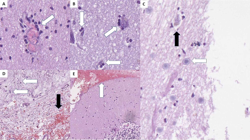
Figure 4. Central nervous system histopathology.
A: inflammatory cell cuffs around small blood vessels (arrow), H&E stain, original magnification 400x; B: neuronophagia - inflammatory cells surrounding degenerative neurons (arrows), H&E stain, original magnification 400x; C: Cowrdy type A body (white arrow) and degenerative neuron with neuronophagia (black arrow), H&E stain, original magnification 400x; D: serous meningitis with focal inflammatory cell infiltration (white arrows) and focal hemorrhages (black arrow), H&E stain, original magnification 100x; E: subarachnoid hemorrhage (arrow) in the posterior fossa, H&E stain, original magnification 100x.
H&E: hematoxylin and eosin.

