Abstract
Objectives
To describe the phenotype variation in Indonesian 46,XX late-identified congenital adrenal hyperplasia (CAH) and the correlation between 17-hydroxyprogesterone (17-OHP) and genital virilization.
Methodology
Retrospective study of 39 cases with five salt-wasting (SW) and 34 simple virilizing (SV) types.
Results
The median age of the patients was 9.83 years (range, 0.58 to 44 years) with Prader score 2 to 5. Clitoromegaly (100%) and skin hyperpigmentation (87%) were the most common features. Lack of breast development (Tanner 1 to 2) and menstrual disorders occurred in 9 patients (teenagers and adults). Short stature (6), low voice (14), prominent Adam’s apple (9) and hirsutism (4) were found only in SV types. Rapid growth (7) and precocious puberty (8) were identified in children. Male gender on admission was found in 13 patients. The mean of 17-OHP level was 304.23 nmol/L [standard deviation (SD) 125.03 nmol/L]. There was no correlation between 17-OHP levels and virilization (r=0.19, p>0.05).
Conclusion
Late-identified CAH showed severe virilization and irreversible sequelae, with clitoromegaly and skin hyperpigmentation as the most commonly seen features. Masculinization of CAH females created uncertainty with regard to sex assignment at birth, resulting in female, male and undecided genders. There is no significant correlation between 17-OHP levels with the degree of virilization in CAH females.
Keywords: CAH, late-identified, phenotype, virilization
INTRODUCTION
Congenital adrenal hyperplasia is a leading cause of 46,XX disorders of sex development (DSD) resulting from a deficiency of enzymes required for cortisol and aldosterone biosynthesis. More than 90% cases have 21-hydroxylase deficiency (21-OHD) and mutations in the CYP21A2 gene, which is located on the short arm of chromosome 6 (6p21.3).1,2 Affected females are born with various degrees of virilization, as a result of prenatal androgen exposure. Aside from 21-OHD, virilization in 46,XX individuals with CAH is also caused by 11β-hydroxylase deficiency (11β-OHD), P450 oxidoreductase deficiency (POR) and 3β-hydroxysteroid dehydrogenase deficiency (3β-HSD).2,3,4
Classic CAH can be further divided into salt-wasting and simple virilizing types. Virilization in a female manifests as clitoromegaly, acne, hirsutism and low voice. At puberty, normal feminization of girls fails to occur, usually presenting with lack of breast development or absence of menstruation.3,4 Elevation of 17-OHP in CAH leads to increased androgen levels, but the relationship between 17-OHP and virilization phenotype has not been consistent as seen in previous studies.2,5
The incidence of CAH in the general population is approximately 1 in 16,000 on screened newborns worldwide, and carriers are present in 1:60.6,7 Data from our center from 2004 to 2016 show that there are 84 patients with CAH, consisting of a wide age range of 3 days to 44 years old. Majority of the patients seen have never received any specific treatment for CAH. Genital ambiguity was the most common reason for physician consult in female patients.
Delayed diagnosis and treatment of CAH in children is common in Indonesia. It is frequent especially for SV or SW types who develop adrenal crises without a definite diagnosis due to the absence of newborn screening.8 In Indonesia, newborn screening for CAH is not yet available, as basic treatment and hormonal tests are not provided in public hospitals with health insurance services. Ethically, it cannot be provided yet because medications such as hydrocortisone and fludrocortisone are not available. In developed countries, a patient with CAH is often diagnosed early in life by neonatal screening programs.9
OBJECTIVES
To describe the phenotype variation in Indonesian 46,XX late-identified CAH, which is rarely reported in other countries, and;
To identify the correlation of 17-OHP and genital virilization.
METHODOLOGY
Study design
This is a descriptive study.
Patients and methods
From 2004 to 2016, we gathered 84 patients with a diagnosis of CAH, evaluated under the clinical management of the multidisciplinary DSD team of the Dr. Kariadi Hospital and Faculty of Medicine Diponegoro University. Thirty-nine patients were enrolled in this study.
CAH was diagnosed based on the clinical manifestations and elevated serum levels of 17-OHP. The inclusion criteria were 46,XX karyotype and age 6 months or older at first visit to our center. Patients who underwent corrective genital surgery before their first visit to our center were excluded.
Medical records were reviewed to gather data including age at diagnosis, anthropometrics, appearance of genitalia, Prader score, Tanner stage, skin hyperpigmentation, acne, hirsutism, and low voice with or without Adam’s apple, menstrual disorders and family history (pedigree). Details and additional information were verified with the patient or parent.
Ethical considerations
This study was approved by the Medical Ethics Committee and informed consent was obtained from all patients and parents.
RESULTS
Clinical findings
Thirty-nine patients (5 SW and 34 SV) fulfilled the inclusion criteria, with a mean age of 9.83 (SD±9.42 years). There were 9 adults and 30 children. Physician-diagnosed SW type was significantly younger compared to SV type (mean age 0.58 versus 7.25 years, p<0.05). The phenotype variation of late identified CAH are shown in Table 1. The most frequent clinical manifestations on both SW and SV types were clitoromegaly (100%) and generalized skin hyperpigmentation mainly around the genital area (87%). Nine patients with ages over 13 years showed lack of breast development (breast Tanner stage 1 to 2). Menstrual disorders included primary amenorrhea (67%) and late menarche occurring at age over 15 years (33%, 15 to 18 years). Precocious puberty (23.3%), defined as premature pubic hair growth, was evident by 3 years old. Fourteen patients had low voice (35.9%), 9 of which had prominent Adam’s apple. Four patients had hirsutism (10.3%), all of whom were SV type. Two patients had acne problems. Eight children had height measurements above the 97th percentiles for age in the Chinese Growth chart (26.7%), the available reference for Asian populations, indicative of rapid growth.10 Short stature, defined as less than the 3rd percentiles of the Chinese Growth Chart, occurred in 6 adult patients (75%). Of this group, one child had accelerated epiphyseal maturation, with bone age at 17 to 18 years.
Table 1.
Phenotypic variation of Indonesian late-identified CAHa patients
| Clinical findings | SWb type (n=5) | SVc type (n=34) | Total | p-value |
|---|---|---|---|---|
| Clitoromegaly (%) | 5/5 (100) | 34/34 (100) | 39/39 (100) | 0 |
| Hyperpigmentation (%) | 5/5 (100) | 29/34 (85.4) | 34/39 (87.2) | 0.6* |
| Hirsutism (%) | 0/5 | 4/34 (11.7) | 4/39 (10.3) | - |
| Low voice (%) | 1/5 (20) | 13/34 (38.2) | 14/39 (35.9) | - |
| With Adam’s apple (%) | 0/5 | 9/13 (69.2) | 9/39 (23.1) | - |
| Acne (%) | 0/5 | 2/34 (5.8) | 2/39 (5.1) | - |
| Short stature | - | |||
| Children | 0/5 | 1/25 (4.0) | 1/30 (3.3) | |
| Adult | 0/5 | 7/9 (77.8) | 7/9 (77.8) | |
| Age 13 years or less | ||||
| Rapid growth | 1/5 (20) | 6/25 (24) | 7/30 (23.3) | - |
| Precocious puberty | 2/5 (40) | 6/25 (24) | 8/30 (26.7) | - |
| Age over 13 years | ||||
| Lack of breast development (Tanner 1 to 2) | 0 | 9/9 (100) | 9/9 (100) | - |
| Menstrual disorders | 0 | 9/9 (100) | 9/9 (100) | - |
| Genitalia status | ||||
| Clitoris/phallus length (cm) | 4.8 ± 2.4 | 4.24 ± 1.2 | 4.3 ± 1.4 | 0.3e |
| Scrotalization | 3/5 (60) | 21/34 (62) | 24/39 (61.5) | 0.6d |
| Complete labial fusion | 1/5 (20) | 6/34 (17.9) | 7/39 (44) | 1d |
| Rudimentary labia minora | 4/5 (80) | 23/34 (67.6) | 27/39 (61.5) | 0.3d |
| Prader score | Stage 3 (60) | Stage 4 (38.2) | Stage 3 (36) | 0.7f |
| Mean serum 17-OHP level, nmol/L | 391.00 | 291.47 | 304.23 | 0.3f |
CAH, congenital adrenal hyperplasia
SW, salt-wasting
SV, simple virilizing
Fisher exact test
T-test
Mann-Whitney test
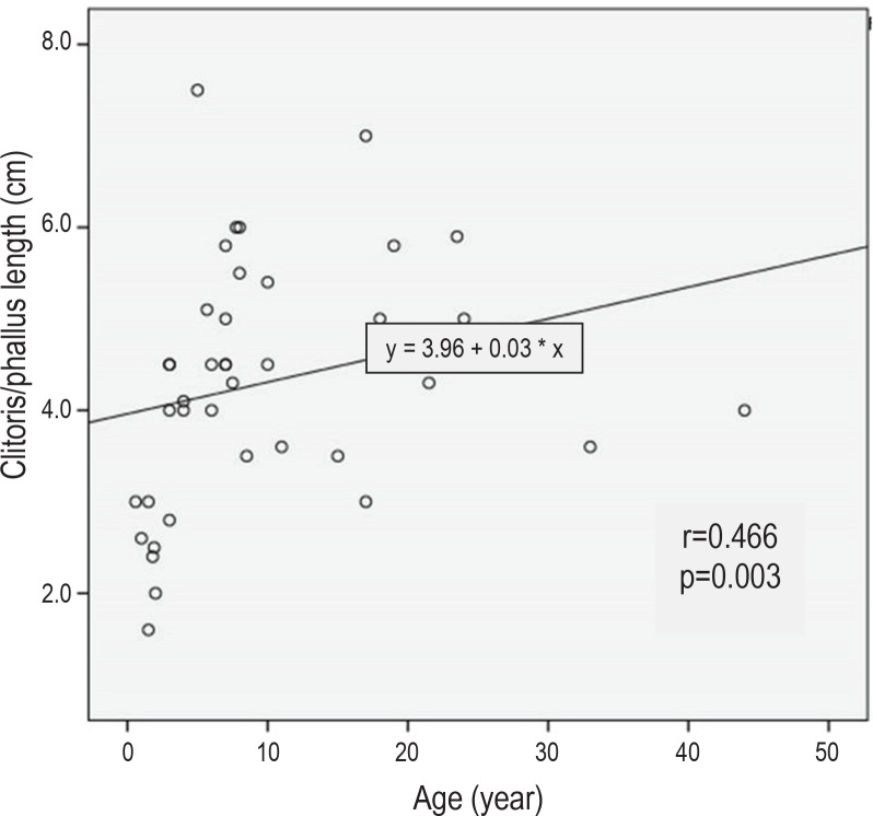
The next most common dysmorphology was rudimentary labia minora, seen in 80% of SW and 67% of SV types (p>0.05). However, clitoral/phallus length was not significantly different between SW and SV types, with mean length 4.8 cm and 4.24 cm, respectively (p>0.05). We found a good correlation between age and phallus length in late-identified CAH (r=0.466, p<0.05) (Figure 1). More than 50% of both SW type and SV type CAH had labial scrotalization. Complete labial fusion was more frequent in SW (20%) compared to SV (17.9%) (p>0.05).
Figure 1.
Relationship between age and phallus length in late-identified CAH.
All patients had genital ambiguity, with a median Prader score of 3 and 4 in SW and SV types, respectively. Thirteen patients (33%) reveal male gender and four patients with male gender of five SW-type on their first visit and one SV type patient had undetermined gender. However, after undergoing thorough physical and laboratory examination and genetic counseling, four patients decided to change their gender from male to female (Table 2).
Table 2.
Gender assignment after final examination
| Patient number | Gender and age on admission | Gender and age after final examination | ||
|---|---|---|---|---|
| Gender | Age (y.o**) | Gender | Age (y.o) | |
| 1 | Male | 17 | Male | 18 |
| 2 | Female | 11 | Female | 12 |
| 3 | Female | 33 | Female | 33.5 |
| 4 | Female | 6 | Female | 6.5 |
| 5 | Female | 7 | Female | 8 |
| 6 | Female | 7 | Female | 8 |
| 7 | Female | 4 | Female | 5 |
| 8 | Male | 3 | Male | 4 |
| 9 | Female | 1.5 | Female | 2 |
| 10 | Female | 1.9 | Female | 2 |
| 11 | Female | 10 | Female | 11 |
| 12 | Female | 3 | Female | 3.5 |
| 13 | Female | 7 | Female | 8 |
| 14 | Female | 6 | Female | 6.5 |
| 15 | Male | 24 | Male | 25 |
| 16 | Female | 7.5 | Female | 8 |
| 17 | Female | 4 | Female | 4.5 |
| 18 | Male* | 7.75 | Male | 8 |
| 19 | Male | 8 | Male | 8.5 |
| 20 | Female | 23.5 | Female | 24 |
| 21 | Female | 18 | Female | 18 |
| 22 | Male | 1.5 | Female | 2 |
| 23 | Female | 15 | Female | 16 |
| 24 | Male | 8.5 | Male | 9.5 |
| 25 | Female | 17 | Female | 17.5 |
| 26 | Female | 3 | Female | 3.5 |
| 27 | undecided | 2 | undecided | 3 |
| 28 | Male | 44 | Male | 46 |
| 29 | Male* | 1 | Female | 1.5 |
| 30 | Male* | 0.58 | Female | 1 |
| 31 | Male | 10 | Male | 10.5 |
| 32 | Female | 3 | Female | 4 |
| 33 | Female | 19 | Female | 20 |
| 34 | Female* | 5.67 | Female | 6.5 |
| 35 | Female | 21.5 | Female | 22 |
| 36 | Male | 8 | Male | 8.5 |
| 37 | Female | 7 | Female | 8 |
| 38 | Male* | 5 | Female | 5.5 |
| 39 | Female | 1.8 | Female | 2 |
Salt Wasting type (*)
y.o = years old (**)
Correlation of 17-OHP with phallus length
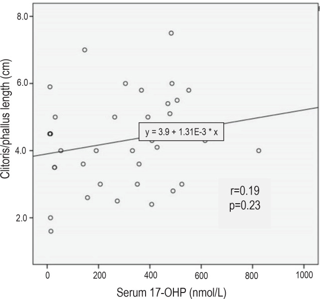
All patients had elevated blood levels of 17-OHP. Based on Spearman’s correlation coefficient test, there was no correlation between 17-OHP level and phallus length (r=0.19, p>0.05) (Figure 2).
Figure 2.
Relationship between serum 17-hydroxyprogesterone (17-OHP) levels and phallus length in late-identified CAH.
DISCUSSION
The mean age of presentation of patients in our study is nine years, quite different from data from Western populations.11 Recognition is frequently late due to lack of knowledge of medical providers, especially in remote regions; lack of awareness of parents to seek medical management; unavailability of neonatal screening in Indonesia; and early death of affected babies before diagnosis.
There are significant differences in the number of patients and their age at diagnosis in SW and SV types in our study, in contrast to other published reports.12 This discrepancy suggests that more of SW type CAH patients died from adrenal crises before being diagnosed. Identifying newborn girls with signs and symptoms of CAH is not difficult because of genital ambiguity and salt wasting symptoms in the SW type. In newborn boys, the diagnosis is challenging because of the absence of ambiguous genitalia, especially in the SV type. Moreover, CAH in Indonesia is still unfamiliar for many medical practitioners, such as general practitioners or midwives. Factors that may lead to late diagnosis and treatment include high cost and limited facilities for hormonal analysis to detect CAH, and the difficult access to appropriate medications.
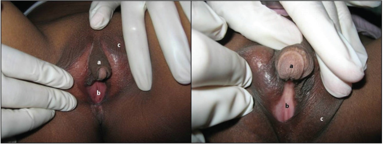
Clinical variations depend on the degree of hormonal disruption, and timeliness and regularity of treatment.13 Virilization is very prominent in late-identified females with CAH due to the untreated condition (Figure 3). There is a good correlation between age and phallus length in late-identified CAH, indicating that older untreated CAH females are more virilized. Androgen exposure in these girls start from the prenatal period and persist until they receive treatment.14 In our study, androgen excess in untreated females resulted to progressive virilization of the clitoris, hyperpigmentation, lack of breast development, primary amenorrhea, delayed menarche, premature pubic hair growth, hirsutism, low voice with or without prominent Adam’s apple and short stature. These observations are comparable with findings in previous studies.15,16
Figure 3.
Virilized external genitalia in 2 female siblings showing clitoromegaly (a), presence of vaginal introitus (b), and hyperpigmentation (c).
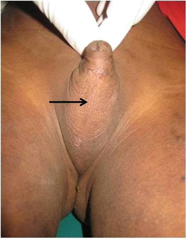
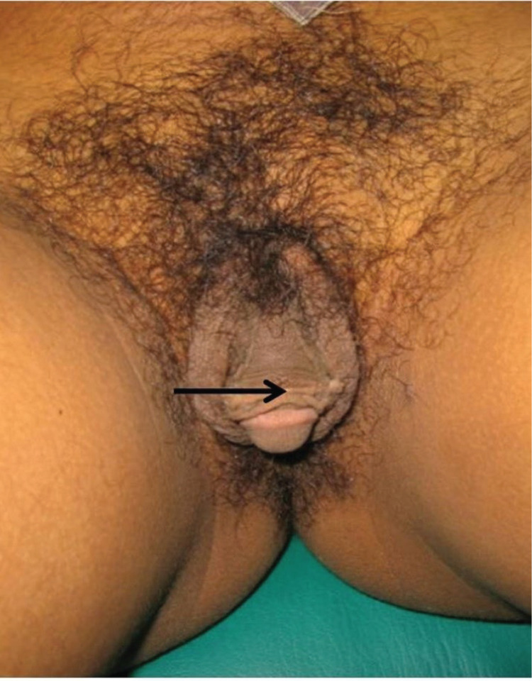
In our study, thirty-three percent of patients were considered male because of the presence of male-looking genitalia (clitoromegaly resembling penis, scrotalization with or without complete labial fusion) and the absence of treatment (Figures 4 and 5). This emphasizes the need for a thorough urogenital examination in the neonatal period for patients with DSD. Chromosomal and hormonal analyses should be performed on neonates with ambiguous genitalia to facilitate gender assignment.17
Figure 4.
An 8.5-year-old child assigned to male gender with 46,XX late-identified CAH. There is complete labial fusion (arrow) resembling a scrotum.
Figure 5.
A 24-year-old adult assigned to male gender with 46,XX late-identified CAH. There is a big phallus (arrow) with scrotalization.
Precocious puberty in female CAH patients are recognized by premature pubic hair growth occurring before 8 years of age and rapid growth.18 In the same study, patients treated after age one year have a higher risk for precocious puberty; for SV type boys, this risk was apparent even in those who received treatment at the age of 6 months.18 We found similar findings with Hargitai et al., our SV type children showed accelerated growth patterns after 3 years old, while the final height of adult patients leading to short stature was reduced compared to the standard population and their respective target heights.19 Bone age determination may be able to show accelerated epiphyseal maturation. Unfortunately, these results were not available in all our CAH children, due to financial constraints and limited facilities.
In our study, all late-identified CAH patients had elevated blood levels of 17-OHP. The 17-OHP levels were weakly correlated with phallus length (r=0.19). However, there was no clear relationship between 17-OHP levels and virilization of external genitalia, similar to findings by Rocha et al. 20
Genetic counseling for families and patients is advised to explain the clinical manifestations, mode of inheritance, recurrence risk and consequences of CAH. Some challenges to our work include varied cultural beliefs within families, patient discrimination and taboos pertaining to their masculinized condition, and the unavailability of a genetic counselor.21 Gender assignment is still a dilemma: some of the children were raised as males, females or left undecided. Late-identified CAH females with progressive virilization should undergo psychological evaluation to assess their emotional condition and gender identity.
CONCLUSION
In our study, the most common clinical features in late-identified Indonesian CAH patients were clitoromegaly and skin hyperpigmentation. Clinical features such as genital virilization, low voice, Adam's apple and short stature in adults were permanent phenotypes, indicating the severity and irreversibility of virilization despite medication. There was no correlation between of 17-OHP levels and virilization. There is a general lack of awareness of medical personnel, family members and patients regarding CAH and its effects. The high cost and limited availability of laboratory tests and difficult access to medications pose significant challenges as well. Improvement in the clinical recognition of suspected CAH patients is important in our Indonesian clinical setting. Genetic counseling is crucial in the management of CAH, especially for gender determination. Newborn screening should be included in the improvement of health policy in Indonesia.
Acknowledgments
We thank the multidisciplinary DSD team of Dr. Kariadi Hospital - FMDU and the staff in CEBIOR for their help with the patients and laboratory work. We would like to thank all the families involved in our study. The author (MU) is a recipient of the Beasiswa Unggulan BPKLN fellowship from the Indonesian Ministry of Education and Culture.
Statement of Authorship
All authors certified fulfillment of ICMJE authorship criteria.
Author Disclosure
The authors declared no conflict of interest.
Funding Source
This study was supported by Competitive Research Grant from the Indonesian Ministry of Research, Technology and Education - DIPA number 023.04.2.673453/2015 and number 022/SP2H/LT/DRPM/II/2016.
Authors are required to accomplish, sign and submit scanned copies of the JAFES Author Form consisting of: (1) the Authorship Certification that the manuscript has been read and approved by all authors, and that the requirements for authorship have been met by each author, (2) the Author Declaration that the article represents original material that is not being considered for publication or has not been published or accepted for publication elsewhere, (3) the Statement of Copyright Transfer[accepted manuscripts become the permanent property of the JAFES and are licensed with an Attribution-Share Alike-Non-Commercial Creative Commons License. Articles may be shared and adapted for non-commercial purposes as long as they are properly cited], (4) the Statement of Disclosure that there are no financial or other relationships that might lead to a conflict of interest. For Original Articles involving human participants, authors are required to submit a scanned copy of the Ethics Review Approval of their research. For manuscripts reporting data from studies involving animals, authors are required to submit a scanned copy of the Institutional Animal Care and Use Committee approval. For Case Reports or Series, and Images in Endocrinology, consent forms, are required for the publication of information about patients; otherwise, authors declared that all means have been exhausted for securing such consent. Articles and any other material published in the JAFES represent the work of the author(s) and should not be construed to reflect the opinions of the Editors or the Publisher.
References
Reference entries #11, 13 and 17 were not cited in the text.
- 1.Ekenze SO, Nwangwu EI, Amah CC, Agugua-Obianyo NE, Onuh AC, Ajuzieogu OV. Disorders of sex development in a developing country: Perspectives and outcome of surgical management of 39 cases. PediatrSurg Int. 2015;31(1):93-9. PMID: https://doi.org/1007/s00383-014-3628-1. [DOI] [PubMed] [Google Scholar]
- 2.Speiser PW, White PC. Congenital adrenal hyperplasia. N Engl J Med. 2003;349(8):776-88. PMID: 10.1056/NEJMra021561 [DOI] [PubMed] [Google Scholar]
- 3.Mooij CF, van Herwaarden AE, Claahsen-van der Grinten HL. Disorders of adrenal steroidogenesis: Impact on gonadal function and sex development. Pediatr Endocrinol Rev. 2016;14(2):109-28. PMID: https://10.17458/PER.2016.MEC.DisordersofAdrenal. [DOI] [PubMed] [Google Scholar]
- 4.Auchus RJ. The classic and nonclassic congenital adrenal hyperplasias. Endocr Pract. 2015;21(4):383-9. PMID: 10.4158/EP14474.RA [DOI] [PubMed] [Google Scholar]
- 5.Sugiyama Y, Mizuno H, Hayashi Y, et al. Severity of virilization of external genitalia in Japanese patients with salt-wasting 21- hydroxylase deficiency. Tohoku J Exp Med. 2008;215(4):341-8. PMID: . [DOI] [PubMed] [Google Scholar]
- 6.van der Kamp HJ, Wit JM. Neonatal screening for congenital adrenal hyperplasia. Eur J Endocrinol. 2004;151(Suppl 3):U71-5. PMID: . [DOI] [PubMed] [Google Scholar]
- 7.Trapp CM, Oberfield SE. Recommendations for treatment of nonclassic congenital adrenal hyperplasia (NCCAH): An update. Steroids. 2012;77(4):342-6. PMID: PMCID: NIHMSID: NIHMS461777. [DOI] [PMC free article] [PubMed] [Google Scholar]
- 8.Juniarto AZ, van der Zwan YG, Santosa A, et al. Application of the new classification on patients with a disorder of sex development in Indonesia. Int J Endocrinol. 2012;2012:237084. PMID: PMCID: 10.1155/2012/237084 [DOI] [PMC free article] [PubMed] [Google Scholar]
- 9.Ediati A, Faradz SM, Juniarto AZ, van der Ende J, Drop SL, Dessens AB. Emotional and behavioral problems in late-identified Indonesian patients with disorders of sex development. J Psychosom Res. 2015;79(1):76-84. PMID: 10.1016/j.jpsychores.2014.12.007 [DOI] [PubMed] [Google Scholar]
- 10.Chang KSF, Lee MMC, Low WD. Standards of height and weight of Southern Chinese Children. Far East Med J. 1965;1:101-9. [Google Scholar]
- 11.Juniarto AZ, van der Zwan YG, Santosa A, et al. Hormonal evaluation in relation to phenotype and genotype in 286 patients with a disorder of sex development from Indonesia. Clin Endocrinol (Oxf). 2016;85(2):247-57. PMID: 10.1111/cen.13051 [DOI] [PubMed] [Google Scholar]
- 12.Dörr HG, Odenwald B, Nennstiel-Ratzel U. Early diagnosis of children with classic congenital adrenal hyperplasia due to 21-hydroxylase deficiency by newborn screening. Int J Neonatal Screen. 2015;1:36-44. 10.3390/ijns1010036 [DOI] [Google Scholar]
- 13.Knowles RL, Khalid JM, Oerton JM, Hindmarsh PC, Kelnar CJ, Dezateux C. Late clinical presentation of congenital adrenal hyperplasia in older children: Findings from nationalpaediatric surveillance. Arch Dis Child. 2014;99(1):30-4. PMID: PMCID: 10.1136/archdischild-2012-303070 [DOI] [PMC free article] [PubMed] [Google Scholar]
- 14.Ambroziak U, Bednarczuk T, Ginalska-Malinowska M, et al. Congenital adrenal hyperplasia due to 21-hydroxylase deficiency - management in adults. Endokrynol Pol. 2010;61(1):142-55. PMID: . [PubMed] [Google Scholar]
- 15.Kulshreshtha B, Eunice M, Ammini AC. Pubertal development among girls with classical congenital adrenal hyperplasia initiated on treatment at different ages. Indian J Endocrinol Metab. 2012;16(4):599-603. PMID: PMCID: 10.4103/2230-8210.98018 [DOI] [PMC free article] [PubMed] [Google Scholar]
- 16.Sahakitrungruang T. Clinical and molecularreview of atypical congenital adrenal hyperplasia. Ann Pediatr Endocrinol Metab. 2015;20(1):1-7. PMID: PMCID: https://doi.org.10.6065/apem.2015.20.1.1. [DOI] [PMC free article] [PubMed] [Google Scholar]
- 17.Moshiri M, Chapman T, Fechner PY, et al. Evaluation and management of disorders of sex development: Multidisciplinary approach to a complex diagnosis. Radiographics. 2012;32(6):1599-618. PMID: 10.1148/rg.326125507 [DOI] [PubMed] [Google Scholar]
- 18.Bonfig W, Schwarz HP. Growth pattern of untreated boys with simple virilizing congenital adrenal hyperplasia indicates relative androgen insensitivity during the first six months of life. Horm Res Paediatr. 2011:75(4):264-8. PMID: 10.1159/000322580 [DOI] [PubMed] [Google Scholar]
- 19.Hargitai B, Solyom J, Battelino T, et al. Growth patterns and final height in congenital adrenal hyperplasia due to classical 21-hydroxylase deficiency. Results of a multicenter Study. Horm Res. 2001;55(4):161-71. PMID: 10.1159/000049990 [DOI] [PubMed] [Google Scholar]
- 20.Rocha RO, Billerbeck AE, Pinto EM, et al. The degree of external genitalia virilization in girls with 21-hydroxylase deficiency appears to be influenced by the CAG repeats in the androgen receptor gene. Clin Endocrinol (Oxf). 2008;68(2):226-32. PMID: 10.1111/j.1365-2265.2007.03023.x [DOI] [PubMed] [Google Scholar]
- 21.Warne GL, Raza J. Disorders of sex development (DSDs), their presentation and management in different cultures. Rev EndocrMetabDisord. 2008;9(3):227-36. PMID: 10.1007/s11154-008-9084-2 [DOI] [PubMed] [Google Scholar]


