Abstract
Background
Cervical esophageal cancer (CEC) is an uncommon malignancy with poor prognosis, and there is no specific model that can be used to accurately predict the survival of patients with CEC.
Methods
The Surveillance, Epidemiology, and End Results (SEER) database was searched for patients with non-metastatic CEC from 2004 to 2015. Overall survival (OS) and disease-specific survival (DSS) rates were calculated using the Kaplan-Meier method. Predictive factors were analyzed by Cox’s proportional hazards regression, and a nomogram was created to predict survival probability using R software.
Results
We identified 601 patients with CEC, 94.3% of whom had squamous cell carcinoma (SCC). The median follow-up time was 71 months. The median OS and DSS for the overall population were 15 and 18 months, respectively. There was a statistically significant decrease in surgical rates over time, from 16.7% in 2004 to 8% in 2015 (P=0.035). Comprehensive strategies consisting of two or three treatment modalities were correlated with significantly better OS and DSS (P<0.001 for both). We randomly assigned half of the patients to the training cohort (n=300) and the other half to the validation cohort (n=301). Multivariate Cox regression analysis was performed using the training cohort. Age, sex, tumor size, stages in the 7th edition of the American Joint Committee on Cancer (AJCC) staging system, and treatment with surgery, radiotherapy, or chemotherapy were identified as independent risk factors for OS. These factors were incorporated into the development of a nomogram for predicting 1-, 3-, and 5-year OS rates. The C-index of the nomogram was 0.743, which was statistically higher than that of the AJCC staging system. The internal validation, using bootstrap resampling and external validation, demonstrated the accuracy of the nomogram.
Conclusions
We developed and validated the first nomogram for CEC. This nomogram could be used to predict the OS of CEC patients with a relatively high accuracy.
Keywords: Cervical esophageal cancer (CEC), nomogram, surgery, comprehensive treatment, prognosis
Introduction
Cervical esophageal cancer (CEC) is a relatively uncommon malignancy, accounting for approximately 5% of all esophageal cancers (1). Squamous cell carcinoma (SCC) is a major histologic type of CEC, accounting for approximately 95% of CEC cases. The 5-year overall survival (OS) of patients with CEC is lower than that of patients with other SCCs of the head and neck region (2), and is more comparable to the 5-year OS of patients with SCC located in other regions of the esophagus, which is approximately 26% (3). However, CEC differs from cancers of the thoracic esophagus in other aspects, such as genetic alterations, prognostic factors, and cancer management (4,5). Therefore, CEC is a unique disease that has specific characteristics.
Nomograms have been widely used for predicting prognoses in a diverse range of cancers with success. Compared to the American Joint Committee on Cancer (AJCC) Tumor-Node-Metastasis (TNM) staging system, nomograms quantify risk by incorporating all clinicopathological variables, allowing for individualized prognostic predictions for various types of cancer (6-11). However, to the best of our knowledge, no specific nomogram has yet been developed for CEC. The present study is the first to develop a prognostic nomogram for CEC based on a large cohort of patients from the Surveillance, Epidemiology, and End Results (SEER) database. The present study was performed in accordance with the STROBE reporting checklist (available at http://dx.doi.org/10.21037/atm-20-2505).
Methods
To identify the population of interest, we collected data from the recent SEER 18 database. Patients with non-metastatic CEC (C150) from 2004 to 2015 were chosen. The following histological subtypes were included: (I) adenocarcinoma (8,050 to 8,052, 8,123, 8,140 to 8,147, 8,210 to 8,211, 8,255, 8,260 to 8,263, 8,310, 8,480 to 8,481, 8,490, 8,550, and 8,570 to 8,575), and (II) SCC (8,032, 8,070 to 8,077, 8,083, and 8,094). Information on patient characteristics (age, sex, race, and year of diagnosis), primary tumor features (histology, grade, T stage, N stage, and tumor size), treatment approaches (surgery, radiation, and chemotherapy), and clinical outcomes (cancer-specific survival and OS) were collected.
Continuous variables were summarized as medium (range), and categorical variables were summarized as number (percentage). Survival was evaluated using the Kaplan-Meier method and the log-rank test. Univariate and multivariate analyses of clinicopathological factors were performed using Cox proportional hazards model to identify risk factors for OS and disease-specific survival (DSS). For statistical testing, we used a two-sided significance level (alpha) of 0.05. We selected the optimum cutoff score for the tumor size using X-tile plots (version 3.6.1; Yale University School of Medicine, New Haven, CT, USA).
For the development of the nomogram, we randomly assigned half of the patients into a training cohort (n=300) and the other half into a validation cohort (n=301). A nomogram was created based on the results of the multivariable analysis. Predictive performance was assessed based on the C-index and external calibration plots with samples in the validation cohort. We compared the nomogram with the TNM stage system using the rcorr.cens function in the R package Hmisc. All statistical analyses were performed using IBM SPSS software (version 23.0; IBM, Armonk, NY, USA) and R software (version 3.1.1; http://www.r-project.org). This study was conducted in accordance with the Declaration of Helsinki (as revised in 2013).
Results
Patient characteristics and treatment
The patient selection process is shown in Figure 1. A total of 601 patients were included in the present study. The demographic and clinicopathological characteristics of the study cohort are provided in Table 1. The median age at diagnosis was 68 years, and 59.4% of the patients were male. SCC was the predominant histological type; 567 (94.3%) patients were diagnosed with SCC, whereas 34 (5.7%) patients had adenocarcinoma (AC). Of the 498 patients with documented tumor size, the median size was 40 mm. The majority of patients presented with locally advanced primary cancer, with 62.1% having a primary tumor classification of T3 or T4. Most of the patients (58.6%) had no nodal involvement.
Figure 1.
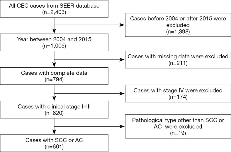
Flow diagram of the patient selection process for the study.
Table 1. Demographics and clinicopathological characteristics of patients with non-metastatic cervical esophageal carcinoma.
| Characteristics | Training set (n=300), n (%) | Validation set (n=301), n (%) | Total (n=601), n (%) |
|---|---|---|---|
| Age (years), median (range) | 67 (25 to 98) | 69 (42 to 99) | 68 (25 to 99) |
| <65 | 131 (43.7) | 115 (38.2) | 246 (40.9) |
| ≥65 | 169 (56.3) | 186 (61.8) | 355 (59.1) |
| Sex | |||
| Male | 178 (59.3) | 179 (59.5) | 357 (59.4) |
| Female | 122 (40.7) | 122 (40.5) | 244 (40.6) |
| Race/region | |||
| White | 227 (75.9) | 242 (80.4) | 469 (78.2) |
| Black | 53 (17.7) | 36 (12.0) | 89 (14.8) |
| Other | 19 (6.3) | 23 (7.6) | 42 (7.0) |
| Year of diagnosis | |||
| 2004 to 2009 | 147 (49.0) | 145 (48.2) | 293 (48.8) |
| 2010 to 2015 | 153 (51.0) | 156 (51.8) | 308 (51.2) |
| Histology | |||
| Squamous | 285 (95.0) | 282 (93.7) | 567 (94.3) |
| Adenocarcinoma | 15 (5.0) | 19 (6.3) | 34 (5.7) |
| Tumor size (mm) | |||
| <55 | 149 (76.4) | 159 (74.6) | 308 (75.5) |
| ≥55 | 46 (23.6) | 54 (25.4) | 100 (24.5) |
| Differentiation | |||
| Well | 15 (6.5) | 12 (5.3) | 27 (5.9) |
| Moderate | 136 (58.9) | 142 (62.8) | 278 (60.8) |
| Poor | 78 (33.8) | 72 (31.9) | 150 (32.8) |
| Undifferentiated | 2 (0.9) | 0 (0.0) | 2 (0.4) |
| T6th stage† | |||
| T1 | 89 (29.7) | 91 (30.2) | 180 (30.0) |
| T2 | 27 (9.0) | 21 (7.0) | 48 (8.0) |
| T3 | 86 (28.7) | 87 (28.9) | 173 (28.8) |
| T4 | 98 (32.7) | 102 (33.9) | 200 (33.3) |
| N6th stage† | |||
| N0 | 169 (57.3) | 178 (59.9) | 347 (58.6) |
| N1 | 126 (42.7) | 119 (40.1) | 245 (41.4) |
| AJCC6th stage† | |||
| I | 68 (22.7) | 70 (23.3) | 138 (23.0) |
| IIa | 59 (19.7) | 64 (21.3) | 123 (20.5) |
| IIb | 31 (10.3) | 25 (8.3) | 56 (9.3) |
| III | 142 (47.3) | 142 (47.2) | 284 (47.3) |
| T7th stage‡ | |||
| T1a | 14 (4.7) | 12 (4.0) | 26 (4.3) |
| T1b | 12 (4.0) | 11 (3.7) | 23 (3.8) |
| T1-NOS | 63 (21.0) | 68 (22.6) | 131 (21.8) |
| T2 | 27 (9.0) | 21 (7.0) | 48 (8.0) |
| T3 | 86 (28.7) | 87 (28.9) | 173 (28.8) |
| T4a | 22 (7.3) | 20 (6.6) | 42 (7.0) |
| T4b | 23 (7.7) | 19 (6.3) | 42 (7.0) |
| T4-NOS | 53 (17.7) | 63 (20.9) | 116 (19.3) |
| N7th stage‡ | |||
| N0 | 169 (67.6) | 179 (70.8) | 348 (69.2) |
| N1 | 64 (25.6) | 62 (24.5) | 126 (25.0) |
| N2 | 13 (5.2) | 8 (3.2) | 21 (4.2) |
| N3 | 4 (1.6) | 4 (1.6) | 8 (1.6) |
| AJCC7th stage‡ | |||
| Ia | 20 (8.8) | 24 (10.8) | 44 (9.8) |
| Ib | 48 (21.2) | 47 (21.2) | 95 (21.2) |
| IIa | 18 (8.0) | 15 (6.8) | 33 (7.4) |
| IIb | 64 (28.3) | 61 (27.5) | 125 (27.9) |
| IIIa | 30 (13.3) | 34 (15.3) | 64 (14.3) |
| IIIb | 4 (1.8) | 5 (2.3) | 9 (2.0) |
| IIIc | 42 (18.6) | 36 (16.2) | 78 (17.4) |
| Surgery | |||
| Yes | 37 (12.3) | 46 (15.3) | 83 (13.8) |
| No | 263 (87.7) | 255 (84.7) | 518 (86.2) |
| Radiation | |||
| Yes | 225 (75.0) | 226 (75.1) | 453 (75.4) |
| No | 75 (25.0) | 75 (24.9) | 148 (24.6) |
| Chemotherapy | |||
| Yes | 199 (66.3) | 205 (68.1) | 404 (67.2) |
| No | 101 (33.7) | 96 (31.9) | 197 (32.8) |
†, from the AJCC 6th edition staging system; ‡, from the AJCC 7th edition staging system. AJCC, American Joint Committee on Cancer; NOS, not otherwise specified.
A total of 83 patients (13.8%) underwent surgery, and 453 patients (75.4%) were treated with radiotherapy (RT). Patients were evaluated to determine whether treatment decisions were related to demographic or clinicopathological factors. We found that patients were more likely to undergo surgery if they were diagnosed before 2009, had AC, had relatively small primary tumors, presented with early-stage disease, or had no nodal involvement (Table 2). We observed a statistically significant decrease in the incidence of surgery between 2004 and 2015, from 16.7% in 2004 to 8% in 2015 (P=0.035) (Figure 2).
Table 2. Correlation between demographic or clinicopathologic factors and treatment decisions.
| Factors | Surgery | Non-surgery | P value | Chemotherapy | Non-chemotherapy | P value | Radiotherapy | Non-radiotherapy | P value |
|---|---|---|---|---|---|---|---|---|---|
| Age (year) | 0.151 | <0.001 | 0.035 | ||||||
| <65 | 206 | 40 | 52 | 194 | 50 | 196 | |||
| ≥65 | 312 | 43 | 145 | 210 | 100 | 255 | |||
| Sex | 0.719 | 0.860 | 0.702 | ||||||
| Male | 212 | 32 | 81 | 163 | 63 | 181 | |||
| Female | 306 | 51 | 116 | 241 | 150 | 451 | |||
| Race/region | 0.036 | 0.930 | 0.680 | ||||||
| White | 397 | 72 | 153 | 316 | 120 | 349 | |||
| Black | 83 | 6 | 31 | 58 | 21 | 68 | |||
| Other | 37 | 5 | 13 | 29 | 9 | 33 | |||
| Year of diagnosis | 0.045 | 0.082 | 0.573 | ||||||
| 2004–2009 | 243 | 49 | 106 | 186 | 76 | 216 | |||
| 2010–2015 | 275 | 34 | 91 | 218 | 74 | 235 | |||
| Histology | 0.017 | 1.000 | 0.157 | ||||||
| Squamous | 494 | 73 | 186 | 381 | 138 | 429 | |||
| Adenocarcinoma | 24 | 10 | 11 | 23 | 12 | 22 | |||
| Tumor size (mm) | 0.014 | 0.542 | 0.894 | ||||||
| <55 | 248 | 60 | 103 | 205 | 77 | 231 | |||
| ≥55 | 91 | 9 | 30 | 70 | 24 | 76 | |||
| Differentiation | 0.657 | 0.742 | 0.828 | ||||||
| Well | 21 | 6 | 10 | 17 | 6 | 21 | |||
| Moderate | 233 | 45 | 89 | 189 | 74 | 204 | |||
| Poor | 129 | 21 | 48 | 102 | 38 | 112 | |||
| Undifferentiated | 2 | 0 | 0 | 2 | 1 | 1 | |||
| T6th stage† | 0.591 | 0.001 | <0.001 | ||||||
| T1 | 159 | 21 | 78 | 102 | 61 | 119 | |||
| T2 | 39 | 9 | 11 | 37 | 8 | 40 | |||
| T3 | 147 | 26 | 42 | 131 | 27 | 146 | |||
| T4 | 173 | 27 | 66 | 134 | 54 | 146 | |||
| N6th stage† | 0.001 | <0.001 | <0.001 | ||||||
| N0 | 285 | 62 | 139 | 208 | 106 | 241 | |||
| N1 | 224 | 21 | 55 | 190 | 42 | 203 | |||
| AJCC6th stage† | 0.024 | <0.001 | 0.025 | ||||||
| I | 119 | 19 | 66 | 72 | 48 | 90 | |||
| IIa | 97 | 26 | 40 | 83 | 28 | 95 | |||
| IIb | 53 | 3 | 14 | 42 | 13 | 43 | |||
| III | 249 | 35 | 77 | 207 | 61 | 223 | |||
| T7th stage‡ | 0.009 | 0.001 | <0.001 | ||||||
| T1a | 19 | 7 | 12 | 14 | 12 | 14 | |||
| T1b | 14 | 9 | 15 | 8 | 14 | 9 | |||
| T2 | 39 | 9 | 11 | 37 | 8 | 40 | |||
| T3 | 147 | 26 | 42 | 131 | 27 | 146 | |||
| T4a | 36 | 6 | 15 | 27 | 8 | 34 | |||
| T4b | 40 | 2 | 13 | 29 | 14 | 28 | |||
| N7th stage‡ | 0.045 | <0.001 | 0.005 | ||||||
| N0 | 284 | 64 | 139 | 209 | 105 | 243 | |||
| N1 | 114 | 12 | 24 | 102 | 20 | 106 | |||
| N2 | 18 | 3 | 6 | 15 | 2 | 19 | |||
| N3 | 5 | 3 | 4 | 4 | 2 | 6 | |||
| AJCC7th stage‡ | 0.011 | <0.001 | <0.001 | ||||||
| Ia | 34 | 10 | 24 | 20 | 20 | 24 | |||
| Ib | 86 | 9 | 42 | 53 | 27 | 68 | |||
| IIa | 31 | 2 | 11 | 22 | 3 | 30 | |||
| IIb | 96 | 29 | 36 | 89 | 33 | 92 | |||
| IIIa | 58 | 6 | 10 | 54 | 6 | 58 | |||
| IIIb | 7 | 2 | 2 | 7 | 1 | 8 | |||
| IIIc | 70 | 8 | 26 | 52 | 20 | 58 |
†, from the AJCC 6th edition staging system; ‡, from the AJCC 7th edition staging system. AJCC, American Joint Committee on Cancer.
Figure 2.
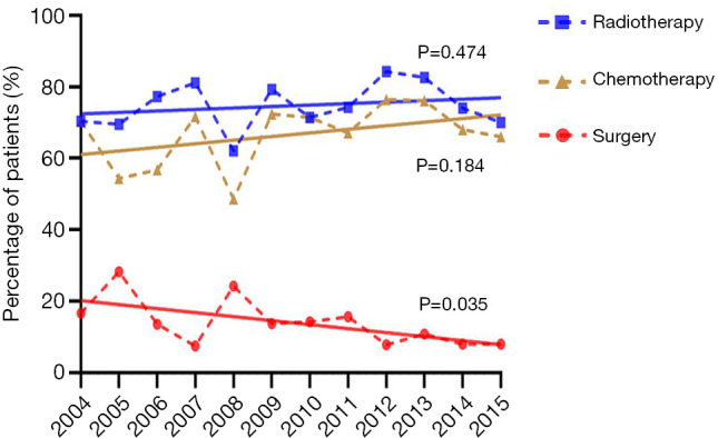
Rates of use of surgery, radiotherapy, and chemotherapy between the years 2004 and 2015 in non-metastatic CEC. P values represent the comparison of the linear regression line and a line with slope equal to 0 for each treatment modality.
Survival analysis
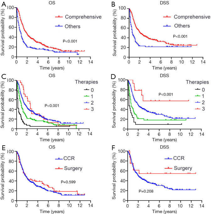
The median follow-up time was 71 months. The median OS and DSS for the overall population were 15 and 18 months, respectively. Most of the patients (64.4%) underwent comprehensive treatment consisting of surgery, RT, or chemotherapy. There was a significant improvement in OS and DSS among patients who underwent comprehensive treatment (Figure 3A,B). In a subgroup of patients with SCC, trimodal therapy consisting of surgery and chemoradiotherapy showed the best DSS, although there was no improvement in OS over dual therapy (Figure 3C,D). Patients who underwent surgery usually had earlier-stage disease and smaller tumor size (Table 3); however, there was no significant difference in OS or DSS between those who underwent surgery only and those who underwent surgery and chemoradiotherapy (Figure 3E,F).
Figure 3.
OS and DSS for patients with non-metastatic CEC. (A,B) OS and DSS among patients who underwent comprehensive treatment and those who did not. (C,D) OS and DSS among patients whose number of treatment modalities was different. (E,F) OS and DSS among patients who underwent surgery alone and those who underwent definitive chemoradiotherapy in the SCC subgroup.
Table 3. Correlation between clinicopathologic factors and treatment decisions.
| Factors | Surgery alone (n) | CCR† (n) | P value |
|---|---|---|---|
| Tumor size (mm) | 0.024 | ||
| <55 | 45 | 177 | |
| ≥55 | 5 | 60 | |
| T7th stage‡ | <0.001 | ||
| T1a | 5 | 9 | |
| T1b | 9 | 7 | |
| T2 | 5 | 31 | |
| T3 | 21 | 121 | |
| T4a | 5 | 25 | |
| T4b | 1 | 24 | |
| N7th stage‡ | 0.007 | ||
| N0 | 49 | 178 | |
| N+ | 10 | 168 | |
| N2 | 2 | 14 | |
| N3 | 1 | 2 | |
| AJCC7th stage‡ | 0.033 | ||
| Ia | 9 | 19 | |
| Ib | 7 | 46 | |
| IIa | 1 | 21 | |
| IIb | 20 | 73 | |
| IIIa | 6 | 53 | |
| IIIb | 1 | 6 | |
| IIIc | 4 | 43 |
†, definitive chemoradiotherapy; ‡, from the AJCC 7th edition staging system. AJCC, American Joint Committee on Cancer.
Prognostic factors for OS and DSS in the overall cohort
Univariate analysis demonstrated that older age (P=0.002), male sex (P=0.006), SCC (vs. AC) (P=0.008), larger tumor size (P<0.046), higher T (7th) stage (P<0.001), higher AJCC (7th) stage (P<0.001) and the absence of RT (P=0.025), chemotherapy (P<0.001), or surgery (P=0.010) were all associated with decreased OS (Table 4).
Table 4. Univariable and multivariable Cox proportional hazards regression for overall survival of the training set.
| Variable | Univariate analysis | Multivariate analysis | |||
|---|---|---|---|---|---|
| HR (95% CI) | P value | HR (95% CI) | P value | ||
| Age ≥65 years | 1.52 (1.17–1.6) | 0.002 | 1.85 (1.13–3.04) | 0.015 | |
| Male vs. female | 1.45 (1.11–1.89) | 0.006 | 1.71 (1.03–2.83) | 0.038 | |
| AC vs. SCC | 0.38 (0.19–0.78) | 0.008 | – | – | |
| Tumor size ≥55 mm | 1.44 (1.01–2.06) | 0.046 | 2.10 (1.20–3.69) | 0.010 | |
| T7th stage† | 1.27 (1.10–1.46) | <0.001 | – | – | |
| AJCC VII stage | 1.16 (1.06–1.26) | <0.001 | 1.34 (1.05–1.71) | 0.017 | |
| Surgery (yes vs. no) | 0.71 (0.55–0.92) | 0.010 | 0.17 (0.08–0.39) | <0.001 | |
| Chemotherapy (yes vs. no) | 0.56 (0.43–0.73) | <0.001 | 0.26 (0.14–0.49) | <0.001 | |
| Radiotherapy (yes vs. no) | 0.72 (0.54–0.95) | 0.025 | 0.49 (0.26–0.93) | 0.030 | |
†, from the AJCC 7th edition staging system. AJCC, American Joint Committee on Cancer; HR, hazard ratio; CI, confidence interval; AC, adenocarcinoma; SCC, squamous cell carcinoma.
Multivariate regression analysis revealed that older age (P=0.015), male sex (P=0.038), larger tumor size (P=0.010), higher AJCC (7th) stage (P=0.017), and the absence of RT (P=0.030), chemotherapy (P<0.001), or surgery (P<0.001) were independent risk factors for decreased OS (Table 4).
Nomogram for predicting locoregional recurrence and validation
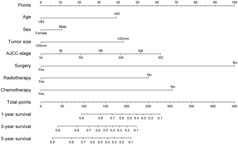
To predict the survival risk for patients with CEC, a nomogram was established by multivariate Cox regression analysis, incorporating all independent factors that were significant for OS (Figure 4). The C-index for the prediction of OS was 0.743, which was significantly higher (P<0.001) than either the 7th edition of the AJCC staging system (C-index =0.559) or the 6th edition of the AJCC staging system (C-index =0.532). Calibration curves demonstrated good agreement between prediction and observation in the probability of 3- and 5-year OS (Figure 5). In the external validation cohort, the C-index of the nomogram was 0.706, indicating that the nomogram demonstrates reasonably good discrimination in prognostic prediction.
Figure 4.
Nomogram for predicting 1-, 3-, and 5-year OS for non-metastatic CEC. To calculate the survival rate of each individual patient, points for each of the factors were first identified on the uppermost point scale, and then the total points from all factors were added up and projected on the bottom point scale to indicate the probability survival.
Figure 5.
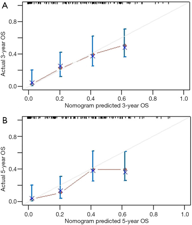
Calibration curve for predicting patient OS at 3 years (A) and 5 years (B) in the training cohort. Nomogram-predicted probability of OS is plotted on the X-axis; actual OS is plotted on the Y-axis.
Discussion
In the present study, we collected data from the SEER database to evaluate prognostic factors for non-metastatic CEC, and then used these risk factors to construct a nomogram to predict the OS of patients with CEC. We included age, sex, tumor size, TNM staging, and treatment modalities when creating the nomogram. The nomogram had a relatively high accuracy which was supported by the C-index (0.743 for the training cohort and 0.706 for the validation cohort, respectively) and calibration plots.
The demographic and clinicopathological characteristics of this cohort resemble those of a previous study, which was also based on the SEER database (12). The median age of the whole group at diagnosis was 68 years, and the proportion of males to females was about 6:4. We set 65 years as the cutoff age because it presented the most significant difference in OS. Most of the cases were moderately differentiated, followed by cases with poor differentiation, whereas only 2 cases were documented as undifferentiated. We found no difference in survival among those who had well-, moderately, or poorly differentiated tumors, although both patients with undifferentiated tumors survived for only 2 months. The majority of patients were stage III (47.3%) and stage II (stage IIA: 20.5%, stage IIB: 9.3%) at diagnosis, which was consistent with reports from other studies (12-16).
SCC and AC represent two primary histological subtypes of thoracic esophageal cancer that are significantly different in clinicopathology and prognosis (17,18). In our cohort of patients with CEC, SCC was the predominant histological type, whereas AC accounted for only 5.7% of patients; these findings are consistent with previously reported data (2). The median OS and DSS for patients with AC were 44 and 84 months, respectively, compared to 15 and 17 months for those with SCC. The 5-year OS for patients with SCC and AC of the cervical esophagus were 19.8% and 46.1%, respectively (data not shown). These results confirmed that patients with AC had a better prognosis compared to those with SCC.
The tumor size of CEC may play a critical role in determining survival; however, the optimal cutoff value has not been established. Performance status and tumor length (≤6 or >6 cm) have previously been described as factors that are significantly related to survival (14). Other cutoff values of tumor length, such as 3 cm or 3.5 cm, have also been reported (19-21). In the present study, a total of 498 patients (82.9%) had documented tumor size. Using Cox regression analysis, we identified tumor size as an independent risk factor for survival. By using X-tile plot software, we set 5.5 cm as the cutoff value, which is close to previously reported values (14). In contrast to breast cancer, tumor size is not currently included in the TNM staging system for esophageal cancer (22,23). Based on our findings, we propose that it be considered for inclusion in future editions.
Historically, surgery has been the preferred treatment for CEC. However, we identified a decreased trend in the implementation of surgery; this may be due to the high risk of major complications and the high rates of morbidity and mortality associated with surgical treatment, although data pertaining to this is not available from the SEER database. In our cohort, 13.8% of patients underwent surgical resection. These patients had significantly longer survival compared to those who did not, which could be attributed to an earlier stage at diagnosis and smaller primary tumors.
Chemoradiotherapy has become the current mainstay for the treatment of CEC. We found that there was no significant difference in prognosis between those who underwent surgery and those who underwent radical chemoradiotherapy, although patients who underwent surgery were more likely to have AC, a smaller tumor size, less lymph node involvement, and lower TNM staging. These results underline the critical role of chemoradiotherapy in CEC, especially among patients who have a greater number of high-risk factors. However, it remains controversial whether OS improves with chemoradiotherapy followed by surgery versus chemoradiotherapy alone for patients with SCC of the esophagus (24-27). Our results showed that trimodal therapy significantly improved DSS when compared with double or single therapy in the SCC subgroup, although no significant difference in OS was found between the trimodal- and dual-therapy groups. This provides favorable evidence for the use of trimodal therapy for CEC patients with SCC.
Nomograms have advantages over the AJCC TNM staging system in predicting patient prognosis, and they have been applied in numerous types of cancers. To the best of our knowledge, no nomogram has been developed specifically for CEC. The present study represents the first effort to develop a prognostic nomogram for CEC, based on a large cohort of patients from the SEER database. The nomogram showed good discrimination in the external validation cohort. In addition, we compared the predictive accuracy of our nomogram with the 7th edition of AJCC TNM staging system, and showed that our nomogram outperformed the TNM staging system in the prognostic prediction of OS in CEC patients. These results suggest that our nomogram has a relatively good discrimination in identifying high-risk populations and predicting prognosis.
The present study has several limitations. First, the SEER database does not include information on treatment toxicities, comorbidities, and failure patterns; therefore, these parameters could not be analyzed in the present study. Second, detailed information about cancer management was not available. We were therefore unable to separate patients who did not undergo surgery, RT, or chemotherapy, or those who underwent these treatments, but were not documented. Information on surgical procedure, radiation dose, and chemotherapy regimens were also not available. Therefore, our nomogram did not include details about treatment. Finally, selection bias and confounding bias should be considered when interpreting the results from the present study based on the SEER database.
Conclusions
We developed a prognostic nomogram to produce an individualized survival prediction for non-metastatic CEC patients. The nomogram had a relatively high accuracy and can likely be used to help identify high-risk patient populations and supplement the current TNM staging system.
Supplementary
The article’s supplementary files as
Acknowledgments
The authors would like to thank SEER for open access to the database, and AME Editing Service (http://editing.amegroups.cn/#editing) for language assistance during the preparation of this manuscript.
Funding: This work was supported by Science Foundation of Peking University Cancer Hospital (No. 18-03); Beijing Municipal Science & Technology Commission (No. Z181100001718192); Beijing Natural Science Foundation (No. 7182028); Clinical Technology Innovation Project of Beijing Hospital Authority (No. XMLX201842); and National Natural Science Foundation of China (No. 81902371).
Ethical Statement: The authors are accountable for all aspects of the work in ensuring that questions related to the accuracy or integrity of any part of the work are appropriately investigated and resolved. The study was conducted in accordance with the Declaration of Helsinki (as revised in 2013).
Footnotes
Reporting Checklist: The authors have completed the STROBE reporting checklist. Available at http://dx.doi.org/10.21037/atm-20-2505
Peer Review File: Available at http://dx.doi.org/10.21037/atm-20-2505
Conflicts of Interest: All authors have completed the ICMJE uniform disclosure form (available at http://dx.doi.org/10.21037/atm-20-2505). WW serves as a current editorial board member for Annals of Translational Medicine (Radiation Medicine) from April 2020 to March 2022. The other authors have no conflicts of interest to declare.
References
- 1.Mendenhall WM, Sombeck MD, Parsons JT, et al. Management of cervical esophageal carcinoma. Seminars in radiation oncology. Semin Radiat Oncol 1994;4:179‐91. 10.1016/S1053-4296(05)80066-9 [DOI] [PubMed] [Google Scholar]
- 2.Popescu CR, Bertesteanu SV, Mirea D, et al. The epidemiology of hypopharynx and cervical esophagus cancer. J Med Life 2010;3:396‐401. [PMC free article] [PubMed] [Google Scholar]
- 3.Cooper JS, Guo MD, Herskovic A, et al. Chemoradiotherapy of locally advanced esophageal cancer: long-term follow-up of a prospective randomized trial (RTOG 85-01). JAMA 1999;281:1623‐7. 10.1001/jama.281.17.1623 [DOI] [PubMed] [Google Scholar]
- 4.Buckstein M, Liu J. Cervical Esophageal Cancers: Challenges and Opportunities. Curr Oncol Rep 2019;21:46. 10.1007/s11912-019-0801-7 [DOI] [PubMed] [Google Scholar]
- 5.Hoeben A, Polak J, Van De Voorde L, et al. Cervical esophageal cancer: a gap in cancer knowledge. Ann Oncol 2016;27:1664-74. 10.1093/annonc/mdw183 [DOI] [PubMed] [Google Scholar]
- 6.Graesslin O, Abdulkarim BS, Coutant C, et al. Nomogram to predict subsequent brain metastasis in patients with metastatic breast cancer. J Clin Oncol 2010;28:2032‐7. 10.1200/JCO.2009.24.6314 [DOI] [PubMed] [Google Scholar]
- 7.Han DS, Suh YS, Kong SH, et al. Nomogram predicting long-term survival after d2 gastrectomy for gastric cancer. J Clin Oncol 2012;30:3834-40. 10.1200/JCO.2012.41.8343 [DOI] [PubMed] [Google Scholar]
- 8.van der Gaag N, Kloek J, de Bakker J, et al. Survival analysis and prognostic nomogram for patients undergoing resection of extrahepatic cholangiocarcinoma. Ann Oncol 2012;23:2642-9. 10.1093/annonc/mds077 [DOI] [PubMed] [Google Scholar]
- 9.Yang L, Shen W, Sakamoto N. Population-based study evaluating and predicting the probability of death resulting from thyroid cancer and other causes among patients with thyroid cancer. J Clin Oncol 2013;31:468-74. 10.1200/JCO.2012.42.4457 [DOI] [PubMed] [Google Scholar]
- 10.Su D, Zhou X, Chen Q, et al. Prognostic Nomogram for Thoracic Esophageal Squamous Cell Carcinoma after Radical Esophagectomy. PLoS One 2015;10:e0124437. 10.1371/journal.pone.0124437 [DOI] [PMC free article] [PubMed] [Google Scholar]
- 11.Xie K, Liu S, Liu J. Nomogram predicts survival benefit for non-metastatic esophageal cancer patients who underwent preoperative radiotherapy. Cancer Manag Res 2018;10:3657-68. 10.2147/CMAR.S165168 [DOI] [PMC free article] [PubMed] [Google Scholar]
- 12.Grass GD, Cooper SL, Armeson K, et al. Cervical esophageal cancer: a population-based study. Head Neck 2015;37:808-14. 10.1002/hed.23678 [DOI] [PubMed] [Google Scholar]
- 13.Huang SH, Lockwood G, Brierley J, et al. Effect of concurrent high-dose cisplatin chemotherapy and conformal radiotherapy on cervical esophageal cancer survival. Int J Radiat Oncol Biol Phys 2008;71:735‐40. 10.1016/j.ijrobp.2007.10.022 [DOI] [PubMed] [Google Scholar]
- 14.Yamada K, Murakami M, Okamoto Y, et al. Treatment results of radiotherapy for carcinoma of the cervical esophagus. Acta Oncol 2006;45:1120‐5. 10.1080/02841860600609768 [DOI] [PubMed] [Google Scholar]
- 15.Burmeister BH, Dickie G, Smithers BM, et al. Thirty-four patients with carcinoma of the cervical esophagus treated with chemoradiation therapy. Arch Otolaryngol Head Neck Surg 2000;126:205‐8. 10.1001/archotol.126.2.205 [DOI] [PubMed] [Google Scholar]
- 16.Uno T, Isobe K, Kawakami H, et al. Concurrent chemoradiation for patients with squamous cell carcinoma of the cervical esophagus. Dis Esophagus 2007;20:12‐8. 10.1111/j.1442-2050.2007.00632.x [DOI] [PubMed] [Google Scholar]
- 17.Hölscher AH, Bollschweiler E, Schneider PM, et al. Prognosis of early esophageal cancer. Comparison between adeno‐and squamous cell carcinoma. Cancer 1995;76:178-86. [DOI] [PubMed] [Google Scholar]
- 18.Bollschweiler E, Metzger R, Drebber U, et al. Histological type of esophageal cancer might affect response to neo-adjuvant radiochemotherapy and subsequent prognosis. Ann Oncol 2009;20:231‐8. 10.1093/annonc/mdn622 [DOI] [PubMed] [Google Scholar]
- 19.Wang BY, Goan YG, Hsu PK, et al. Tumor length as a prognostic factor in esophageal squamous cell carcinoma. Ann Thorac Surg 2011;91:887‐93. 10.1016/j.athoracsur.2010.11.011 [DOI] [PubMed] [Google Scholar]
- 20.Griffiths EA, Brummell Z, Gorthi G, et al. Tumor length as a prognostic factor in esophageal malignancy: univariate and multivariate survival analyses. J Surg Oncol 2006;93:258-67. 10.1002/jso.20449 [DOI] [PubMed] [Google Scholar]
- 21.Yendamuri S, Swisher SG, Correa AM, et al. Esophageal tumor length is independently associated with long‐term survival. Cancer 2009;115:508-16. 10.1002/cncr.24062 [DOI] [PubMed] [Google Scholar]
- 22.Edge SB, Compton CC. The American Joint Committee on Cancer: the 7th edition of the AJCC cancer staging manual and the future of TNM. Ann Surg Oncol 2010;17:1471-4. [DOI] [PubMed] [Google Scholar]
- 23.Amin MB, Edge SB, Greene FL, et al. AJCC Cancer Staging Manual. 8th ed. New York: Springer, 2017. [Google Scholar]
- 24.Stahl M, Stuschke M, Lehmann N, et al. Chemoradiation with and without surgery in patients with locally advanced squamous cell carcinoma of the esophagus. J Clin Oncol 2005;23:2310-7. 10.1200/JCO.2005.00.034 [DOI] [PubMed] [Google Scholar]
- 25.Bedenne L, Michel P, Bouché O, et al. Chemoradiation followed by surgery compared with chemoradiation alone in squamous cancer of the esophagus: FFCD 9102. J Clin Oncol 2007;25:1160-8. 10.1200/JCO.2005.04.7118 [DOI] [PubMed] [Google Scholar]
- 26.van Hagen P, Hulshof M, Van Lanschot J, et al. Preoperative chemoradiotherapy for esophageal or junctional cancer. N Engl J Med 2012;366:2074-84. 10.1056/NEJMoa1112088 [DOI] [PubMed] [Google Scholar]
- 27.Yang H, Liu H, Chen Y, et al. Neoadjuvant chemoradiotherapy followed by surgery versus surgery alone for locally advanced squamous cell carcinoma of the esophagus (NEOCRTEC5010): a phase III multicenter, randomized, open-label clinical trial. J Clin Oncol 2018;36:2796. 10.1200/JCO.2018.79.1483 [DOI] [PMC free article] [PubMed] [Google Scholar]
Associated Data
This section collects any data citations, data availability statements, or supplementary materials included in this article.
Supplementary Materials
The article’s supplementary files as


