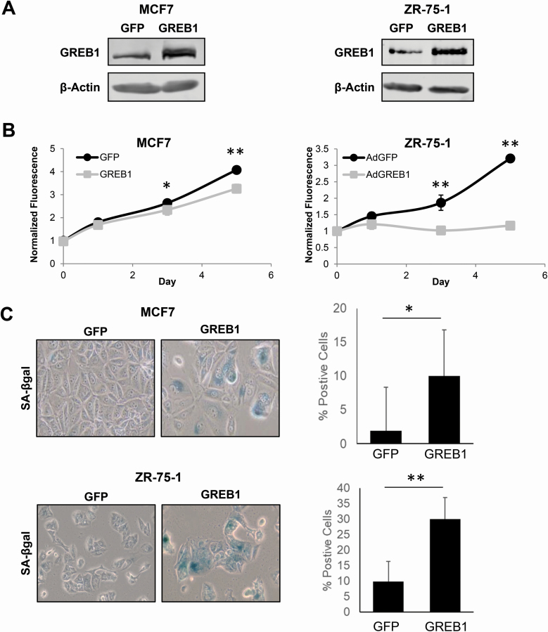
Figure 1.
Exogenous GREB1 initiates cellular senescence. MCF7 or ZR-75-1 cells were transduced with adenovirus expressing GFP or GREB1. (A) Immunoblot depicting the relative overexpression of GREB1 in cells transduced with GREB1 adenovirus compared with GFP control. (B) Proliferation of transduced cells was measured by alamar blue assay. Data are plotted as mean fluorescence normalized to day 0 ± SD; n = 3 for each cell line. *P ≤ 0.05, **P ≤ 0.005. (C) Cells were fixed and stained for SA-β-galactosidase activity 7 days post-transduction. Data are plotted as percent of SA-β-galactosidase stained cells ± SD. *P ≤ 0.05, **P ≤ 0.01.

