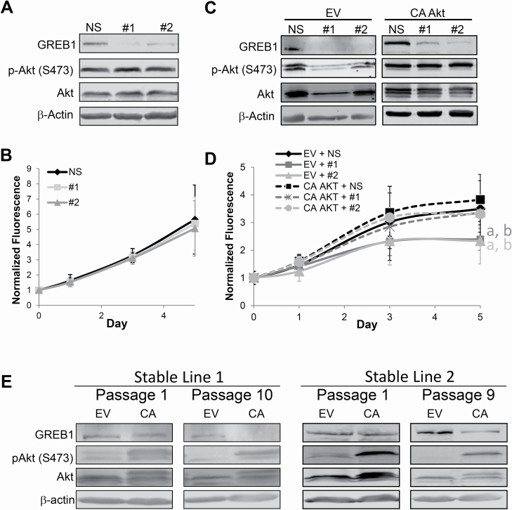
Figure 6.
GREB1 regulates breast cancer proliferation through activation of the PI3K/Akt pathway. (A) T47D cells were transduced with lentivirus expressing NS shRNA or shRNA targeted to GREB1 (#1 or #2). Immunoblot depicting the expression of indicated proteins. (B) Proliferation was measured via alamar blue assay. Data are plotted as mean fluorescence normalized to day 0 ± SD; n = 3. (C) MCF7 cells were transduced with lentivirus expressing empty vector (EV) or myristoylated Akt (CA AKT) and either NS shRNA NS or shRNA targeted to GREB1 (#1 or #2). Representative immunoblot depicting expression of indicated proteins. (D) Proliferation was measured via alamar blue assay. Data are plotted as mean fluorescence normalized to day 0 ± SD; n = 3; aP < 0.05 versus EV + NS; bP < 0.05 versus CA AKT + NS. (E) Following transduction with control (EV) or constitutively active Akt (CA) expressing lentivirus, MCF7 stable lines were generated by placing cells on selection. Lysates from different passages were probed for GREB1 expression and Akt activation. Two distinct stable lines are depicted (left and right panels, respectively).

