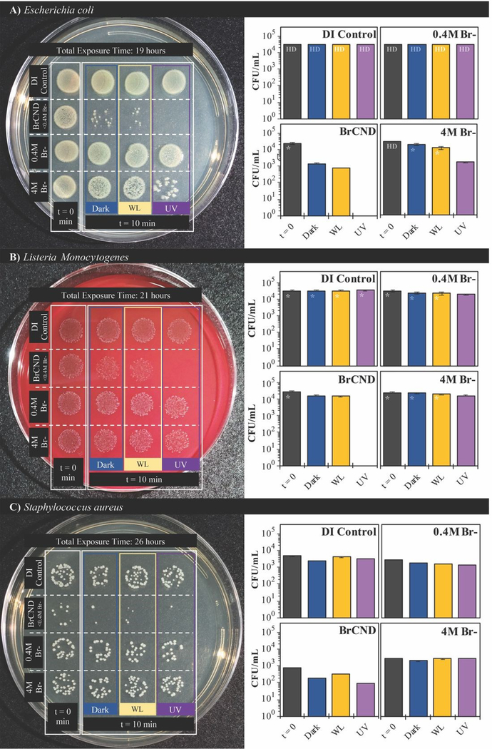
Figure 5.
Growth of A) Escherichia coli, B) Listeria monocytogenes, and C) Staphylococcus aureus with exposure to brominated carbon nanodots (“BrCND”) at pH 3.5 under UV (λexposure = 365 nm, 40 J•cm−2) and white light (λmax = 572 nm, 300 J•cm−2) exposure conditions versus dark exposure to BrCND. Real-color photographs of labeled plates after overnight bacterial incubation (left) and corresponding colony counts for each sample condition (right). Error is from counts by 3x individuals to reduce bias in counting. Colony growth too dense for adequate counting is indicated as “HD”; high density estimates are indicated by “*”.

