

A 32-year-old woman presented with worsening dyspnea lasting several months. Chest radiogram revealed a prominent pulmonary conus (Fig. 1). She was hospitalized for further assessment after undergoing computed tomography (CT) of the thorax (Fig. 2a, 2b). Her echocardiogram revealed an estimated systolic pulmonary artery pressure of 28 mm Hg with mild right heart chamber dilatation. A loud P2 with a soft holosystolic murmur in the tricuspid area and a long diastolic murmur in the pulmonary area were noted on auscultation.
Figure 1.
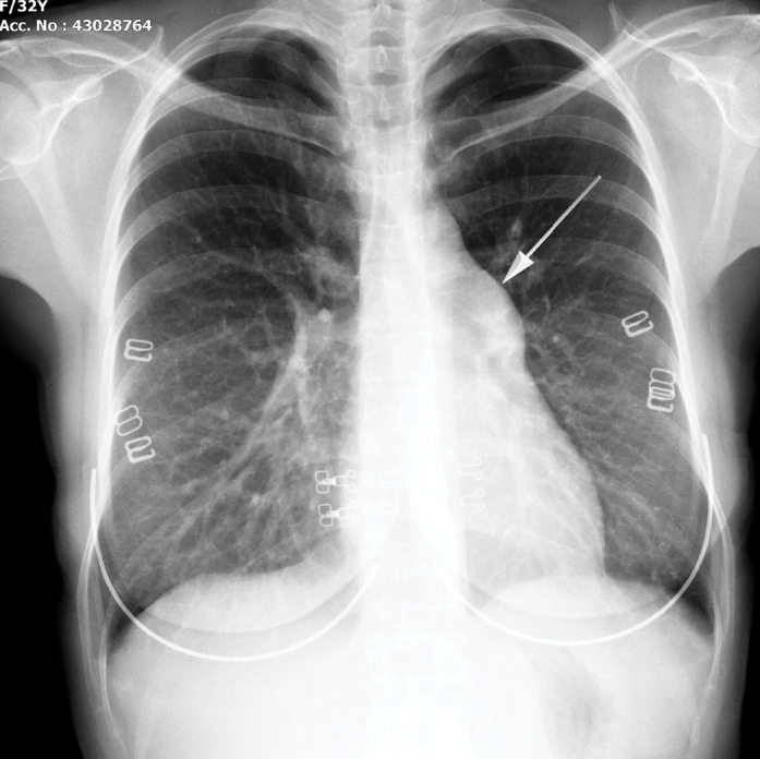
Chest radiography showing prominent pulmonary conus (arrow)
Figure 2.

(a) Sagittal computed tomography (CT) image showing the inferior vena cava (IVC, black arrow) and an anomalous connection to IVC (gray arrow). (b) Coronal CT image showing the anomalous connection (gray arrow)
What is your diagnosis?
Sinus venosus defect
Aortopulmonary window
Scimitar syndrome
Patent ductus arteriosus
Answer: p. 59