Abstract
Purpose
To investigate the ocular and systemic factors related to glaucoma and to be adjusted for interindividual comparison of ocular blood flow measurement results by laser speckle flowgraphy (LSFG) obtained from the optic nerve head (ONH) in normal Japanese individuals.
Methods
A multicenter, prospective cross-sectional study was conducted. The ONH tissue-area and vessel-area mean blur rate (MT and MV) were evaluated using LSFG and ONH structural parameters using planimetric methods. Multivariate linear mixed-effects modeled regression analysis was used to identify the contributing factors to the MT and MV. The explanatory variables were age; gender; smoking history; body mass index; mean arterial pressure (MAP); heart rate; intraocular pressure; axial length (AL); disc, rim, cup, and β-peripapillary atrophy (β-PPA) areas; and central retinal artery and vein equivalents.
Results
In total, 195 eyes of 126 healthy individuals with an average age of 48.1 years were included. Multivariate analysis showed that MAP and disc area had a negative (P < 0.001) correlation, whereas β-PPA area had a positive correlation with MT (P = 0.010). Age and AL had a negative correlation (P = 0.001 and P = 0.011, respectively), whereas cup area had a positive correlation (P = 0.012) with MV.
Conclusions
Interindividual comparison of MT or MV must be adjusted for both systemic factors (blood pressure or age) and local ocular factors (AL and disc, cup, or β-PPA area).
Translational Relevance
Our results provided reference data on the LSFG measurement and are important in comparing ocular blood flow between individuals using LSFG.
Keywords: blood flow, optic nerve head, laser speckle flowgraphy, normal subjects, disc parameter
Introduction
Laser speckle flowgraphy (LSFG) uses the laser speckle phenomenon in estimating ocular circulation in living eyes in a noninvasive manner.1 The current LSFG instrument yields the mean blur rate (MBR), which is a quantitative index of blood flow velocity in target tissues. For the optic nerve head (ONH), the ONH tissue-area MBR (MT) corresponding to the ONH microcirculation and the ONH vessel-area MBR (MV) corresponding to the blood velocity through large visible vessels in the ONH area are provided.2–5 The MT is significantly correlated with blood flow values determined using the microsphere method or hydrogen gas clearance method in primates and rabbits, respectively, regardless of the presence or absence of choroidal pigmentation or ONH atrophy.6–8 These animal studies suggested that MT also has potential to be used as a quantitative index of the ONH tissue blood flow in humans. In fact, many studies have compared the MT between normal and glaucoma eyes or glaucoma eyes with different stages of the disease.9–17 With regard to MV, in vitro experiments and comparison with laser Doppler velocimetry indicate that MV is a quantitative parameter of blood velocity in the large retinal vessels at the ONH.18–20
In performing comparative clinical studies, it is important not only to establish the reference data of the MT or MV values in healthy eyes but also to identify the factors influencing the MT or MV measurement results, which should be adjusted for interindividual comparison. Luft et al.2 in European normal white individuals and Yanagida et al.,3 Iwase et al.,4 and Aizawa et al.5 in Japanese normal individuals, respectively, report normal values of MT and MV in each ethnicity and suggest that age,2,4 gender,3,4 blood pressure,4 axial length (AL),4 and intraocular pressure (IOP)5 correlate with MT and that age,2,5 gender,3–5 blood pressure,3,4 and IOP5 correlate with MV, which should be adjusted for interindividual comparison. Because a laser speckle phenomenon is an interference event observed when lasers are scattered in a diffusing surface,21 the LSFG-measured parameters such as MT should also be influenced by the reflection, absorption, and penetration depth of the laser in the target tissue.1,20 Because the ONH of human eyes has a large structural variation even in normal individuals,22,23 it is important to study whether the structural variation in the human ONH has significant influences on the LSFG-measured MT or MV values as systemic factors.2–5 To date, no studies have examined the relationship between the measurement results of MT or MV and the ONH structural parameters such as disc or cup area, and there is no available information on which the ONH structural parameter must be adjusted for interindividual comparison of MT or MV. In the current study, we aimed to measure the MT and MV in normal Japanese individuals according to the predetermined uniform measurement protocol and to determine not only the effects of systemic factors but also those of the ONH structural parameters on the MT or MV measurement results.
Methods
Study Participants
This prospective cross-sectional study was conducted in multiple facilities. The participating research facilities were as follows (all in Japan): Fukui-ken Saiseikai Hospital (Fukui), Kanazawa University Hospital (Ishikawa), Kitasato University Hospital (Kanagawa), Tajimi Iwase Eye Clinic (Gifu), Toho University Ohashi Medical Center (Tokyo), Toho University Omori Medical Center (Tokyo), and Tohoku University Hospital (Miyagi). This study was approved by the institutional review boards of each facility, and all studies conducted adhered to the tenets of the Declaration of Helsinki. Written informed consent was obtained from all participants.
All participants underwent a comprehensive screening examination, including a slit-lamp examination with indirect fundoscopy, IOP measurement using a Goldmann applanation tonometer and a standard automated perimetry using the Humphrey visual field analyzer (Carl Zeiss Meditec, Dublin, CA, USA) using the Swedish interactive threshold algorithm-standard strategy of the 24–2 program. Healthy individuals aged 30 to 80 years on completion of the consent form who do not meet the exclusion criteria described below during the ophthalmic examination were included in the study. By contrast, participants with best-corrected visual acuity of <20/40, spherical refractive errors exceeding ±6.0 diopters (D), refractive cylindrical errors of >2.0 D, AL of >26.5 mm, IOP of >21 mm Hg, gonioscopically narrow and occludable angles, significant opacities of the optical media (e.g., corneal scars and cataract with Emery-Little classification grading ≥3), any abnormal ophthalmoscopic findings in the fundus, any findings suggestive of the presence of glaucoma or anomaly in the disc and peripapillary retina, an abnormal visual field test or an unreliable visual field test (false positives or false negatives >20% or fixation losses >30%), history of intraocular eye diseases and intraocular surgery, history of diabetes mellitus and cardiovascular disease, systolic blood pressure (SBP) of >150 mm Hg or diastolic blood pressure (DBP) of >90 mm Hg, and intake of oral medications that may affect blood flow (calcium antagonist, α-1 blocker, β-1 blocker, or sildenafil) were excluded.24,25
Measurement Protocol
The LSFG measurement protocol was as follows:
-
1.
On measurement days, smoking and caffeine-containing beverages (coffee, green tea, black tea, etc.) were prohibited.
-
2.
An interview regarding the patients’ medical history, including oral medication, and smoking history was conducted to ensure that they do not meet the exclusion criteria.
-
3.
Height and weight were measured.
-
4.
The following ocular examinations were performed: refraction, visual acuity, corneal curvature, ALs, IOP, standard automated perimetry, optical coherence tomography (OCT), and color fundus photography.
-
5.
The pupils were dilated by topical instillation of 0.4% tropicamide 30 minutes before LSFG examination.2 Measurements were taken in the afternoon between 2:00 PM and 6:00 PM at least two hours after taking a meal.
-
6.
Blood pressure and heart rate measurements were performed after a 10-minute resting period. After a further 10-minute resting period in a dark room, three consecutive LSFG measurements were performed. During the time of measurement, the participants were encouraged to keep the breathing constant. If the tear film was unstable because of dry eyes, artificial teardrops were instilled.
Laser Speckle Flowgraphy
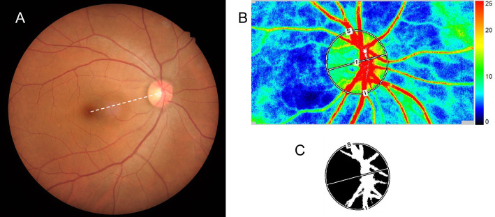
ONH blood flow was evaluated using LSFG (LSFG-NAVI; Softcare Ltd., Fukuoka, Japan). The principle and methods of LSFG have been previously described.1 Briefly, the instrument consists of a fundus camera equipped with a diode laser (wavelength, 830 nm) and an ordinary charge-coupled device camera (resolution, 750 × 360 pixels). The ONH margin was manually drawn with an ellipsoidal band, and the position of the ONH was saved in the system software. The accompanying LSFG software automatically divided the ONH area into the large visible vessels and capillary (tissue) area and provided MT (Fig. 1), MV, and all-area MBR (MA) as the sum of MT and MV. After collecting the LSFG data from each facility, the determination of the ONH margins with the ellipsoidal bands for all participants was made by a single experienced operator (TS) while referring to the fundus photograph. The average of the three measurements of MT or MV was used in the analysis.
Figure 1.
(A) Color fundus photograph showing the fovea–disc center axis (white dotted line). (B) Laser speckle flowgraphy false-color map. High numbers indicate faster blood flow. The ONH margin was manually drawn with an ellipsoidal band while referring to the fovea–disc center axis of a color fundus photograph. (C) A binary format image for segmentation between the tissue area (black area) and the vessel area (white area) on the ONH.
Measurements of Clinical Parameters
The AL was measured using an optical biometer (IOLMaster; Carl Zeiss Meditec) or OA-2000 (Tomey, Nagoya, Japan). The data from OA-2000 were converted into values yielded by the IOLMaster according to the previous studies.26,27 Color fundus photographs were taken using one of the following instruments: TRC-50DX (Topcon, Tokyo, Japan), TRC-NW7SF (Topcon), or Kowa Nonmyd WX (Kowa, Tokyo, Japan). The OCT measurements were performed using one of the following instruments: RS-3000 (Nidek, Tokyo, Japan), 3D OCT-2000 (Topcon), DRI OCT Triton (Topcon), and RTVue-XR Avanti (Optovue Inc., Fremont, CA, USA). With regard to the OCT measurements, only the values of the cup/disc area ratio were used in the analysis. The mean arterial blood pressure (MAP) was calculated as follows: MAP = diastolic blood pressure + 1/3 (systolic blood pressure − diastolic blood pressure) using the result obtained as described.
Disc Area and β-PPA Area Measurements
The details of the current planimetric method using a Topcon fundus camera were reported previously.22,23,28 An experienced ophthalmologist (AI) examined all color fundus photographs. After correcting for magnification using the modified version of Littman's method provided by the manufacturer (Topcon), planimetric parameters and disc and β-PPA areas were calculated using an image analysis software. One institute used Kowa Nonmyd WX as a fundus camera, which also allowed the calculation of the disc and β-PPA areas with magnification correction on the basis of the modified version of Littman's method provided by the manufacturer (Kowa).29 In the study participants, the OCT measurements were carried out using one of the following OCT instruments: RS-3000 (Nidek), 3D OCT-2000 (Topcon), DRI OCT Triton (Topcon), or RTVue-XR Avanti (Optovue Inc.), which was available in each facility. Because the methods to correct the magnification of the fundus image may not be the same among each OCT instrument, we adopted the disc area of each participant's eye determined using a fundus photograph and only used the cup/disc area ratio obtained using each OCT instrument to calculate the cup and rim areas of each participant's eye by use of the following formula: cup area = disc area provided by the fundus photograph × cup/disc area ratio provided by each OCT instrument; rim area = disc area − cup area of the same eye. In eyes photographed using Topcon fundus cameras, central retinal artery equivalent (CRAE) and central retinal vein equivalent (CRVE) were also calculated according to the method reported previously.30
Statistical Analysis
Spearman's correlation coefficients were used to evaluate the correlation between MT or MV and clinical parameters. A multivariate linear mixed-effect modeled regression analysis was used to detect the factors contributing to MT or MV adjusting for the confounding effects of other factors, and to adjust the correlation between two eyes of a subject. Variables that had an effect on MT or MV with P < 0.2 in the univariate analysis were used in the multivariate analysis after confirming that no explanatory variables included showed high inter-correlations (Spearman's correlation coefficient > 0.65). Because the disc area is the sum of the cup area and rim area, two of them were separately included in the analysis. In the analysis of MV, the quantitative index of blood velocity in the large retinal vessels in the ONH, CRAE, and CRVE were included as explanatory variables. A P value of <0.05 was considered significant. The data analysis was performed using SPSS version 24.0 for Windows (IBM, Tokyo, Japan).
Results
In total, 195 eyes of 126 healthy individuals (63 male and 63 female) were included in the study. The average age was 48.1 ± 11.6 years. Table 1 summarizes the demographic and ocular characteristics of the study participants. The CRAE and CRVE were measured in 111 eyes (86 participants).
Table 1.
Demographics and Ocular Characteristics of the Study Population
| Parameter | Mean ± SD |
|---|---|
| Number of eyes (number of individuals) | 195 (126) |
| Age (years) | 48.1 ± 11.6 |
| Gender (male/female) | 96/99 |
| Spherical equivalent (diopters) | −1.6 ± 2.1 |
| Axial length (mm) | 24.2 ± 1.1 |
| Intraocular pressure (mm Hg) | 14.3 ± 2.2 |
| Disc area (mm2) | 2.31 ± 0.42 |
| Rim area (mm2) | 1.51 ± 0.43 |
| Cup area (mm2) | 0.80 ± 0.46 |
| β-PPA area (mm2) | 0.52 ± 0.61 |
| Central retinal artery equivalent (µm)* | 141.5 ± 17.6 |
| Central retinal vein equivalent (µm)* | 222.8 ± 24.8 |
| Mean arterial pressure (mm Hg) | 88.9 ± 10.1 |
| Heart rate (bpm) | 72.3 ± 9.4 |
| Body mass index (kg/m2) | 22.1 ± 3.0 |
| Smoking history (yes/no) | 25/170 |
111 eyes of 86 individuals.
A preliminary study in 20 normal eyes confirmed that disc and β-PPA area measurements, calculated from photographs taken using TRC-NW7SF (Topcon) and Kowa Nonmyd WX showed no significant difference (P > 0.10), with a Pearson's correlation coefficient of 0.89 and 0.99 (P < 0.001), respectively. Another preliminary study in 30 normal eyes indicated that the MT and MV measurement results by TS yielded intraclass correlation coefficients of 0.947 (95% confidence interval [CI]: 0.892–0.974) for MT measurement results and 0.923 (0.846–0.962) for MV measurement results.
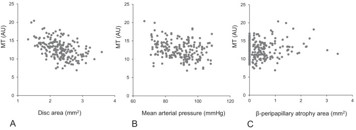
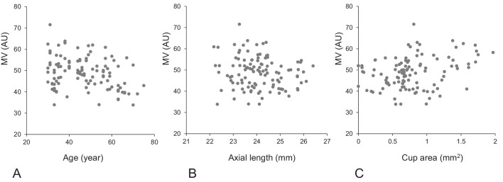
The MT had an average of 12.6 ± 2.5 (standard deviation) arbitrary units (AU), whereas the MV had an average of 48.3 ± 7.0 AU. The intraclass correlation coefficients among the three measurements were 0.943 (95% CI: 0.927–0.955) for MT measurement and 0.908 (0.883– 0.928) for MV measurement. In 69 individuals whose both eyes met the inclusion criteria, the MT values were 12.7 ± 2.5 and 12.4 ± 2.5 AU (paired t-test, P = 0.225) and the MV values were 48.8 ± 7.4 and 47.8 ± 6.3 AU (P = 0.231) for the right and left eyes, respectively. A simple correlation analysis showed a significant negative correlation between MT and gender (greater in female), MAP, disc area, and cup area; a significant negative correlation between MV and age, gender (greater in female), MAP, and AL; and a significant positive correlation between MV and heart rate and cup area (Table 2). Tables 3A and 3B show the results of the multivariate linear mixed-effects modeled linear regression analysis assessing the contribution of each factor to MT and MV. MAP showed a significant negative correlation (P < 0.001), whereas β-PPA area showed a significant positive correlation with MT (P = 0.010). Meanwhile, disc area showed a significant negative correlation with MT (P < 0.001) when disc and rim areas or disc and cup areas were adopted as explanatory variables, whereas both rim and cup areas showed negative correlations (P < 0.001) when rim and cup areas were adopted as explanatory variables. Because the disc area is the sum of the rim and cup areas, the correlation of the disc structural parameters with MT is summarized as a significant negative correlation between disc area and MT (P < 0.001) (Table 3A). Age and AL showed a significant negative correlation (P = 0.001 and P = 0.011, respectively), whereas disc area showed a significant positive correlation with MV (P = 0.012) when disc and rim areas were adopted as explanatory variables. Cup area showed a significant positive correlation with MV (P = 0.012) when rim and cup areas were adopted as explanatory variables. On the basis of the theory above and considering that the correlation of rim and cup areas (P = 0.065) were reversed, when disc and rim or cup were adopted as explanatory variables, the correlation of the disc structural parameters with MV is summarized as a significant positive correlation of cup area (P = 0.012) (Table 3B). Figures 2A and 2B show the scatterplots of the relationships between those contributing factors and MT or MV.
Table 2.
Results of Spearman's Rank Correlation Coefficient Between Clinical Parameters and MT and MV
| MBR | Age | Gender | Smoking History | BMI | MAP | Heart Rate | IOP | AL | Disc Area | Rim Area | Cup Area | β-PPA Area | CRAE | CRVE |
|---|---|---|---|---|---|---|---|---|---|---|---|---|---|---|
| MT | 0.113 | −0.190† | −0.043 | −0.069 | −0.227† | −0.080 | 0.073 | −0.135 | −0.480* | −0.135 | −0.268* | 0.029 | — | — |
| MV | −0.238† | −0.147‡ | −0.024 | −0.106 | −0.157‡ | 0.142‡ | 0.016 | −0.187† | 0.098 | −0.074 | 0.156‡ | −0.072 | 0.124 | 0.142 |
MA, all-area MBR; BMI, body mass index.
P < 0.001.
P < 0.01.
P < 0.05.
Table 3A.
Results of Linear Mixed-Effects Modeled Regression Analysis Assessing the Contribution of Each Factor on MT in 195 Eyes of 126 Individuals
| Univariate | Multivariate 1* | Multivariate 2† | Multivariate 3‡ | |||||
|---|---|---|---|---|---|---|---|---|
| Variables | β | P Value | β | P Value | β | P Value | β | P Value |
| Age | 0.022 | 0.218 | ||||||
| Gender (male = 1, female = 0) | −0.639 | 0.123 | −0.311 | 0.379 | −0.311 | 0.379 | −0.311 | 0.379 |
| Smoking history | −0.192 | 0.768 | ||||||
| BMI (kg/m2) | −0.079 | 0.260 | ||||||
| MAP (mm Hg) | −0.059 | 0.003 | −0.067 | <0.001 | −0.067 | <0.001 | −0.067 | <0.001 |
| Heart rate (beats/min) | −0.027 | 0.212 | ||||||
| IOP (mm Hg) | 0.056 | 0.519 | ||||||
| Axial length (mm) | −0.120 | 0.531 | ||||||
| Disc area (mm2) | −2.744 | <0.001 | −2.933 | <0.001 | −2.923 | <0.001 | ||
| Rim area (mm2) | −0.833 | 0.072 | 0.010 | 0.982 | −2.923 | <0.001 | ||
| Cup area (mm2) | −1.589 | <0.001 | −0.010 | 0.982 | −2.933 | <0.001 | ||
| β-PPA area (mm2) | 0.600 | 0.041 | 0.658 | 0.010 | 0.658 | 0.010 | 0.658 | 0.010 |
BMI, body mass index.
Disc area and rim area were adopted as explanatory variables.
Disc area and cup area were adopted as explanatory variables.
Cup area and rim area were adopted as explanatory variables.
Table 3B.
Results of Linear Mixed-Effects Modeled Regression Analysis Assessing the Contribution of Each Factor on MV in 111 Eyes of 86 Individuals
| Univariate | Multivariate 1* | Multivariate 2† | Multivariate 3‡ | |||||
|---|---|---|---|---|---|---|---|---|
| Variables | β | P Value | β | P Value | β | P Value | β | P Value |
| Age | −0.197 | 0.002 | −0.207 | 0.001 | −0.207 | 0.001 | −0.207 | 0.001 |
| Gender (male = 1, female = 0) | −1.153 | 0.464 | ||||||
| Smoking history | −0.346 | 0.878 | ||||||
| BMI (kg/m2) | 0.037 | 0.896 | ||||||
| MAP (mm Hg) | −0.080 | 0.302 | ||||||
| Heart rate (beats/min) | 0.040 | 0.668 | ||||||
| IOP (mm Hg) | 0.253 | 0.478 | ||||||
| Axial length (mm) | −1.103 | 0.117 | −1.991 | 0.011 | −1.991 | 0.011 | −1.991 | 0.011 |
| Disc area (mm2) | 4.230 | 0.028 | 5.083 | 0.012 | 1.682 | 0.421 | ||
| Rim area (mm2) | −1.155 | 0.528 | −3.401 | 0.065 | 1.682 | 0.421 | ||
| Cup area (mm2) | 4.510 | 0.008 | 3.401 | 0.065 | 5.083 | 0.012 | ||
| β-PPA area (mm2) | −1.121 | 0.452 | ||||||
| CRAE (µm) | 0.043 | 0.272 | ||||||
| CRVE (µm) | 0.036 | 0.215 | ||||||
BMI, body mass index.
Disc area and rim area were adopted as explanatory variables.
Disc area and cup area were adopted as explanatory variables.
Cup area and rim area were adopted as explanatory variables.
Figure 2A.
Scatterplots showing the relationships between ONH MT and disc area (A), MAP (B), and β-PPA (C).
Figure 2B.
Scatterplots showing the relationships between ONH MV and age (A), axial length (B), and cup area (C).
Discussion
The LSFG used a laser speckle phenomenon, an interference event observed when a laser is scattered in a diffusing surface, and the LSFG measurement results are influenced by the optical characteristics of the target tissue.1,20,21 The appearance of the human ONH greatly varies even in normal Japanese individuals,22,23 which would be greater between normal and glaucoma eyes, and it is possible that the optical characteristics of the laser beam delivered to the cup and rim areas differ from each other. Thus it is important to study the effects of the ONH structural factors, as well as systemic ones on the LSFG measurement results such as MT or MV. Needless to say, the factors significantly affecting the LSFG measurement results in normal eyes, if they existed, should be adjusted for interindividual or intergroup comparison. In the current study, we found that MAP, disc area, and β-PPA area had significant impacts on the measurement results of MT. Meanwhile, age, AL, and cup area had significant impacts on the measurement results of MV in normal Japanese eyes.
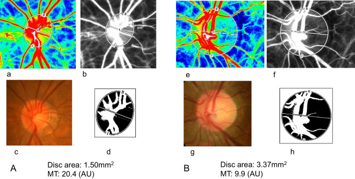
Iwase et al.4 reported that the MT had a significant negative correlation with AL. Since disc area had a positive correlation with AL in the current cohort, the negative correlation between disc area and MT currently found is not incompatible with this previous result. Among the possible explanations of the disc area dependency of the MT measurement results, instrumental characteristics are the most plausible. The software divides the ONH area into the vessel area and area free from large visible vessels (tissue area) to calculate MV and MT separately (Fig. 3) by automatically identifying the major blood vessels in the ONH area. The examples of the binary format image for segmentation are presented in Figures 3A-d and 3B-h. In determining the side wall of the vessel, pixels lying on the border between the visible vessel-free tissue area and visible large vessels were included in the tissue area, but not in the vessel area in the current binary format image. The positive influence of MBR obtained from the border area between the visible vessel-free tissue area and visible large retinal vessels on the MT measurement results is relatively more significant in a relatively smaller-sized, vessel-crowded disc than in a relatively larger-sized, less vessel-crowded disc. Furthermore, relatively small vessels as seen in the grayscale map (Figs. 3A-b and 3B-f) were generally less visible on the binary format image (Figs. 3A-d and 3B-h) and were included in the visible vessel-free tissue area, which would increase the MT measurement results. Thus the MT measurement results in a smaller-sized disc are more influenced by such an effect. These mechanisms would result in a tendency to obtain relatively higher (lower) MT measurement results from smaller-sized (larger-sized) discs. On the contrary, the possibility that a larger disc has a relatively lower blood flow rate per unit area may not be completely excluded. The superficial region of the ONH is mostly nourished by retinal circulation, and deeper regions mainly receive blood supply from the peripapillary choroid through the branches of the short posterior ciliary arteries.31–33 Histologic studies demonstrated that the optic nerve fiber count significantly increased with the enlargement of the optic disc size, but the nerve fiber density per disc area decreased when the disc area increased.34,35 If local circulation is associated with the density of nerve fibers, the MBR measurement results from a unit area (in this case, one pixel) of the ONH, such as MT, would tend to be lower in a larger disc. However, the effect of this mechanism would be minor, if it existed, because the adjusted correlation between MT measurement results and rim or cup area was not significant.
Figure 3.
(A) An example of a relatively small disc with high MT. (B) An example of a relatively large disc with low MT: (a) and (e) are laser speckle flowgraphy false-color maps; (b) and (f) are laser speckle flowgraphy grayscale maps; (c) and (g) are color fundus photographs; (d) and (h) are binary format images for segmentation between the tissue area (black area) and the vessel area (white area) on the ONH.
The β-PPA is characterized by marked atrophy of the retinal pigment epithelium and choriocapillaris, visible large choroidal vessels, and sclera adjacent to the clinically visible disc margin.36,37 Moreover, the β-PPA area had a significant positive correlation with the MT measurement results in the current normal individuals. If β-PPA could affect laser absorption or reflection in the ONH area adjacent to it, the presence of β-PPA might influence the MT measurement results. However, this possibility does not seem to be the primary reason, because it was confirmed in an animal experiment using albino and pigmented rabbits that the presence or absence of pigmentation in the choroid adjacent to the ONH had little effect on the MT measurement results.8 As discussed above, deeper regions of the ONH mainly receive blood supply from the branches of the short posterior ciliary arteries.31–33 Histologically, the choroid is thin or absent in the β-PPA area,37 resulting in the compromised circulation in this area.38–40 Therefore, in a normal condition, the disc area adjacent to a greater β-PPA area nourished by the same short ciliary artery might have a relatively greater blood supply, because the blood to be supplied to the peripapillary choroid could not enter the β-PPA area.
In the current study on normal Japanese individuals with a mean age of 48 years, only MAP showed a significant negative impact on the MT among the systemic factors examined. In normal Japanese individuals with a mean age of 63.5 years, Iwase et al.4 found a significant negative correlation between MT and systolic and diastolic blood pressure. In normal Japanese individuals with a mean age of 45 years, Aizawa et al.5 reported a negative correlation between MT and systolic blood pressure (P = 0.08). The current result agreed with those of previous studies, suggesting that a higher blood pressure, per se, was not always advantageous for the ONH tissue. On the contrary, age showed no significant impacts on MT in the current study population. This result also agreed with the findings obtained in normal Japanese individuals with a mean age of 39.33 or 45 years.5 By contrast, a significant negative correlation between age and MT was reported in normal Japanese individuals with a mean age of 63.5 years4 and in normal European white individuals with a mean age of 48.9 years.2 Taken together, age has little effects on the MT in normal Japanese individuals aged 50 years or younger, and its negative impact on the MT was evident in those aged 60 years or older. The ethnical difference may be partly responsible for the discrepancy between the results obtained in normal Japanese individuals3,5 and those obtained in normal European white individuals with a similar age range.2 Females were reported to have greater MT than males.3,4 The current result was not incompatible with those of previous studies, because a simple correlation analysis of the current results showed a significant correlation between MT and gender, with females having a greater MT.
MV represents retinal blood velocity in the large vessels measured in the ONH area.18–20 This implied that it would be better to include the retinal vessel caliber, CRAE, and CRVE in the analysis as one of the explanatory variables.41 In the simple correlation analyses of the current cohort, MV was found to show significant negative correlations with age, MAP, AL, and gender (greater values in female) and significant positive correlations with heart rate and cup area. The negative correlation of MV with blood pressure,3,4 age,2,5 or greater value in female3–5 has been reported previously, consistent with the results of the current study. In the multivariate analysis, MV showed a significant negative correlation with age and AL, whereas MV showed a positive correlation with cup area. The negative correlation between MV and age agreed well with the previous MV measurement results.2,5 An eye with a smaller cup area is more likely to develop nonarteritic ischemic optic neuropathy.42–44 Although not always confirmed,45 an eye with a smaller cup area was more likely to develop branch vein occlusion.46 These clinical findings suggest that blood flow is relatively more stagnant in the ONH with a smaller cup area and vice versa. In accordance with these results, a greater cup area was reportedly associated with decreased CRVE, that is, less stagnant venous flow, in healthy children free from any pathologic changes in the tissue.47 Taken together, an ONH with a greater cup area is believed to provide less resistance to blood vessels passing through it, resulting in less stagnant flow. Less stagnant blood flow in the retinal vein at the ONH would yield a higher blood velocity and thus higher MV measurement results.
A previous study using laser Doppler velocimetry reported that retinal blood flow was decreased in individuals with mild and high myopia compared with those with emmetropia. Moreover, they found that the retinal arterial diameter of eyes with high-grade myopia was significantly smaller than that of eyes with emmetropia and mild myopia, but there was no significant difference between mild myopia and emmetropia.48 In the current study, AL showed a significant negative correlation with MV, being compatible with the result of the above study.48
Whatever the causes of the significant correlation between MT or MV measurement results and the ONH structural parameters or AL, the current result demonstrated that the interindividual or intergroup comparison of MT or MV must be adjusted not only for systemic factors but also for local ocular factors such as disc and β-PPA area for MT and cup area and AL for MV measurement results.
The current study has several limitations. First, we used disc parameters that were evaluated using the planimetric methods. Current photographically determined β-PPA area included γ-zone PPA, and the disc area would have been evaluated better using spectral-domain OCT.49,50 However, to date, the effects of β-PPA area on glaucoma were still investigated using photographs in many clinical studies. Moreover, it is not common to measure β-PPA area using OCT in routine clinical practice, but rather to evaluate β-PPA area with photographs or ophthalmoscopy. Therefore we believe that the current findings regarding the photographically determined β-PPA have clinical and practical significance. Second, the ellipsoidal bands needed to be fitted to determine the ONH margins. Thus the current study cohort did not include those of which the contour deviated from ellipsoid, such as eyes with highly myopic discs. Therefore this result should not be used especially for cases with high-grade myopia. Finally, the average age of the current cohort was relatively young. Therefore the influence of age or blood pressure might not have been evaluated sensitively in the current study. Thus the absence of apparent adjusted correlation between MT measurement results and age and between MV measurement results and blood pressure might not be applicable to more aged normal Japanese individuals.
In conclusion, we investigated the factors related to ONH circulation measured by LSFG in normal individuals. Results showed that MAP and disc area had a significant negative correlation whereas β-PPA area had a significant positive correlation with MT measurements. Age and AL showed a significant negative correlation, whereas cup area had a positive correlation with MV measurements. These results were thought to have a special implication in applying LSFG for interindividual comparison of MT or MV measurement results, indicating the specific factors that must be adjusted.
Acknowledgments
The authors thank Mari S. Oba for assistance with statistical analyses.
Disclosure: A. Anraku, None; N. Enomoto, None; G. Tomita, None; A. Iwase, Topcon (P); T. Sato, None; N. Shoji, None; T. Shiba, None; T. Nakazawa, None; K. Sugiyama, None; K. Nitta, None; M. Araie, Topcon (C, P, R)
References
- 1. Sugiyama T, Araie M, Riva CE, Schmetterer L, Orgul S.. Use of laser speckle flowgraphy in ocular blood flow research. Acta Ophthalmol. 2010; 88: 723–729. [DOI] [PubMed] [Google Scholar]
- 2. Luft N, Wozniak PA, Aschinger GC, et al.. Ocular blood flow measurements in healthy white subjects using laser speckle flowgraphy. PLoS One . 2016; 11: e0168190. [DOI] [PMC free article] [PubMed] [Google Scholar]
- 3. Yanagida K, Iwase T, Yamamoto K, et al.. Sex-related differences in ocular blood flow of healthy subjects using laser speckle flowgraphy. Invest Ophthalmol Vis Sci. 2015; 56: 4880–4890. [DOI] [PubMed] [Google Scholar]
- 4. Iwase T, Yamamoto K, Yanagida K, et al.. Investigation of causes of sex-related differences in ocular blood flow in healthy eyes determined by laser speckle flowgraphy. Sci Rep. 2017; 7: 13878. [DOI] [PMC free article] [PubMed] [Google Scholar]
- 5. Aizawa N, Kunikata H, Nitta F, et al.. Age- and sex-dependency of laser speckle flowgraphy measurements of optic nerve vessel microcirculation. PLoS One. 2016; 11: e0148812. [DOI] [PMC free article] [PubMed] [Google Scholar]
- 6. Wang L, Cull GA, Piper C, Burgoyne CF, Fortune B.. Anterior and posterior optic nerve head blood flow in nonhuman primate experimental glaucoma model measured by laser speckle imaging technique and microsphere method. Invest Ophthalmol Vis Sci. 2012; 53: 8303–8309. [DOI] [PMC free article] [PubMed] [Google Scholar]
- 7. Takahashi H, Sugiyama T, Tokushige H, et al.. Comparison of CCD-equipped laser speckle flowgraphy with hydrogen gas clearance method in the measurement of optic nerve head microcirculation in rabbits. Exp Eye Res. 2013; 108: 10–15. [DOI] [PubMed] [Google Scholar]
- 8. Aizawa N, Nitta F, Kunikata H, et al.. Laser speckle and hydrogen gas clearance measurements of optic nerve circulation in albino and pigmented rabbits with or without optic disc atrophy. Invest Ophthalmol Vis Sci. 2014; 55: 7991–7996. [DOI] [PubMed] [Google Scholar]
- 9. Aizawa N, Kunikata H, Nakazawa T.. Diagnostic power of laser speckle flowgraphy-measured optic disc microcirculation for open-angle glaucoma: Analysis of 314 eyes. Clin Exp Ophthalmol. 2019; 47: 680–683. [DOI] [PubMed] [Google Scholar]
- 10. Kiyota N, Shiga Y, Yasuda M, et al.. Sectoral differences in the association of optic nerve head blood flow and glaucomatous visual field defect severity and progression. Invest Ophthalmol Vis Sci. 2019; 60: 2650–2658. [DOI] [PubMed] [Google Scholar]
- 11. Yamada Y, Higashide T, Udagawa S, et al.. The relationship between interocular asymmetry of visual field defects and optic nerve head blood flow in patients with glaucoma. J Glaucoma. 2019; 28: 231–237. [DOI] [PubMed] [Google Scholar]
- 12. Kuroda F, Iwase T, Yamamoto K, Ra E, Terasaki H.. Correlation between blood flow on optic nerve head and structural and functional changes in eyes with glaucoma. Sci Rep. 2020; 10: 729. [DOI] [PMC free article] [PubMed] [Google Scholar]
- 13. Shiga Y, Kunikata H, Aizawa N, et al.. Optic nerve head blood flow, as measured by laser speckle flowgraphy, is significantly reduced in preperimetric glaucoma. Curr Eye Res. 2016; 41: 1447–1453 [DOI] [PubMed] [Google Scholar]
- 14. Kiyota N, Kunikata H, Shiga Y, Omodaka K, Nakazawa T.. Ocular microcirculation measurement with laser speckle flowgraphy and optical coherence tomography angiography in glaucoma. Acta Ophthalmol. 2018; 96: e485–e492. [DOI] [PubMed] [Google Scholar]
- 15. Gardiner SK, Cull G, Fortune B, Wang L.. Increased optic nerve head capillary blood flow in early primary open-angle glaucoma. Invest Ophthalmol Vis Sci. 2019; 60: 3110–3118. [DOI] [PMC free article] [PubMed] [Google Scholar]
- 16. Takeyama A, Ishida K, Anraku A, Ishida M, Tomita G.. Comparison of optical coherence tomography angiography and laser speckle flowgraphy for the diagnosis of normal-tension glaucoma . J Ophthalmol. 2018; 2018: 1751857. [DOI] [PMC free article] [PubMed] [Google Scholar]
- 17. Mursch-Edlmayr AS, Luft N, Podkowinski D, Ring M, Schmetterer L, Bolz M.. Laser speckle flowgraphy derived characteristics of optic nerve head perfusion in normal tension glaucoma and healthy individuals: A pilot study. Sci Rep. 2018; 8: 5343. [DOI] [PMC free article] [PubMed] [Google Scholar]
- 18. Shiga Y, Asano T, Kunikata H, et al.. Relative flow volume, a novel blood flow index in the human retina derived from laser speckle flowgraphy. Invest Ophthalmol Vis Sci. 2014; 55: 3899–3904. [DOI] [PubMed] [Google Scholar]
- 19. Luft N, Wozniak PA, Aschinger GC, et al.. Measurements of retinal perfusion using laser speckle flowgraphy and doppler optical coherence tomography. Invest Ophthalmol Vis Sci. 2016; 57: 5417–5425. [DOI] [PubMed] [Google Scholar]
- 20. Nagahara M, Tamaki Y, Tomidokoro A, Araie M.. In vivo measurement of blood velocity in human major retinal vessels using the laser speckle method. Invest Ophthalmol Vis Sci . 2011; 52: 87–92. [DOI] [PubMed] [Google Scholar]
- 21. Françon M. Laser speckle and application in optics. (Translated by Arsenault HH.). New York, San Francisco London: Academic Press;1979: 1–72. [Google Scholar]
- 22. Mataki N, Tomidokoro A, Araie M, Iwase A.. Morphology of the optic disc in the Tajimi Study population. Jpn J Ophthalmol. 2017; 61: 441–447. [DOI] [PubMed] [Google Scholar]
- 23. Iwase A, Sawaguchi S, Sakai H, Tanaka K, Tsutsumi T, Araie M.. Optic disc, rim and peripapillary chorioretinal atrophy in normal Japanese eyes: the Kumejima Study. Jpn J Ophthalmol. 2017; 61: 223–229. [DOI] [PubMed] [Google Scholar]
- 24. Shiba C, Shiba T, Takahashi M, Matsumoto T, Hori Y.. Relationship between glycosylated hemoglobin A1c and ocular circulation by laser speckle flowgraphy in patients with/without diabetes mellitus. Graefes Arch Clin Exp Ophthalmol. 2016; 254: 1801–1809. [DOI] [PubMed] [Google Scholar]
- 25. Shiba T, Takahashi M, Matsumoto T, Shirai K, Hori Y.. Arterial stiffness shown by the cardio-ankle vascular index is an important contributor to optic nerve head microcirculation. Graefes Arch Clin Exp Ophthalmol. 2017; 255: 99–105. [DOI] [PMC free article] [PubMed] [Google Scholar]
- 26. Hua Y, Qiu W, Xiao Q, Wu Q.. Precision (repeatability and reproducibility) of ocular parameters obtained by the Tomey OA-2000 biometer compared to the IOLMaster in healthy eyes. PloS One. 2018; 13: e0193023. [DOI] [PMC free article] [PubMed] [Google Scholar]
- 27. Goebels S, Pattmöller M, Eppig T, Cayless A, Seitz B, Langenbucher A.. Comparison of 3 biometry devices in cataract patients. J Cataract Refract Surg. 2015; 41: 2387–2393. [DOI] [PubMed] [Google Scholar]
- 28. Saito H, Tsutsumi T, Iwase A, Tomidokoro A, Araie M.. Correlation of disc morphology quantified on stereophotographs to results by Heidelberg Retina Tomograph II, GDx variable corneal compensation, and visual field tests. Ophthalmology. 2010; 117: 282–289. [DOI] [PubMed] [Google Scholar]
- 29. Yokoyama Y, Tanito M, Nitta K, et al.. Stereoscopic analysis of optic nerve head parameters in primary open angle glaucoma: The glaucoma stereo analysis study. PloS One. 2014; 9: e99138. [DOI] [PMC free article] [PubMed] [Google Scholar]
- 30. Iwase A, Sekine A, Suehiro J, et al.. A new method of magnification correction for accurately measuring retinal vessel calibers from fundus photographs. Invest Ophthalmol Vis Sci. 2017; 58: 1858–1864. [DOI] [PubMed] [Google Scholar]
- 31. Anderson DR, Braverman S.. Reevaluation of the optic disk vasculature. Am J Ophthalmol. 1976; 82;165–174. [DOI] [PubMed] [Google Scholar]
- 32. Hayreh SS. The 1994 Von Sallman Lecture. The optic nerve head circulation in health and disease. Exp Eye Res. 1995; 61;259–272. [DOI] [PubMed] [Google Scholar]
- 33. Olver JM, Splaton DJ, McCartney ACE.. Microvascular study of the retrolaminar optic nerve in man: The possible significance in anterior ischaemic optic neuropathy. Eye. 1990; 4: 7–24. [DOI] [PubMed] [Google Scholar]
- 34. Jonas JB, Schmidt AM, Muller-Bergh JA, Schlotzer-Schrehardt UM, Naumann GO.. Human optic nerve fiber count and optic disc size. Invest Ophthalmol Vis Sci. 1992; 33: 2012–2018. [PubMed] [Google Scholar]
- 35. Jonas JB, Schmidt AM, Muller-Bergh JA, Naumann GO.. Optic nerve fiber count and diameter of the retrobulbar optic nerve in normal and glaucomatous eyes. Graefes Arch Clin Exp Ophthalmol. 1995; 233: 421–424. [DOI] [PubMed] [Google Scholar]
- 36. Jonas JB. Clinical implications of peripapillary atrophy in glaucoma. Curr Opin Ophthalmol. 2005; 16: 84–88. [DOI] [PubMed] [Google Scholar]
- 37. Fantes FE, Anderson DR.. Clinical histologic correlation of human peripapillary anatomy. Ophthalmology. 1989; 96: 20–25. [DOI] [PubMed] [Google Scholar]
- 38. Sung MS, Heo H, Park SW.. Microstructure of parapapillary atrophy is associated with parapapillary microvasculature in myopic Eyes. Am J Ophthalmol. 2018; 192: 157–168. [DOI] [PubMed] [Google Scholar]
- 39. Hu X, Shang K, Chen X, Sun X, Dai Y.. Clinical features of microvasculature in subzones of parapapillary atrophy in myopic eyes: an OCT-angiography study. Eye. 2020, doi: 10.1038/s41433-020-0872-6. [DOI] [PMC free article] [PubMed] [Google Scholar]
- 40. Kiyota N, Kunikata H, Takahashi S, Shiga Y, Omodaka K, Nakazawa T.. Factors associated with deep circulation in the peripapillary chorioretinal atrophy zone in normal-tension glaucoma with myopic disc. Acta Ophthalmol. 2018; 96: e290–e297. Online ahead of print. [DOI] [PubMed] [Google Scholar]
- 41. Sun C, Wang JJ, Mackey DA, Wong TY.. Retinal vascular caliber: systemic, environmental, and genetic associations. Surv Ophthalmol. 2009; 54: 74–95. [DOI] [PubMed] [Google Scholar]
- 42. Beck RW, Savino PJ, Repka MX, Schatz NJ, Sergott RC.. Optic disc structure in anterior ischemic optic neuropathy. Ophthalmology . 1984; 91: 1334–1337. [DOI] [PubMed] [Google Scholar]
- 43. Beck RW, Servais GE, Hayreh SS.. Anterior ischemic optic neuropathy. IX. Cup-to-disc ratio and its role in pathogenesis. Ophthalmology. 1987; 94: 1503–1508. [PubMed] [Google Scholar]
- 44. Hayreh SS, Zimmerman MB.. Nonarteritic anterior ischemic optic neuropathy: refractive error and its relationship to cup/disc ratio. Ophthalmology. 2008; 115: 2275–2281. [DOI] [PubMed] [Google Scholar]
- 45. Chan EW, Wong TY, Liao J, et al.. Branch retinal vein occlusion and optic nerve head topographic parameters: the Singapore Indian eye study. Br J Ophthalmol. 2013; 97: 611–616. [DOI] [PubMed] [Google Scholar]
- 46. Citirik M, Sonmez K, Simsek T, Unal M.. Optic disk analysis with Heidelberg retina tomography in patients with branch retinal vein occlusion. Retina. 2012; 32: 985–989. [DOI] [PubMed] [Google Scholar]
- 47. Lim LS, Saw SM, Cheung N, Mitchell P, Wong TY.. Relationship of retinal vascular caliber with optic disc and macular structure. Am J Ophthalmol. 2009; 148: 368–375. [DOI] [PubMed] [Google Scholar]
- 48. Shimada N, Ohno-Matsui K, Harino S, et al.. Reduction of retinal blood flow in high myopia. Graefes Arch Clin Exp Ophthalmol. 2004; 242: 284–288. [DOI] [PubMed] [Google Scholar]
- 49. Chauhan BC, Burgoyne CF.. From clinical examination of the optic disc to clinical assessment of the optic nerve head: a paradigm change. Am J Ophthalmol. 2013; 156: 218–227.e2. [DOI] [PMC free article] [PubMed] [Google Scholar]
- 50. Jonas JB, Wang YX, Zhang Q, et al.. Parapapillary gamma zone and axial elongation-associated optic disc rotation: the Beijing eye study. Invest Ophthalmol Vis Sci. 2016; 57: 396–402. [DOI] [PubMed] [Google Scholar]


