. 2020 Dec 14;9:e62614. doi: 10.7554/eLife.62614

Figure 4. DolP specifically recognises anionic phospholipid via BON2:α1.

(A) Histograms showing the normalised CSP values observed in 15N-labelled DolP (300 μM) amide signals in the presence of 20 mM 1,2,-dihexanoyl-sn-glycero-3-phosphethanolamine, 20 mM 1,2-dihexanoyl-sn-glycero-3-phospho-(1'-rac-glycerol) and 5 mM cardiolipin.( B) Mutagenesis of the BON2:α1 helix residues identified by CSPs. The positions of W127 and L137 are indicated as sticks. Western blots of total protein extracts show plasmid-mediated expression of DolP in E. coli ΔdolP after site-directed mutation of amino acid residues. The empty vector (EV) control is labelled and WT represents wild-type DolP. Colony growth assays of E. coli ΔdolP complemented with DolP mutants reveal which residues are critical for the maintenance of OM barrier function. The presence of the protein PqiB was used as a control. (C) Histograms showing the normalised CSP values observed in 15N-labelled DolPWT or DolPW127E mutant (300 μM) amide signals in the presence of 40 mM 1,2-dihexanoyl-sn-glycero-3-phospho-(1'-rac-glycerol).

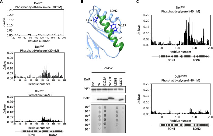
Figure 4—source data 1. Effect of site-directed mutations on DolP function.

Figure 4.

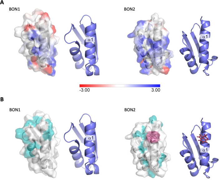
Figure 4—figure supplement 1. Electrostatic analysis of DolP.

Figure 4—figure supplement 1.

(A) Electrostatic surface map of DolP BON domains 1 and 2 calculated using DelPhi (Li et al., 2012) at a pH of 6 and 0.05M ionic strength (which approximates the experimental conditions). The −3kT/e surface is shown in red and the +3kT/e surface is shown in blue. A formal charge library was used, with a dielectric of 2 assigned to the protein interior and a dielectric of 80 assigned to the exterior. Cartoon representations of the BON structures are shown to the right of each surface to more clearly highlight the orientations of the protein. The BON1:α1 and BON2:α1 helices show clear differences, with BON1:α1 being predominantly neutral with an electronegative patch towards its N-terminus, whilst BON2:α2 shows no electronegatively at all, but rather has a large electropositive patch towards the centre of this helix presumably explaining its specificity for the electropositive surface of phosphatidylglycerol. (B) Hydrophobic surface map of DolP BON domains 1 and 2, hydrophobic residues (A, G, V, I, L, F, M) are shown in cyan, W127 (Red) is shown exposed on the surface of the BON2:α1 helix. Cartoon representations of the BON structures are shown to the right of each surface to more clearly highlight the orientations of the protein.
Figure 4—figure supplement 2. Analysis of DolP mutants.

Figure 4—figure supplement 2.

(A) E. coli BW25113 ∆dolP mutants were complemented with plasmids expressing a wild-type copy of DolP or a mutant version. Each strain was serially diluted and plated on LB-agar containing either vancomycin (100 μg/ml) or SDS (4.8% wt/vol), and growth was observed after overnight incubation. The W127E and L137E mutants failed to grow. (B) Western immunoblotting of whole cell lysates derived from overnight cultures of mutants highlighted in the top panel. Blots were probed with antibodies to the outer-membrane lipoprotein BamB and to DolP.