Abstract
Maternal undernutrition has detrimental effects on fetal development and adult health. Total caloric restriction during early pregnancy followed by adequate nutrition for the remainder of gestation, is particularly linked to cardiovascular and metabolic disease risks during adulthood. The placenta is responsible for transport of nutrients from the maternal to fetal circulation, and the efficiency with which it does so can be adjusted to the maternal nutrient supply. There is evidence that placental adaptations to nutrient restriction in early pregnancy may be retained even when adequate nutrition is restored later in pregnancy, leading to a potential mismatch between placental efficiency and maternal nutrient supplies. However, in the mouse, 50% caloric restriction from days 1.5–11.5 of gestation, while temporarily altering placental structure and gene expression, had no significant effect on day 18.5. The periconceptional period, during which oocyte maturation, fertilization, and preimplantation development occur may be especially critical in creating lasting impact on the placenta. Here, mice were subjected to 50% caloric restriction from 3 weeks prior to pregnancy through d11.5, and then placental structure, the expression of key nutrient transporters, and global DNA methylation levels were examined at gestation d18.5. Prior exposure to caloric restriction increased maternal blood space area, but decreased expression of the key System A amino acid transporter Slc38a4 at d18.5. Neither placental and fetal weights, nor placental DNA methylation levels were affected. Thus, total caloric restriction beginning in the periconceptional period does have a lasting impact on placental development in the mouse, but without changing placental efficiency.
Introduction
Chronic diseases are leading causes of morbidity and mortality. For example, in the United States more than 29.1 million people (9.3%) are diabetic, and at least one-third of these cases in adults and 17% in children are associated with obesity [1]. Heart disease is the leading cause of death, accounting for 1 in every 4 deaths in the U.S. [2]. Though genetics, improper diet and lack of exercise play roles in predisposing adults to these chronic diseases, a growing body of evidence links their incidence to prenatal, environmental insults. In particular, maternal nutrient restriction, whether in the form of total caloric restriction, protein deficiency, or micronutrient deficiencies, predisposes offspring to heart disease [3–5] and obesity [6] in adulthood in both humans and animal models. One interpretation of these findings is that organisms may undergo adaptive responses to their environment (e.g. nutritional scarcity) that may prove maladaptive when the environment changes, but can no longer be reversed once sensitive developmental windows have closed [7].
In the classic Dutch Hunger Winter Study, higher rates of obesity and cardiovascular disease were found in adult sons of mothers that received only about 50% of normal calories during the first trimester, followed by an adequate diet later in gestation [8, 9]. In contrast, those subjected to the famine in later gestation did not have elevated rates of these diseases, though they were more susceptible to airway disease, and diabetes risk was increased by exposure at any fetal stage [10, 11]. First trimester exposure to the Chinese Great Famine was also particularly highly associated with hypertension in adulthood [12]. In sheep, nutrient restriction during early-mid gestation likewise increases adiposity [13, 14] and cardiovascular dysfunction [15, 16], and alters hypothalamic-pituitary-adrenal axis function in offspring [17, 18].
The mechanisms by which the particular combination of early gestation undernutrition and adequate nutrition in mid-late gestation programs poor adult outcomes remain poorly understood. The placenta is the organ responsible for the transfer of nutrients from the mother to the fetus. It is also in direct contact with both the maternal and fetal circulations. The placenta therefore can respond to maternal environmental stimuli to in turn affect the growing fetus [19]. There is evidence that the placenta itself may adapt to undernutrition during early gestation, and in some cases if adequate nutrition is restored, it may even grow larger than normal. For example, women that were exposed to the Dutch Hunger Famine during the first trimester and received an adequate diet in later pregnancy delivered significantly larger placentas than control mothers [20]. Similarly, placentas of ewes born to mothers who were undernourished through mid-pregnancy were larger than normal at birth [21–23]. Interestingly, in these cases, offspring were normal weight at term, with greater adipose tissue in the lamb, suggesting that placental adaptations compensated or even overcompensated for early nutrient restriction [20, 23]. On the other hand, maternal food restriction prior to and during the first half of gestation in guinea pigs reduced placental surface area and labyrinth zone size [24]. In the mouse, 50% caloric restriction from days 1.5–11.5 of gestation initially reduced placental weight and the size of junctional zone, but after restoration of adequate nutrition from days 11.5–18.5, placental size and structure recovered [25, 26]. These findings suggest that differences in the timing of caloric restriction interact with species differences to determine long-term impacts on placental development.
Here, we test the hypothesis that caloric restriction will permanently impact the mouse placenta if it occurs over the periconceptional period. Periconception is defined as the period from oocyte maturation and selection through early embryo development [27]. Particularly critical events during this time include the establishment of parent-of-origin specific DNA methyation, the erasure of most DNA methylation in non-imprinted regions, the separation of embryonic and extraembryonic lineages, and the beginnings of lineage-specific DNA methylation [27, 28]. Multiple studies show that exposing gametes and embryos to an adverse environment in vitro, prior to implantation in a normal uterine environment, is sufficient to alter embryo development and adult health [29]. In vivo, feeding mice a low protein diet only during the periconceptional period led to smaller, more efficient placentas at d17 [30], and to greater glucose and System A amino acid transport activity on d19 [31]. However, the long-term placental effects of total caloric restriction during the periconceptional period, such as that experienced in the Dutch and Chinese famines, have not been examined. As we have previously demonstrated that placental effects of food restriction from days 1.5–11.5 are reversible [25], here, mice were subjected to 50% caloric restriction beginning 3 weeks prior to mating, when oocyte maturation begins. Mice were returned to unlimited access to control diet for the latter half of gestation (d11.5), and then placental structure, nutrient transporter expression and global DNA methylation levels were determined near term (d18.5).
Materials and methods
Animals
All animal procedures were approved by University of Missouri Institutional Animal Care and Use Committee. Swiss Webster mice 6–8 weeks old and ≥ 25g were obtained from Envigo (Indianapolis, IN). They were individually housed in a 12hr dark, 12hr light cycle and fed an AIN-93G (Research Diets, Inc) growing rodent diet ad libitum for one week to acclimatize them to their surroundings. Specialized feeding containers were used so that daily food consumption could be recorded [32]. The experimental timeline is shown in Fig 1. Beginning three weeks prior to mating, female mice were randomly separated into two groups: a control group fed ad libitum and a restricted group fed 50% of previous average daily food consumption for each individual. Mice were fed at 2 pm each day. During the mating period, males were placed in the female cages each night at 8 pm and returned to their own cages each morning, with free access to food until a copulatory plug was detected for up to five nights. Females on the nutrient restricted diet consumed their entire food allotment each day before males were added to the cage. The day a copulatory plug was observed was termed d0.5 of gestation. Dietary treatments continued until d11.5, and then all mice were fed ad libitum from d11.5 to d18.5. All animals were allowed free access to water. At d18.5, mice were sacrificed, and placentae collected to examine placental morphology and gene expression. In total, tissues were collected from five mice in the control group and six in the restricted group.
Fig 1. Experimental timeline.

After one week of acclimation and measurement of consumption, female mice were randomized to ad libitum feeding (control), or fed 50% of previous consumption levels. Three weeks later, females were bred, and all were returned to ad libitum feeding at gestation d11.5 before tissue collection at d18.5.
Tissue collection
At d18.5, pregnant females were euthanized by CO2 asphyxiation and cervical dislocation, and fetuses were euthanized by decapitation. Placentae were collected, and placental and fetal weights were recorded. One half of each placenta was fixed in 4% paraformaldehyde in PBS overnight for histological analysis. The second half of each placenta was divided into two pieces, one for gene expression analysis and the second for DNA methylation analysis. Both were snap frozen in liquid nitrogen and stored at -80°C until needed.
Placental morphology
Fixed placentae were dehydrated and embedded in paraffin. Placentae were cut into 6 serial mid- sagittal sections, 5um thick, 50um apart, mounted on glass slides and stained with hematoxylin and eosin (H&E) (IDEXX Bioresearch, Columbia, MO). The largest cross-section on each slide was chosen for morphological analysis. For determination of total cross-sectional area and that of each of the major zones, overlapping images were photographed with a 4x objective lens on a Zeiss Axiovert 200M microscope, and arranged into a single, high-resolution image of the entire placental cross-section. Junctional zone and labyrinth zone areas were measured by manual outlining using the freehand selection tool in ImageJ software (NIH) by an operator blinded to treatment groups. Three placentas were chosen at random from each litter for the morphometric analysis by drawing three tubes from a bag.
To identify fetal blood vessels in the labyrinth zone, cross sections were immunohistochemically stained for PECAM1 (CD31) by using VECTASTAIN Elite ABC HRP Kit (Peroxidase, Rabbit IgG). Briefly, sections were rehydrated in a graded alcohol series, and antigen retrieval was performed by boiling in sodium citrate buffer (10mM Na Citrate, 0.05% Tween 20, pH 6.0) for 3 minutes in a pressure cooker, and then incubated in normal goat serum diluted according to manufacturer’s instructions to block non-specific binding. Placental sections were then incubated with anti-PECAM1 (Abcam ab28364) overnight at 4C. Secondary antibody and color development were carried out according to Vectastain instructions, and nuclei were counterstained with hematoxylin. Five representative images were photographed per placenta with a 40x objective on an Olympus IX81 microscope. The total cross-sectional areas of maternal blood sinuses and fetal blood vessels were determined by using the freehand selection tool within ImageJ software.
Real time RT-PCR
Three placentas were chosen at random from each litter for analysis of gene expression. Placental tissue was homogenized in Trizol Reagent (Sigma Aldrich) with an Omni GLH Homogenizer. After phase separation, RNA was further purified using Qiagen RNeasy kit (QIAGEN, Valencia, CA) according to manufacturer’s protocol. RNA concentration was determined by absorbance using the Take3 Plate (Bio-Tek Instruments). Two μg of total RNA was reversed transcribed with SuperScript III First-Strand Synthesis System (Invitrogen/Life Technologies, Grand Island, NY), using random hexamer primers, according to manufacturer’s protocol.
Real-time RT-PCR was used to measure relative transcript levels of genes specific to the major trophoblast lineages: heart and neural crest derivative 1 (Hand1) was selected as a marker for trophoblast giant cells [33]; trophoblast specific protein alpha (Tpbpa) for spongiotrophoblast and glycogen cells [34, 35]; extraembryonic spermatogenesis homeobox 1 homolog (Esx1) for synctiotrophoblast cells [36]; and platelet and endothelial cell adhesion molecule 1 (Pecam1, CD31) for endothelial cells [37]. Placental lactogen 2 (Prl3b1) marks spongiotrophoblast, parietal and sinusoidal trophoblast giant cells, and has been found to be regulated by fasting at the protein, but not mRNA level [38–40]. Cathepsin Q (Ctsq) is a marker of sinusoidal trophoblast giant cells [38, 41].
Additionally, messenger RNAs encoding six key placental nutrient transporters were examined. The placenta expresses Cd36 (FAT), a key fatty acid transporter, and Srebp1c¸ (Sterol regulatory element binding transcription factor 1), a regulator of fatty acid synthesis [42, 43]. Slc2a3 (GLUT3) is the major placental glucose transporter and has previously been shown to be regulated by maternal nutrition [44–46], as have the amino acid transporters Slc7a5 (LAT1) and Slc38a4 (SNAT4) [44, 47, 48].
Primers (Table 1) were synthesized by Integrated DNA Technologies. Primers for Slc7a5 were predesigned by IDT (Mm.PT.58.10563809). The specificity and efficiency of each reaction was validated by using serial dilutions of placental cDNA. PCR was conducted in triplicate using RT2 SYBR Green ROX qPCR Master Mix (Qiagen) in a final volume of 12μl on a CFX Connect Thermocycler (Bio-Rad). Cycling conditions were as follows: 95°C for 10 minutes, 40 cycles of 95°C for 15 secs, 60°C for 1 minute. No-template and no reverse transcriptase controls were performed. Data from each gene of interest were normalized to the average values of housekeeping genes hypoxanthine phosphoribosyltransferase 1 (Hprt) and Glyceraldehyde-3-Phosphate Dehydrogenase (Gapdh), which were determined not to change with nutrient restriction. Fold change differences in gene expression and error bars were calculated using the ΔΔCt method [49].
Table 1. PCR primers.
| Gene | Forward primer | Reverse primer | Source |
|---|---|---|---|
| Cd36 | GGTCCTTACACTACAGAGTTCGTTA | CATTGGGCTGTACAAAAGACACA | [25] |
| Ctsq | CATTGCCAGTTGACAACACAAG | ATAGCCTTCATTTCGCCAATCA | [50] |
| Esx1 | CCCATGCATCCTCAAATGATG | GCCTAAATGGTGGAGGCATTC | [51] |
| Gapdh | TGCACCACCAACTGCTTAGC | GGCATGGACTGTGGTCATGAG | [52] |
| Hand1 | GAGATGTATACCTGAGAGCAACAGGCATGATAGGTAG | CTTCTCCTTCATTTCTTTCCTTTTCCTTC | [53] |
| Hprt | TGACACTGGCAAAACAATGCA | GGTCCTTTTCACCAGCAAGCT | [26] |
| Prl3b1 | AGCAGCCTTCTGGTGTTGTC | TGTGACACCACAATCACACG | [54] |
| Pecam1 | AGGCTTGCATAGAGCTCCAG | TTCTTGGTTTCCAGCTATGG | [55] |
| Rbm31xy | CACCTTAAGAACAAGCCAATACA | GGCTTGTCCTGAAAACATTTGG | [56] |
| Slc2a3 | CTCTTCAGGTCACCCAACTACGT | CCGCGTCCTTGAAGATTCC | [57] |
| Slc7a5 | GGCGTACATCAGCGTCAT | GTTCACGTCCTCAAGACTGTT | IDT |
| Slc38a4 | TCACACTGCTGTTTCCAAGG | CAGCCGGAAGAATGAAAATC | [58] |
| Srebp1c | TGGTGGGCACTGAAGCAAA | GCAAGAAGCGGATGTAGTCGAT | [25] |
| Tpbpa | GCCAGTTGTTGATGACCCTGA | CCCATCGCCACTCTCTGTGT | [59] |
DNA isolation and methylation assay
Genomic DNA was isolated as previously described [60] from three placentas per dam, chosen by using a random number generator. Briefly, tissues were lysed in lysis buffer with proteinase K followed by separation using Phenol:Chloroform:Isoamyl Alcohol. Genomic DNA samples were stored at -20⁰C until use. DNA concentration was measured using an Epoch Microplate Spectrophotometer (Bio-Tek Instruments).
The level of global DNA methylation was measured using the 5-mC DNA ELISA kit (Zymo Research) according to the manufacturer's protocol, with the following exceptions: (1) Antibody incubation was performed overnight at 4⁰C; (2) 200ng of DNA was used per standard and sample well. To construct a standard curve, methylated DNA (Zymo Research) and unmethylated Lambda DNA (New England BioLabs) were mixed in various proportions (12.5%, 6.25%, 3.125%, 1.56%, 0.78%, and 0% methylated DNA). Intra-assay coefficient of variation was 5.41%. Samples were run in duplicate. To calculate the percentage of CpG that are methylated, the %5-methylcytosine (mC) calculated from the standard curve was corrected for the differences in average CpG density between mouse and E.Coli (used for the standard curve) genomic DNA, according to manufacturer’s instructions.
Sex determination
RNA was isolated and reverse transcribed or DNA was isolated, as described above for each placenta used for the morphometry, RT-PCR, or methylation analyses. Sex was then determined by amplification of a 269 bp region of Rbm31x and the corresponding 353 bp region of Rbm31y by PCR (Table 1) [56].
Statistical analysis
Fetal and placental weights were averaged for each litter, and these averages were compared between control and restricted groups by using Student’s t-test in GraphPad Prism software. Morphological features were analyzed by using the Mixed Procedure in SAS, with treatment group and placental sex as independent variables and litter nested within treatment group as a covariate, to account for the interdependence of placentas from the same litter. The same method was used to compare delta Ct values for analysis of real-time RT-PCR data. Data were termed statistically significant if p< 0.05.
Results
Placental and fetal growth
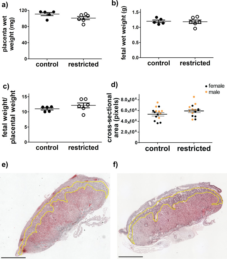
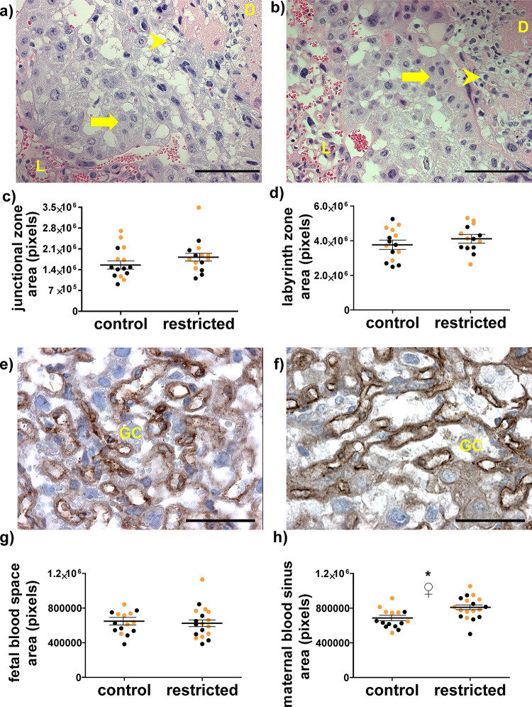
The overall size of the placenta near term, measured either by weight or cross-sectional area, was not different in dams fed an adequate diet throughout pregnancy and dams restricted during the periconceptional period-mid gestation (Fig 2A–2D). No structural abnormalities, hemorrhaging, or necrosis were observed (Fig 2E and 2F). Similarly, there were no significant differences in the appearance or cross-sectional areas of the junctional zone (Fig 3A–3C). While the total cross-sectional area of the labyrinth zone also was not affected (Fig 3D), the structure of blood spaces within it was altered (Fig 3E–3H). Specifically, there was an approximately 20% increase in the cross-sectional area of maternal blood sinuses per unit area following periconceptional exposure to food restriction (Fig 3H). Maternal blood sinus area also differed by sex, with male placentas having greater area than female placentas, but both sexes responded similarly to food restriction.
Fig 2.
There were no significant differences between control fed and food restricted dams on d18.5 in (a) placental wet weights (b) fetal wet weights (c) placental efficiency (fetal weight/placental weight) or d) total cross-sectional area. a-c) Each marker represents the mean from one litter. Horizontal lines are the means for each maternal treatment group (control or restricted dams), and bars represent SEM. In d) each marker represents one placenta and lines represent least square means and SEM for each maternal treatment group. For a-d, N = 5 control, 6 restricted dams. Representative images of cross-sections of the placenta from e) control and f) food restricted. Junctional zones are outlined in yellow, with the decidua above and to the right, and labyrinth below left. Scale bar represents 1 mm. No grossly abnormal pathology was observed.
Fig 3. Placental histomorphometry at day 18.5.
Representative images of hematoxylin and eosin stained sections of the junctional zone were captured with a 20x objective from control (a) and previously food-restricted dams (b). L = labyrinth zone, D = decidua, arrow indicates spongiotrophoblast cells, arrowhead indicates glycogen cells. Scale bars are 100 μm. Total cross-sectional areas of the (c) junctional and (d) labyrinth zone Each marker is one placenta, horizontal lines represent least square means of each maternal treatment group, and error bars are SEM. Representative images of the labyrinth zone were captured with a 40x objective from placentas carried (e) control and (f) food-restricted dams. Immunoperoxidase staining (brown) indicates endothelial marker PECAM1, showing fetal blood vessels, and nuclei are counterstained with hematoxylin (purple). GC = trophoblast giant cell in maternal sinus. Scale bars are 50 μm. Quantification of (g) fetal and (h) maternal blood spaces was performed in five sections each from three placentas per dam. Each marker is one placenta, horizontal lines represent least square means of each maternal treatment group, and error bars are SEM.* p <0.05 control vs. restricted. ♀ p<0.05 female vs. male. Black dots are female and gold dots are male.
Trophoblast differentiation
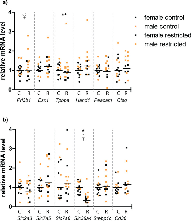
In order to further characterize the permanent impacts of periconceptional food restriction on placental development, semi-quantitative real-time RT-PCR was used to compare steady-state mRNA levels of transcription factors that are specifically expressed in the major trophoblast sublineages within the placenta (Fig 4A). There were no significant differences in relative mRNA levels of placental lineage markers Esx1 (synctiotrophoblast), Tpbpa (spongiotrophoblast and glycogen cells), Hand1(trophoblast giant cells), Prl3b1(spongiotrophoblast, parietal and sinusoidal trophoblast giant cells) [61], Ctsq (sinusoidal trophoblast giant cells) [41] or Pecam1(fetal endothelial cells) [37]. However, Prl3b1 expression did differ with placental sex, with males expressing more than females (p = 0.01). Tpbpa expression also was significantly higher in males than in females, but only in the restricted group (p = 0.003) (Fig 4A).
Fig 4.
Relative steady-state mRNA levels encoding (a) selected trophoblast lineage markers and (b) key placental nutrient transporters. Relative levels are calculated by the 2ΔΔCt method, using the least square mean of the control group as the reference value, such that the control group = 1. Each symbol represents one placenta. Black horizontal lines represent the 2ΔΔCt for the least square mean of each treatment group. * C = Control, R = restricted. * p<0.05 control vs restricted; ♀ = p<0.05 male vs female; ** = p<0.05 sex x treatment interaction. Placentas were obtained from five dams each in the control and restricted groups.
Placental nutrient transporters
Real-time PCR was also used to compare expression of key placental nutrient transporters at d18.5 following periconceptional food restriction (Fig 4B). In the placenta from previously food restricted dams, there was a three-fold decrease in mRNA encoding Slc38a4, which transports neutral amino acids such as glutamine, glycine, serine and alanine [62]. Slc38a4 mRNA levels were also higher in males than females, although food restriction had the same effect regardless of sex. In contrast, there were no differences in mRNA levels for the L-type, branched-chain amino acid transporters Slc7a5 and Slc7a8 (also known as LAT1 and LAT2). Levels of mRNA for fatty acid transporter Cd36 and Srebp1c, a transcription factor that regulates genes involved in sterol biosynthesis, were not changed, nor were there differences in expression of glucose transporter Slc2a3 between control and restricted placentae.
Placental DNA methylation
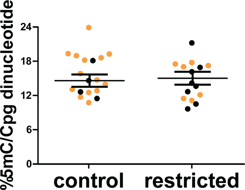
An ELISA was used to quantify the effects of periconceptional food restriction on the total level of placental DNA methylation (Fig 5). There was no significant difference in the overall percentage of methylated CpGs between placentas from control dams and those exposed to periconceptional undernutrition (p = 0.8), or between male and female placentas (p = 0.4).
Fig 5. The overall percentage of CpG methylation (%5mC/CpG sites) in placentas from control and periconceptionally food-restricted dams was determined by ELISA.
Each marker represents an individual placenta. Black dots are female and gold dots are male. Horizontal lines represent least square means of each maternal treatment group, and error bars are SEM. N = 5 control dams and 6 restricted dams.
Discussion
The goal of this study was to determine whether the periconceptional period is a critical time in which maternal total caloric restriction can permanently alter placental development and function. Whereas studies in humans and sheep found that total caloric restriction during early pregnancy leads to placental overgrowth near term [20, 22, 23], we previously found that food restriction from days 1.5–11.5 of pregnancy did not change placental characteristics in late pregnancy [21, 22, 63]. In contrast, here we show that food restriction from three weeks prior to mating through d11.5 does alter the structure of the labyrinth zone and the expression of System A neutral amino acid transporter Slc38a4 (SNAT4), even in late pregnancy.
These results are consistent with the hypothesis that adverse exposures during the periconceptional period, including balanced, total, caloric restriction, have irreversible effects on the placenta. Exposure to a low protein diet from conception to implantation results in a smaller placenta, with greater glucose transport and reduced amino acid transport near term [30, 31]. Alcohol exposure from days -4 to 4 post-conception reduced placental volume and maternal blood space area at day 15 in rats [64], and in sheep, inclusion of serum in the culture medium for the first two days, but not the last two days of preimplantation embryo culture resulted in larger placentas at term [65]. The importance of the periconceptional period has been less clear following total dietary restriction. In “natural” experiments in humans, like the Dutch and Chinese famines, it isn’t possible to distinguish between the periconceptional period and the rest of early pregnancy. In sheep, where this distinction is possible, the periconceptional period appears to be important, but not uniquely important. Restricting ewes to 50% of requirements from days -60 to 30 increased placental vascularity at day 80 [66], although it did not significantly impact placental size or placentome number at day 131 [67]. Nutrient restriction outside the periconceptional period, from days 28–80 of pregnancy was also effective, resulting in a larger placenta, greater placental glucose transporter expression, and greater number of placentomes near term, at day 140–145 (term = 147 days) [22, 68].
The current study, combined with our previous work, shows that in the mouse a period of nutrient restriction that includes periconception (-3 weeks to d11.5) but not the period immediately following (d1.5–11.5), is sufficient for lasting effects on the placenta, strongly suggesting the importance of periconception. However, one limitation to this study is that nutrient restriction was not limited to the periconception period. We cannot rule out the possibility that nutrient restriction both before and after d1.5 are needed. Furthermore, the overall length of the nutrient restriction was longer than in than in the previous study, which could contribute to the more long-lasting effects. Future experiments targeted to just -3 weeks through d1.5, or an even narrower window, would help to more precisely pinpoint the period of susceptibility.
Nonetheless, the window of susceptibility identified in the present study provides some insight into possible mechanisms of action. From -3 weeks to d1.5, the oocyte, and then the zygote, undergo major rewriting of DNA methylation. Maternal imprints are established in the oocyte, and methylation of most non-imprinted genes is erased shortly following fertilization. Evidence from monozygotic twins further shows that many DNA methylation marks are established very early in embryonic development, prior to twinning [69]. A diet deficient in methyl donors (folate, vitamin B12, methionine) during the periconceptional period caused global hypomethylation in fetal sheep in mid-gestation [70]. Caloric restriction throughout gestation is associated with global hypomethylation in mouse placenta at day 19 [71]. This led us to hypothesize that maternal nutrition could alter later placental phenotypes through global DNA methylation differences. However, the overall percentage of methylated CpGs in the d18.5 placentas of control mothers and those that had previously been nutrient restricted were not different. Thus, neither the 50% reduction in macronutrients, nor the accompanying decrease in methyl donors like folate, in the periconceptional period prevented normal overall methylation levels in the late pregnancy placenta. It must be noted that the absence of differences in DNA methylation levels does not rule out differences in individual DNA regions. For example, survivors of the Dutch Hunger Winter show several specific methylation changes in whole blood without differences in global DNA methylation [72]. Additional studies, for example by whole genome bisulfite sequencing, are needed to determine whether any individual differences in placental DNA methylation follow periconceptional undernutrition in the mouse.
Another unknown variable is the effect of refeeding on the growth and function of the placenta. Very little is known about the dynamics of refeeding after nutrient restriction in pregnant mice, and here food consumption was not determined after the resumption of ad libitum feeding in the calorie restricted mice. However, it is known that caloric restriction can induce later hyperphagia in non-pregnant mice, and that estrogen plays a role in acute refeeding after fasting [73, 74]. Thus, it is possible that after the food restricted mice were returned to ad libitum feeding at day 11.5, that they consumed more calories than the control mice. Additional experiments, including pair-feeding, would be needed to determine whether post-restriction eating plays a role in the placental phenotype on day 18.5.
Placental adaptations in response to maternal undernutrition that outlast the period of undernutrition are potentially harmful because they produce a mismatch between the placenta and the environment. The placenta is “nutrient sensing,” adjusting structure and nutrient transport to preserve fetal growth in the face of reduced maternal nutrient supplies [48]. For example, in the mouse, while the placenta is smaller during maternal food restriction, the size of the labyrinth zone, where nutrients are exchanged, is preserved in comparison to that of the junctional zone [26, 75]. Similarly, regulation of glucose and amino acid transport can compensate for lower nutrient availability, to preserve either maternal or fetal supplies, but if these changes outlast the period of nutrient restriction, they can continue to alter fetal body composition and development. Early pregnancy exposures can alter placental glucose transporter expression later in pregnancy. Slc2a3 is reduced in the late gestation mouse placenta following assisted reproduction procedures in the periconceptional period [58, 76]. Caloric restriction throughout pregnancy has been shown both to reduce [77] and stimulate [78] placental Slc2a3 expression in rodents. However, here no significant differences in late gestation gene expression of Slc2a3 were detected in placentae from mothers that were nutrient restricted during early pregnancy. Along with the previous observation that placental Slc2a3 levels are acutely regulated by maternal glucose concentrations [79], these results suggest that its expression can change in response to the restoration of food following nutrient restriction.
Amino acid transport is also regulated by maternal nutritional status during pregnancy [80]. The System A neutral amino acid transporter SLC38A4 is a major contributor to fetal growth [47, 81]. Its mRNA, protein and activity levels have been shown to fall in response to various models of nutrient restriction. For example, in rats, a maternal low protein diet throughout gestation reduced its activity [82] and in the baboon, ongoing nutrient restriction reduced microvillous membrane levels of the protein in mid-pregnancy [45]. In contrast, Slc38a4 mRNA levels increase with periconceptional exposure to assisted reproduction procedures [76]. In the present study, even on d18.5, expression of Slc38a4 in placentas from previously nutrient restricted dams was only one-third that of controls. This reduction would be predicted to decrease amino acid transport and thereby limit fetal growth. Conversely, placentas from dams that had been nutrient restricted also had increased maternal blood space area within the sampled regions of the labyrinth zone, providing a greater surface area for nutrient exchange between the maternal blood and surrounding syncytiotrophoblast; this would support greater growth. Thus, the overall consequences of the combined placental adaptations for fetal growth and development are difficult to predict, but the observed lack of change in fetal weights could be a result of these placental alterations canceling one another. While both alterations were present at d18.5, it is possible that one occurred first, triggering a compensatory response to maintain consistent amino acid transport.
Offspring exposed to undernutrition during early pregnancy have greater chronic disease risk as adults, even without changes in birth weight [10]. Our data support the idea that maternal food deprivation during the periconceptional period is particularly important for placental function throughout pregnancy even if nutrition is later restored [83–85]. Additionally, the findings suggest that careful examination of the term placenta may provide evidence of adverse exposures in the periconception period, perhaps even serving as an indicator of future health.
Data Availability
All relevant data are within the manuscript.
Funding Statement
This work was funded by the National Institutes of Health IMSD R25GM056901 to GVGC, and University of Missouri Mission Enhancement to LCS. The funders had no role in study design, data collection and analysis, decision to publish or preparation of the manuscript.
References
- 1.Ogden C.L., Carroll M.D., Kit B.K., Flegal K.M., Prevalence of childhood and adult obesity in the United States, 2011–2012, JAMA 311(8) (2014) 806–14. 10.1001/jama.2014.732 [DOI] [PMC free article] [PubMed] [Google Scholar]
- 2.Benjamin E.J., Muntner P., Alonso A., Bittencourt M.S., Callaway C.W., Carson A.P., et al. American Heart Association Council on, C. Prevention Statistics, S. Stroke Statistics, Heart Disease and Stroke Statistics-2019 Update: A Report From the American Heart Association, Circulation 139(10) (2019) e56–e528. 10.1161/CIR.0000000000000659 [DOI] [PubMed] [Google Scholar]
- 3.Barker D.J., The developmental origins of chronic adult disease, Acta Paediatr Suppl 93(446) (2004) 26–33. 10.1111/j.1651-2227.2004.tb00236.x [DOI] [PubMed] [Google Scholar]
- 4.Frankel S., Elwood P., Sweetnam P., Yarnell J., Smith G.D., Birthweight, body-mass index in middle age, and incident coronary heart disease, Lancet 348(9040) (1996) 1478–80. 10.1016/S0140-6736(96)03482-4 [DOI] [PubMed] [Google Scholar]
- 5.Rich-Edwards J.W., Stampfer M.J., Manson J.E., Rosner B., Hankinson S.E., Colditz G.A., et al. , Birth weight and risk of cardiovascular disease in a cohort of women followed up since 1976, BMJ 315(7105) (1997) 396–400. 10.1136/bmj.315.7105.396 [DOI] [PMC free article] [PubMed] [Google Scholar]
- 6.Curhan G.C., Chertow G.M., Willett W.C., Spiegelman D., Colditz G.A., Manson J.E., et al. , Birth weight and adult hypertension and obesity in women, Circulation 94(6) (1996) 1310–5. 10.1161/01.cir.94.6.1310 [DOI] [PubMed] [Google Scholar]
- 7.Gluckman P.D., Hanson M.A., Low F.M., Evolutionary and developmental mismatches are consequences of adaptive developmental plasticity in humans and have implications for later disease risk, Philos Trans R Soc Lond B Biol Sci 374(1770) (2019) 20180109 10.1098/rstb.2018.0109 [DOI] [PMC free article] [PubMed] [Google Scholar]
- 8.Ravelli A.C., Bleker O.P., Roseboom T.J., van Montfrans G.A., Osmond C., Barker D.J., Cardiovascular disease in survivors of the Dutch famine, Nestle Nutr Workshop Ser Pediatr Program 55 (2005) 183–91; discussion 191–5. 10.1159/000082602 [DOI] [PubMed] [Google Scholar]
- 9.Ravelli A.C., van Der Meulen J.H., Osmond C., Barker D.J., Bleker O.P., Obesity at the age of 50 y in men and women exposed to famine prenatally, Am J Clin Nutr 70(5) (1999) 811–6. 10.1093/ajcn/70.5.811 [DOI] [PubMed] [Google Scholar]
- 10.Roseboom T., de Rooij S., Painter R., The Dutch famine and its long-term consequences for adult health, Early Hum Dev 82(8) (2006) 485–91. 10.1016/j.earlhumdev.2006.07.001 [DOI] [PubMed] [Google Scholar]
- 11.Schulz L.C., The Dutch Hunger Winter and the developmental origins of health and disease, Proc Natl Acad Sci U S A 107(39) (2010) 16757–8. 10.1073/pnas.1012911107 [DOI] [PMC free article] [PubMed] [Google Scholar]
- 12.Wang P.X., Wang J.J., Lei Y.X., Xiao L., Luo Z.C., Impact of fetal and infant exposure to the Chinese Great Famine on the risk of hypertension in adulthood, PLoS One 7(11) (2012) e49720 10.1371/journal.pone.0049720 [DOI] [PMC free article] [PubMed] [Google Scholar]
- 13.Ojha S., Symonds M.E., Budge H., Suboptimal maternal nutrition during early-to-mid gestation in the sheep enhances pericardial adiposity in the near-term fetus, Reprod Fertil Dev 27(8) (2015) 1205–12. 10.1071/RD14007 [DOI] [PubMed] [Google Scholar]
- 14.Bispham J., Gopalakrishnan G.S., Dandrea J., Wilson V., Budge H., Keisler D.H., et al. , Maternal endocrine adaptation throughout pregnancy to nutritional manipulation: consequences for maternal plasma leptin and cortisol and the programming of fetal adipose tissue development, Endocrinology 144(8) (2003) 3575–85. 10.1210/en.2003-0320 [DOI] [PubMed] [Google Scholar]
- 15.Dong F., Ford S.P., Fang C.X., Nijland M.J., Nathanielsz P.W., Ren J., Maternal nutrient restriction during early to mid gestation up-regulates cardiac insulin-like growth factor (IGF) receptors associated with enlarged ventricular size in fetal sheep, Growth Horm IGF Res 15(4) (2005) 291–9. 10.1016/j.ghir.2005.05.003 [DOI] [PubMed] [Google Scholar]
- 16.Gopalakrishnan G.S., Gardner D.S., Rhind S.M., Rae M.T., Kyle C.E., Brooks A.N., et al. , Programming of adult cardiovascular function after early maternal undernutrition in sheep, Am J Physiol Regul Integr Comp Physiol 287(1) (2004) R12–20. 10.1152/ajpregu.00687.2003 [DOI] [PubMed] [Google Scholar]
- 17.Poore K.R., Boullin J.P., Cleal J.K., Newman J.P., Noakes D.E., Hanson M.A., et al. , Sex- and age-specific effects of nutrition in early gestation and early postnatal life on hypothalamo-pituitary-adrenal axis and sympathoadrenal function in adult sheep, J Physiol 588(Pt 12) (2010) 2219–37. 10.1113/jphysiol.2010.187682 [DOI] [PMC free article] [PubMed] [Google Scholar]
- 18.Gardner D.S., Van Bon B.W., Dandrea J., Goddard P.J., May S.F., Wilson V., et al. , Effect of periconceptional undernutrition and gender on hypothalamic-pituitary-adrenal axis function in young adult sheep, J Endocrinol 190(2) (2006) 203–12. 10.1677/joe.1.06751 [DOI] [PubMed] [Google Scholar]
- 19.Dimasuay K.G., Boeuf P., Powell T.L., Jansson T., Placental Responses to Changes in the Maternal Environment Determine Fetal Growth, Front Physiol 7 (2016) 12 10.3389/fphys.2016.00012 [DOI] [PMC free article] [PubMed] [Google Scholar]
- 20.Lumey L.H., Compensatory placental growth after restricted maternal nutrition in early pregnancy, Placenta 19(1) (1998) 105–11. 10.1016/s0143-4004(98)90105-9 [DOI] [PubMed] [Google Scholar]
- 21.McCrabb G.J., Egan A.R., Hosking B.J., Maternal undernutrition during mid-pregnancy in sheep. Placental size and its relationship to calcium transfer during late pregnancy, Br J Nutr 65(2) (1991) 157–68. 10.1079/bjn19910077 [DOI] [PubMed] [Google Scholar]
- 22.Dandrea J., Wilson V., Gopalakrishnan G., Heasman L., Budge H., Stephenson T., et al. , Maternal nutritional manipulation of placental growth and glucose transporter 1 (GLUT-1) abundance in sheep, Reproduction 122(5) (2001) 793–800. [PubMed] [Google Scholar]
- 23.Symonds M.E., Gopalakrishnan G., Bispham J., Pearce S., Dandrea J., Mostyn A., et al. , Maternal nutrient restriction during placental growth, programming of fetal adiposity and juvenile blood pressure control, Arch Physiol Biochem 111(1) (2003) 45–52. 10.1076/apab.111.1.45.15141 [DOI] [PubMed] [Google Scholar]
- 24.Roberts C.T., Sohlstrom A., Kind K.L., Earl R.A., Khong T.Y., Robinson J.S., et al. , Maternal food restriction reduces the exchange surface area and increases the barrier thickness of the placenta in the guinea-pig, Placenta 22(2–3) (2001) 177–85. 10.1053/plac.2000.0602 [DOI] [PubMed] [Google Scholar]
- 25.Harper J.L., Caesar G.A., Pennington K.A., Davis J.W., Schulz L.C., Placental changes caused by food restriction during early pregnancy in mice are reversible, Reproduction (2015). 10.1530/REP-15-0010 [DOI] [PMC free article] [PubMed] [Google Scholar]
- 26.Schulz L.C., Schlitt J.M., Caesar G., Pennington K.A., Leptin and the placental response to maternal food restriction during early pregnancy in mice, Biol Reprod 87(5) (2012) 120 10.1095/biolreprod.112.103218 [DOI] [PMC free article] [PubMed] [Google Scholar]
- 27.Velazquez M.A., Fleming T.P., Watkins A.J., Periconceptional environment and the developmental origins of disease, J Endocrinol 242(1) (2019) T33–T49. 10.1530/JOE-18-0676 [DOI] [PubMed] [Google Scholar]
- 28.Li E., Chromatin modification and epigenetic reprogramming in mammalian development, Nat Rev Genet 3(9) (2002) 662–73. 10.1038/nrg887 [DOI] [PubMed] [Google Scholar]
- 29.Feuer S.K., Rinaudo P.F., Physiological, metabolic and transcriptional postnatal phenotypes of in vitro fertilization (IVF) in the mouse, J Dev Orig Health Dis 8(4) (2017) 403–410. 10.1017/S204017441700023X [DOI] [PubMed] [Google Scholar]
- 30.Watkins A.J., Lucas E.S., Marfy-Smith S., Bates N., Kimber S.J., Fleming T.P., Maternal nutrition modifies trophoblast giant cell phenotype and fetal growth in mice, Reproduction 149(6) (2015) 563–75. 10.1530/REP-14-0667 [DOI] [PMC free article] [PubMed] [Google Scholar]
- 31.Coan P.M., Vaughan O.R., McCarthy J., Mactier C., Burton G.J., Constancia M., et al. , Dietary composition programmes placental phenotype in mice, J Physiol 589(Pt 14) (2011) 3659–70. 10.1113/jphysiol.2011.208629 [DOI] [PMC free article] [PubMed] [Google Scholar]
- 32.Townsend K.L., Lorenzi M.M., Widmaier E.P., High-fat diet-induced changes in body mass and hypothalamic gene expression in wild-type and leptin-deficient mice, Endocrine 33(2) (2008) 176–88. 10.1007/s12020-008-9070-1 [DOI] [PubMed] [Google Scholar]
- 33.El-Hashash A.H., Warburton D., Kimber S.J., Genes and signals regulating murine trophoblast cell development, Mech Dev 127(1–2) (2010) 1–20. 10.1016/j.mod.2009.09.004 [DOI] [PMC free article] [PubMed] [Google Scholar]
- 34.Natale D.R., Starovic M., Cross J.C., Phenotypic analysis of the mouse placenta, Methods Mol Med 121 (2006) 275–93. 10.1385/1-59259-983-4:273 [DOI] [PubMed] [Google Scholar]
- 35.Cowden Dahl K.D., Fryer B.H., Mack F.A., Compernolle V., Maltepe E., Adelman D.M., et al. , Hypoxia-inducible factors 1alpha and 2alpha regulate trophoblast differentiation, Mol Cell Biol 25(23) (2005) 10479–91. 10.1128/MCB.25.23.10479-10491.2005 [DOI] [PMC free article] [PubMed] [Google Scholar]
- 36.T.M. Plant, A.J. Zeleznik, Knobil and Neill's Physiology of Reproduction, Elsevier Science2014.
- 37.Shafiee A., Patel J., Hutmacher D.W., Fisk N.M., Khosrotehrani K., Meso-Endothelial Bipotent Progenitors from Human Placenta Display Distinct Molecular and Cellular Identity, Stem Cell Reports 10(3) (2018) 890–904. 10.1016/j.stemcr.2018.01.011 [DOI] [PMC free article] [PubMed] [Google Scholar]
- 38.Simmons D.G., Fortier A.L., Cross J.C., Diverse subtypes and developmental origins of trophoblast giant cells in the mouse placenta, Dev Biol 304(2) (2007) 567–78. 10.1016/j.ydbio.2007.01.009 [DOI] [PubMed] [Google Scholar]
- 39.Rawn S.M., Huang C., Hughes M., Shaykhutdinov R., Vogel H.J., Cross J.C., Pregnancy Hyperglycemia in Prolactin Receptor Mutant, but Not Prolactin Mutant, Mice and Feeding-Responsive Regulation of Placental Lactogen Genes Implies Placental Control of Maternal Glucose Homeostasis, Biol Reprod 93(3) (2015) 75 10.1095/biolreprod.115.132431 [DOI] [PMC free article] [PubMed] [Google Scholar]
- 40.Fielder P.J., Ogren L., Edwards D., Talamantes F., Effects of fasting on serum lactogenic hormone concentrations during mid- and late pregnancy in mice, Am J Physiol 253(1 Pt 1) (1987) E40–4. 10.1152/ajpendo.1987.253.1.E40 [DOI] [PubMed] [Google Scholar]
- 41.Outhwaite J.E., McGuire V., Simmons D.G., Genetic ablation of placental sinusoidal trophoblast giant cells causes fetal growth restriction and embryonic lethality, Placenta 36(8) (2015) 951–5. 10.1016/j.placenta.2015.05.013 [DOI] [PubMed] [Google Scholar]
- 42.Duttaroy A.K., Transport of fatty acids across the human placenta: a review, Prog Lipid Res 48(1) (2009) 52–61. 10.1016/j.plipres.2008.11.001 [DOI] [PubMed] [Google Scholar]
- 43.Ganguly A., Devaskar S.U., High-fat diet affects pregestational adiposity and glucose tolerance perturbing gestational placental macronutrient transporters culminating in an obese offspring in wild-type and glucose transporter isoform 3 heterozygous null mice, J Nutr Biochem 62 (2018) 192–201. 10.1016/j.jnutbio.2018.09.001 [DOI] [PMC free article] [PubMed] [Google Scholar]
- 44.Ganguly A., Touma M., Thamotharan S., De Vivo D.C., Devaskar S.U., Maternal Calorie Restriction Causing Uteroplacental Insufficiency Differentially Affects Mammalian Placental Glucose and Leucine Transport Molecular Mechanisms, Endocrinology 157(10) (2016) 4041–4054. 10.1210/en.2016-1259 [DOI] [PMC free article] [PubMed] [Google Scholar]
- 45.Kavitha J.V., Rosario F.J., Nijland M.J., McDonald T.J., Wu G., Kanai Y., et al. , Down-regulation of placental mTOR, insulin/IGF-I signaling, and nutrient transporters in response to maternal nutrient restriction in the baboon, FASEB J 28(3) (2014) 1294–305. 10.1096/fj.13-242271 [DOI] [PMC free article] [PubMed] [Google Scholar]
- 46.Coan P.M., Angiolini E., Sandovici I., Burton G.J., Constancia M., Fowden A.L., Adaptations in placental nutrient transfer capacity to meet fetal growth demands depend on placental size in mice, J Physiol 586(Pt 18) (2008) 4567–76. 10.1113/jphysiol.2008.156133 [DOI] [PMC free article] [PubMed] [Google Scholar]
- 47.Matoba S., Nakamuta S., Miura K., Hirose M., Shiura H., Kohda T., et al. , Paternal knockout of Slc38a4/SNAT4 causes placental hypoplasia associated with intrauterine growth restriction in mice, Proc Natl Acad Sci U S A (2019). 10.1073/pnas.1907884116 [DOI] [PMC free article] [PubMed] [Google Scholar]
- 48.Vaughan O.R., Rosario F.J., Powell T.L., Jansson T., Regulation of Placental Amino Acid Transport and Fetal Growth, Prog Mol Biol Transl Sci 145 (2017) 217–251. 10.1016/bs.pmbts.2016.12.008 [DOI] [PubMed] [Google Scholar]
- 49.Livak K.J., Schmittgen T.D., Analysis of relative gene expression data using real-time quantitative PCR and the 2(-Delta Delta C(T)) Method, Methods 25(4) (2001) 402–8. 10.1006/meth.2001.1262 [DOI] [PubMed] [Google Scholar]
- 50.Yang Y., Liu B., Xu J., Wang J., Wu J., Shi C., et al. , Derivation of Pluripotent Stem Cells with In Vivo Embryonic and Extraembryonic Potency, Cell 169(2) (2017) 243–257 e25. 10.1016/j.cell.2017.02.005 [DOI] [PMC free article] [PubMed] [Google Scholar]
- 51.Schaiff W.T., Knapp F.F. Jr., Barak Y., Biron-Shental T., Nelson D.M., Sadovsky Y., Ligand-activated peroxisome proliferator activated receptor gamma alters placental morphology and placental fatty acid uptake in mice, Endocrinology 148(8) (2007) 3625–34. 10.1210/en.2007-0211 [DOI] [PubMed] [Google Scholar]
- 52.Talton O.O., Pennington K.A., Pollock K.E., Bates K., Ma L., Ellersieck M.R., et al. , Maternal Hyperleptinemia Improves Offspring Insulin Sensitivity in Mice, Endocrinology 157(7) (2016) 2636–48. 10.1210/en.2016-1039 [DOI] [PubMed] [Google Scholar]
- 53.Carter M.G., Sharov A.A., VanBuren V., Dudekula D.B., Carmack C.E., Nelson C., et al. , Transcript copy number estimation using a mouse whole-genome oligonucleotide microarray, Genome Biol 6(7) (2005) R61 10.1186/gb-2005-6-7-r61 [DOI] [PMC free article] [PubMed] [Google Scholar]
- 54.Kaiser S., Koch Y., Kuhnel E., Sharma N., Gellhaus A., Kuckenberg P., et al. , Reduced Gene Dosage of Tfap2c Impairs Trophoblast Lineage Differentiation and Alters Maternal Blood Spaces in the Mouse Placenta, Biol Reprod 93(2) (2015) 31 10.1095/biolreprod.114.126474 [DOI] [PubMed] [Google Scholar]
- 55.Li J., Qu X., Bertram J.F., Endothelial-myofibroblast transition contributes to the early development of diabetic renal interstitial fibrosis in streptozotocin-induced diabetic mice, Am J Pathol 175(4) (2009) 1380–8. 10.2353/ajpath.2009.090096 [DOI] [PMC free article] [PubMed] [Google Scholar]
- 56.Tunster S.J., Genetic sex determination of mice by simplex PCR, Biol Sex Differ 8(1) (2017) 31. 10.1186/s13293-017-0154-6 [DOI] [PMC free article] [PubMed] [Google Scholar]
- 57.Feil D., Lane M., Roberts C.T., Kelley R.L., Edwards L.J., Thompson J.G., et al. , Effect of culturing mouse embryos under different oxygen concentrations on subsequent fetal and placental development, J Physiol 572(Pt 1) (2006) 87–96. 10.1113/jphysiol.2005.102681 [DOI] [PMC free article] [PubMed] [Google Scholar]
- 58.Bloise E., Lin W., Liu X., Simbulan R., Kolahi K.S., Petraglia F., et al. , Impaired placental nutrient transport in mice generated by in vitro fertilization, Endocrinology 153(7) (2012) 3457–67. 10.1210/en.2011-1921 [DOI] [PMC free article] [PubMed] [Google Scholar]
- 59.Sferruzzi-Perri A.N., Macpherson A.M., Roberts C.T., Robertson S.A., Csf2 null mutation alters placental gene expression and trophoblast glycogen cell and giant cell abundance in mice, Biol Reprod 81(1) (2009) 207–21. 10.1095/biolreprod.108.073312 [DOI] [PMC free article] [PubMed] [Google Scholar]
- 60.Li Y., Hagen D.E., Ji T., Bakhtiarizadeh M.R., Frederic W.M., Traxler E.M., et al. , Altered microRNA expression profiles in large offspring syndrome and Beckwith-Wiedemann syndrome, Epigenetics 14(9) (2019) 850–876. 10.1080/15592294.2019.1615357 [DOI] [PMC free article] [PubMed] [Google Scholar]
- 61.Simmons D.G., Rawn S., Davies A., Hughes M., Cross J.C., Spatial and temporal expression of the 23 murine Prolactin/Placental Lactogen-related genes is not associated with their position in the locus, BMC Genomics 9 (2008) 352 10.1186/1471-2164-9-352 [DOI] [PMC free article] [PubMed] [Google Scholar]
- 62.Cleal J.K., Lewis R.M., The mechanisms and regulation of placental amino acid transport to the human foetus, J Neuroendocrinol 20(4) (2008) 419–26. 10.1111/j.1365-2826.2008.01662.x [DOI] [PubMed] [Google Scholar]
- 63.Harper J.L., Caesar G.A., Pennington K.A., Davis J.W., Schulz L.C., Placental changes caused by food restriction during early pregnancy in mice are reversible, Reproduction 150(3) (2015) 165–72. 10.1530/REP-15-0010 [DOI] [PMC free article] [PubMed] [Google Scholar]
- 64.Kalisch-Smith J.I., Steane S.E., Simmons D.G., Pantaleon M., Anderson S.T., Akison L.K., et al. , Periconceptional alcohol exposure causes female-specific perturbations to trophoblast differentiation and placental formation in the rat, Development 146(11) (2019). 10.1242/dev.172205 [DOI] [PubMed] [Google Scholar]
- 65.Rooke J.A., McEvoy T.G., Ashworth C.J., Robinson J.J., Wilmut I., Young L.E., et al. , Ovine fetal development is more sensitive to perturbation by the presence of serum in embryo culture before rather than after compaction, Theriogenology 67(3) (2007) 639–47. 10.1016/j.theriogenology.2006.09.040 [DOI] [PubMed] [Google Scholar]
- 66.Zhu M.J., Du M., Hess B.W., Nathanielsz P.W., Ford S.P., Periconceptional nutrient restriction in the ewe alters MAPK/ERK1/2 and PI3K/Akt growth signaling pathways and vascularity in the placentome, Placenta 28(11–12) (2007) 1192–9. 10.1016/j.placenta.2007.05.004 [DOI] [PubMed] [Google Scholar]
- 67.Oliver M.H., Hawkins P., Harding J.E., Periconceptional undernutrition alters growth trajectory and metabolic and endocrine responses to fasting in late-gestation fetal sheep, Pediatr Res 57(4) (2005) 591–8. 10.1203/01.PDR.0000155942.18096.9C [DOI] [PubMed] [Google Scholar]
- 68.Heasman L., Clarke L., Stephenson T.J., Symonds M.E., The influence of maternal nutrient restriction in early to mid-pregnancy on placental and fetal development in sheep, Proc Nutr Soc 58(2) (1999) 283–8. 10.1017/s0029665199000397 [DOI] [PubMed] [Google Scholar]
- 69.Van Baak T.E., Coarfa C., Dugue P.A., Fiorito G., Laritsky E., Baker M.S., et al. , Epigenetic supersimilarity of monozygotic twin pairs, Genome Biol 19(1) (2018) 2. 10.1186/s13059-017-1374-0 [DOI] [PMC free article] [PubMed] [Google Scholar]
- 70.Sinclair K.D., Allegrucci C., Singh R., Gardner D.S., Sebastian S., Bispham J., et al. , DNA methylation, insulin resistance, and blood pressure in offspring determined by maternal periconceptional B vitamin and methionine status, Proc Natl Acad Sci U S A 104(49) (2007) 19351–6. 10.1073/pnas.0707258104 [DOI] [PMC free article] [PubMed] [Google Scholar]
- 71.Chen P.Y., Ganguly A., Rubbi L., Orozco L.D., Morselli M., Ashraf D., et al. , Intrauterine calorie restriction affects placental DNA methylation and gene expression, Physiol Genomics 45(14) (2013) 565–76. 10.1152/physiolgenomics.00034.2013 [DOI] [PMC free article] [PubMed] [Google Scholar]
- 72.Tobi E.W., Goeman J.J., Monajemi R., Gu H., Putter H., Zhang Y., et al. , DNA methylation signatures link prenatal famine exposure to growth and metabolism, Nat Commun 5 (2014) 5592 10.1038/ncomms6592 [DOI] [PMC free article] [PubMed] [Google Scholar]
- 73.Pankevich D.E., Teegarden S.L., Hedin A.D., Jensen C.L., Bale T.L., Caloric restriction experience reprograms stress and orexigenic pathways and promotes binge eating, J Neurosci 30(48) (2010) 16399–407. 10.1523/JNEUROSCI.1955-10.2010 [DOI] [PMC free article] [PubMed] [Google Scholar]
- 74.Yu K., He Y., Hyseni I., Pei Z., Yang Y., Xu P., et al. , 17beta-estradiol promotes acute refeeding in hungry mice via membrane-initiated ERalpha signaling, Mol Metab 42 (2020) 101053 10.1016/j.molmet.2020.101053 [DOI] [PMC free article] [PubMed] [Google Scholar]
- 75.Coan P.M., Vaughan O.R., Sekita Y., Finn S.L., Burton G.J., Constancia M., et al. , Adaptations in placental phenotype support fetal growth during undernutrition of pregnant mice, J Physiol 588(Pt 3) (2010) 527–38. 10.1113/jphysiol.2009.181214 [DOI] [PMC free article] [PubMed] [Google Scholar]
- 76.de Waal E., Vrooman L.A., Fischer E., Ord T., Mainigi M.A., Coutifaris C., et al. , The cumulative effect of assisted reproduction procedures on placental development and epigenetic perturbations in a mouse model, Hum Mol Genet 24(24) (2015) 6975–85. 10.1093/hmg/ddv400 [DOI] [PMC free article] [PubMed] [Google Scholar]
- 77.Belkacemi L., Jelks A., Chen C.H., Ross M.G., Desai M., Altered placental development in undernourished rats: role of maternal glucocorticoids, Reprod Biol Endocrinol 9 (2011) 105 10.1186/1477-7827-9-105 [DOI] [PMC free article] [PubMed] [Google Scholar]
- 78.Ganguly A., Collis L., Devaskar S.U., Placental glucose and amino acid transport in calorie-restricted wild-type and Glut3 null heterozygous mice, Endocrinology 153(8) (2012) 3995–4007. 10.1210/en.2011-1973 [DOI] [PMC free article] [PubMed] [Google Scholar]
- 79.Boileau P., Mrejen C., Girard J., Hauguel-de Mouzon S., Overexpression of GLUT3 placental glucose transporter in diabetic rats, J Clin Invest 96(1) (1995) 309–17. 10.1172/JCI118036 [DOI] [PMC free article] [PubMed] [Google Scholar]
- 80.McGivan J.D., Pastor-Anglada M., Regulatory and molecular aspects of mammalian amino acid transport, Biochem J 299 (Pt 2) (1994) 321–34. [DOI] [PMC free article] [PubMed] [Google Scholar]
- 81.Jansson T., Powell T.L., Role of the placenta in fetal programming: underlying mechanisms and potential interventional approaches, Clin Sci (Lond) 113(1) (2007) 1–13. 10.1042/CS20060339 [DOI] [PubMed] [Google Scholar]
- 82.Malandro M.S., Beveridge M.J., Kilberg M.S., Novak D.A., Effect of low-protein diet-induced intrauterine growth retardation on rat placental amino acid transport, Am J Physiol 271(1 Pt 1) (1996) C295–303. 10.1152/ajpcell.1996.271.1.C295 [DOI] [PubMed] [Google Scholar]
- 83.Gardner D.S., Pearce S., Dandrea J., Walker R., Ramsay M.M., Stephenson T., et al. , Peri-implantation undernutrition programs blunted angiotensin II evoked baroreflex responses in young adult sheep, Hypertension 43(6) (2004) 1290–6. 10.1161/01.HYP.0000126991.67203.7b [DOI] [PMC free article] [PubMed] [Google Scholar]
- 84.Fleming T.P., Kwong W.Y., Porter R., Ursell E., Fesenko I., Wilkins A., et al. , The embryo and its future, Biol Reprod 71(4) (2004) 1046–54. 10.1095/biolreprod.104.030957 [DOI] [PubMed] [Google Scholar]
- 85.Edwards L.J., McMillen I.C., Periconceptional nutrition programs development of the cardiovascular system in the fetal sheep, Am J Physiol Regul Integr Comp Physiol 283(3) (2002) R669–79. 10.1152/ajpregu.00736.2001 [DOI] [PubMed] [Google Scholar]
Associated Data
This section collects any data citations, data availability statements, or supplementary materials included in this article.
Data Availability Statement
All relevant data are within the manuscript.


