

Abstract
Giant cell tumor (GCT) of bone is a benign, locally aggressive lytic lesion in the subarticular location occurring in skeletally mature individuals. Acute hemarthrosis from pathological fracture of GCT should be considered in the differential diagnosis for acute swelling of the knee. Here we describe the imaging appearances of GCT on conventional radiography and magnetic resonance imaging (MRI).
Keywords: Giant cell tumor of bone, Magnetic resonance imaging, Acute hemarthrosis
Case report
A 36-year-old male patient presented with pain and swelling in the left knee since 4 months. Radiographs revealed a large subarticular lytic lesion of size 4.7 × 6.1 × 6.3 cm (anteroposterior x mediolateral x craniocaudal) in the distal femur with minimal/absent surrounding sclerosis and narrow zone of transition (Fig. 1, Fig. 2). On magnetic resonance imaging (MRI), the lesion demonstrated multiseptated bubbly appearance on T2W/PD images with multiple fluid–fluid levels. This lesion is isointense in T1W images with multiple areas of blooming on GRE sequence representing blood products. There was a linear breach in the articular cortex in the intercondylar region representing pathological fracture and communication with the joint space lead to moderate hemarthrosis with blood/fluid-fluid levels (Fig. 3, Fig. 4, Fig. 5, Fig. 6).
Fig. 1.
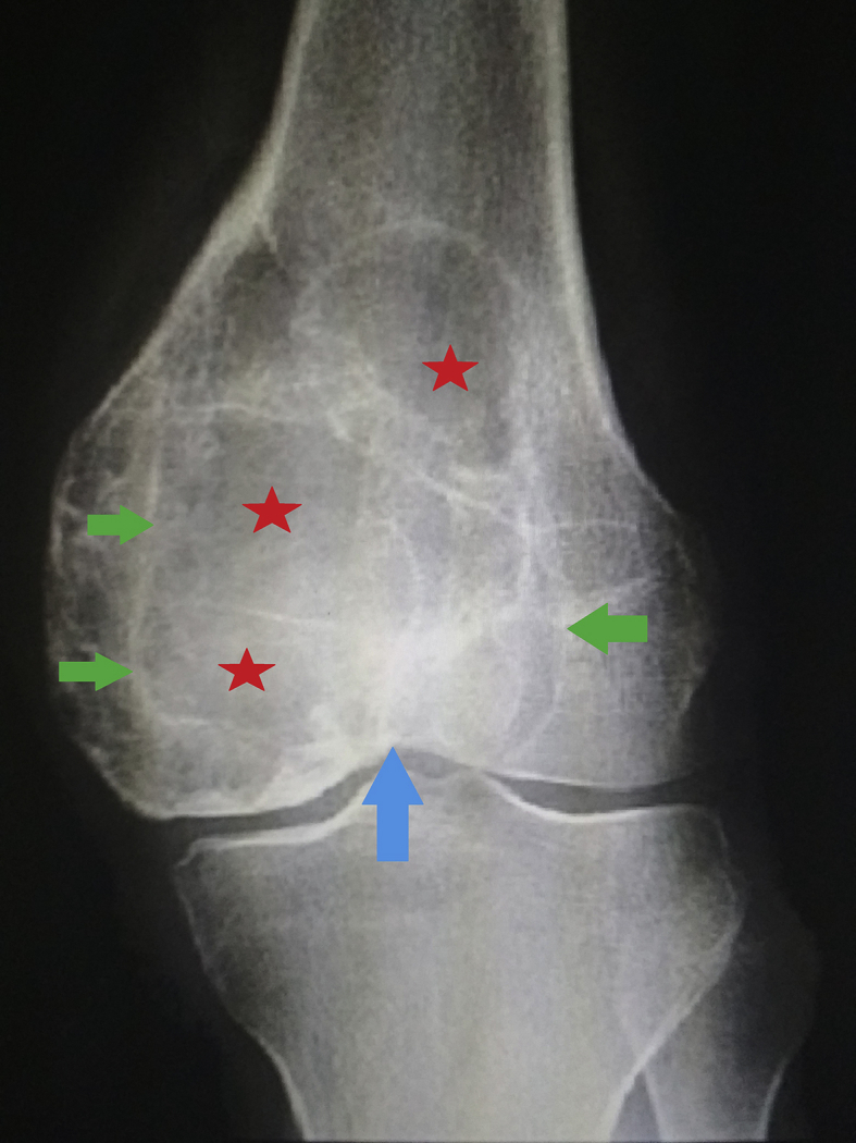
Antero-posterior radiograph of the left knee joint demonstrating GCT of distal femur at first presentation. Note the expansile lytic lesion (stars), with sharp well defined margins (green arrows), narrow zone of transition and extensive subchondral lysis (blue arrow).
Fig. 2.
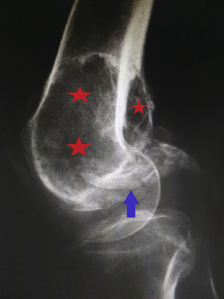
Lateral radiograph of the knee joint taken 4 months later demonstrating expansile lytic lesion of the distal femur (stars) with pathological fracture (blue arrow).
Fig. 3.

Coronal T1W-- MR image of GCT 4 months prior to the current acute presentation demonstrating an eccentric lesion of intermediate signal intensity in the distal femur. Note the low signal periphery due to hemosiderin deposition (red arrows) and the subarticular location of the tumor (blue arrow).
Fig. 4.

Sagittal T2W -- MR image 4 months prior to the current acute presentation demonstrating typical subarticular location of multi-septated GCT with bubbly appearance and multiple fluid-fluid levels (red arrow). Note the minimal suprapatellar fossa effusion (blue arrow).
Fig. 5.

Axial T2W -- MR image of GCT at current presentation demonstrating breach in the articular cortex of distal femur in the intercondylar region (red arrow) with the lesion communicating with joint space. Hemarthrosis with blood /fluid- fluid levels is also demonstrated (blue arrow).
Fig. 6.

Sagittal T2W -- MR image of GCT at current presentation demonstrating moderate hemarthrosis with fluid-blood interface sign (red arrow) seen secondary to pathological fracture of GCT. Multiple areas of cortical breach posteriorly (blue arrows) are seen with adjacent hematoma (green arrow).
Discussion
Giant cell tumor (GCT) of bone is an aggressive primary benign bone tumor with peak incidence between 3rd and 5th decades.1 It has epimetaphyseal region preponderance of the long bones and constitutes 5% of primary bone tumors.2 GCT being an aggressive osteolytic lesion is prone to pathologic fractures and high local recurrence rates after surgery.3 On radiographs, GCT has an appearance of a lytic lesion with circumscribed borders, absent sclerotic rim, and geographical pattern of destruction, centered in the epiphysis but involving the metaphysis and extending to the subarticular cortex. In the long bones such as the femur and tibia, GCT has an eccentric and intramedullary location with a symmetric and centrally located growth pattern. Early lesions are contained within the original bone contours that have a propensity to abut margins of the subchondral bone in one or more planes. There is no mineralized tumor matrix and no evident periosteal reaction unless a pathological fracture is present. MRI with its superior contrast resolution and multiplanar imaging capabilities is the best modality for imaging GCT and provides accurate tumor delineation of intraosseous and intramedullary lesions, articular surface involvement, and extraosseous extent of the tumor.4 On MRI, GCT appears hypointense on T1-weighted images and has heterogeneous high intensity on T2-weighted images. Extraosseous involvement is best appreciated on T2-weighted images and intramedullary tumor expansion on T1-weighted images. Hemosiderin deposition within the tumor is a feature of GCT, and hence demonstrates low signal intensity on all pulse sequences.5 By contrast, GCT shows areas of hypervascularity and enhancement with heterogeneous signal pattern. A cystic appearance with fluid–fluid levels is present in 10%–14% of GCT.6 Dynamic contrast-enhanced MRI demonstrates early and progressive enhancement of the tumor followed by rapid contrast washout. Aneurysmal bone cyst, telangiectatic osteosarcoma, geode, brown tumor in hyperparathyroidism, plasmacytoma, osteolytic metastasis, and intraosseous ganglion constitute differential diagnoses of GCT.7 Geodes, also known as subchondral cysts, are well-defined lytic lesions at the periarticular surfaces. Traumatic causes of acute hemarthrosis include meniscal injuries, ligamentous injuries, overuse syndrome, and fractures. Non-traumatic causes of acute hemarthrosis include crystal deposition disease, bleeding disorders, infection, arthritis, and tumors. In conclusion, acute hemarthrosis from pathological fracture of GCT should be considered in the differential diagnosis for acute swelling of the knee.
Conflict of interest
The authors have none to declare.
References
- 1.Sobti A., Agrawal P., Agarwala S., Agarwal M. Giant cell tumor of bone - an overview. Arch Bone Jt Surg. 2016;4:2–9. [PubMed] [PMC free article] [PubMed] [Google Scholar]
- 2.Campanacci M., Baldini N., Boriani S. Giant-cell tumor of bone. J Bone Joint Surg Am. 1987;69:106–114. [PubMed] [PubMed] [Google Scholar]
- 3.Rigollino A.V., Fernando T.S., Tanaka M.H., Souza M.M. Giant cell tumor locally advanced around the knee: treatment and literature review. Revista Brasileira de Ortopedia. 2017;52:473–478. doi: 10.1016/j.rboe.2017.06.009. [PubMed] [DOI] [PMC free article] [PubMed] [Google Scholar]
- 4.Purohit S., Pardiwala D.N. Imaging of giant cell tumor of bone. Indian J Orthop. 2007;41:91–96. doi: 10.4103/0019-5413.32037. [PubMed] [DOI] [PMC free article] [PubMed] [Google Scholar]
- 5.Aoki J., Moriya K., Yamashita K. Giant cell tumors of bone containing large amounts of hemosiderin: MR-pathologic correlation. J Comput Assist Tomogr. 1991;15:1024–1027. doi: 10.1097/00004728-199111000-00023. [PubMed] [DOI] [PubMed] [Google Scholar]
- 6.Resnik C.S., Steffe J.W., Wang S.E. Case report 353: giant cell tumor of distal end of the femur, containing a fluid level as demonstrated by computed tomography. Skelet Radiol. 1986;15:175–177. doi: 10.1007/BF00350215. [PubMed] [DOI] [PubMed] [Google Scholar]
- 7.Wilson J.N. Spontaneous hemarthrosis in the osteoarthritis of the knee. A report of five cases. Br Med J. 1959;23:1327–1328. doi: 10.1136/bmj.1.5133.1327. [PubMed] [DOI] [PMC free article] [PubMed] [Google Scholar]