PLOS ONE logoLink to PLOS ONE
. 2021 Jan 15;16(1):e0240641. doi: 10.1371/journal.pone.0240641

GABA quantification in human anterior cingulate cortex

Jan Weis 1,*, Jonas Persson 2, Andreas Frick 3, Fredrik Åhs 4, Maarten Versluis 5, Daniel Alamidi 6
Editor: Peter Lundberg7
PMCID: PMC7810278  PMID: 33449935

Abstract

γ-Aminobutyric acid (GABA) is a primary inhibitory neurotransmitter in the human brain. It has been shown that altered GABA concentration plays an important role in a variety of psychiatric and neurological disorders. The main purpose of this study was to propose a combination of PRESS and MEGA-PRESS acquisitions for absolute GABA quantification and to compare GABA estimations obtained using total choline (tCho), total creatine (tCr), and total N-acetyl aspartate (tNAA) as the internal concentration references with water referenced quantification. The second aim was to demonstrate the fitting approach of MEGA-PRESS spectra with QuasarX algorithm using a basis set of GABA, glutamate, glutamine, and NAA in vitro spectra. Thirteen volunteers were scanned with the MEGA-PRESS sequence at 3T. Interleaved water referencing was used for quantification, B0 drift correction and to update the carrier frequency of RF pulses in real time. Reference metabolite concentrations were acquired using a PRESS sequence with short TE (30 ms) and long TR (5000 ms). Absolute concentration were corrected for cerebrospinal fluid, gray and white matter water fractions and relaxation effects. Water referenced GABA estimations were significantly higher compared to the values obtained by metabolite references. We conclude that QuasarX algorithm together with the basis set of in vitro spectra improves reliability of GABA+ fitting. The proposed GABA quantification method with PRESS and MEGA-PRESS acquisitions enables the utilization of tCho, tCr, and tNAA as internal concentration references. The use of different concentration references have a good potential to improve the reliability of GABA estimation.

Introduction

Two important neurotransmitters in the mammalian brain are glutamate (Glu) and γ-aminobutyric acid (GABA). It should be noted, that both these compounds are not only neurotransmitters. They have other metabolic functions as well. In fact, only a small portion of the Glu and GABA are neurotransmitters. Glu is the major excitatory transmitter in the brain [1]. Glu serves as a metabolic precursor for GABA which is the primary inhibitory neurotransmitter. GABA plays a crucial role in shaping and regulating neuronal activity [2]. Changes in GABA concentrations have been associated with a variety of neuropsychiatric disorders, such as depression, anxiety, epilepsy, schizophrenia, ADHD, chronic pain, etc. [3,4].

Glu can be quantified with good accuracy by short echo time (TE < 40 ms) magnetic resonance spectroscopy using 3, 4 and 7T scanners [57]. However, the quantification of GABA is challenging because GABA spectral lines centered at 1.89, 2.28 and 3.01 ppm are overlapped with the strong signals of total creatine (tCr) (creatine and phosphocreatine), Glu, glutamine (Gln), total N-acetylaspartate (tNAA) (NAA and N-acetylaspartylglutamate (NAAG)), macromolecules (MM) and others. The most widely used approach for GABA detection at 3T is a J-difference Mescher-Garwood (MEGA) spectral editing technique incorporated within a point resolved spectroscopy (PRESS) sequence [8]. MEGA-PRESS exploits the scalar (J) coupling between GABA C4 protons (4CH2) at 3.01 ppm and C3 protons (3CH2) at 1.89 ppm. J-difference editing involves the acquisition of two spectra measured with TE 68 ms. The first (ON) spectrum is acquired by applying a pair of frequency-selective GABA-editing RF pulses (center frequency 1.89 ppm). The second (OFF) spectrum is measured with editing frequency-selective pulses at center frequency 7.46 ppm, which are not expected to have an impact on the spectrum. It should be noted that MM resonances at ~1.7 ppm are also inverted by editing frequency-selective RF pulses and coupled to MM protons at 3 ppm. These co-edited MM signals overlap with GABA C4 resonances. The resulting GABA peak is therefore referred to as GABA+ to point out the summation of GABA with MM signals. Recent studies showed that approximately 50% of GABA+ intensity originates from MM [3].

Cerebral GABA content is most often expressed as a spectral intensity ratio of GABA+/tCr. A disadvantage of such an approach is the fact that it is difficult to determine whether the alteration is caused by the numerator, denominator, or both. A suitable example is the tCr concentration that is subject to change in patients with schizophrenia, Alzheimer’s and Parkinsson diseases [9,10]. This problem can be minimized by the evaluation of GABA+ spectral intensity ratio to the intensity of other metabolites or to the intensity of the water. The alternative to the unitless spectral intensity ratio is absolute quantification. The most common method for absolute GABA quantification utilizes tissue water as an internal concentration reference [1113]. However, the quantification is not straightforward because the brain water originates from three tissue compartments: cerebrospinal fluid (CSF), gray (GM) and white matter (WM). Each compartment has different MR-visible water fraction and is weighted by different T1 and T2 relaxation times.

Recently, GABA estimation using tCr as an internal concentration reference was suggested as an alternative to water reference [14,15]. This approach benefits from the fact, that partial volume and relaxation corrections are unnecessary because metabolites originate only from GM and WM compartments and the relaxation times of metabolites are approximately equal in both compartments. Grewal et al [15] assumed tCr to be 7.1 mM. This concentration was estimated for WM using water referenced spectroscopic imaging (PRESS, TR/TE 1500/135 ms) [16]. Similarly, Bhattacharyya et al [14] applied the value 9.22 mM measured by single-voxel PRESS technique (TR/TE 2700/68 ms). A short TR and long TE caused these approaches to be sensitive to the inaccuracies of tCr and water relaxation times in WM, GM, and CSF.

The goal of this study was threefold: 1) to demonstrate a new fitting approach of MEGA-PRESS spectra using QuasarX algorithm as implemented in jMRUI 6.0 software package [17]. A basis set of GABA, Glu, Gln, and NAA in vitro spectra were measured for this purpose; 2) to quantify GABA using water as the internal concentration reference; and 3) to quantify GABA using total choline (tCho: free choline, phosphocholine, and glycerophosphocholine), tCr, tNAA as the internal concentration references. Contrary to previous studies [14,15], the reliability of reference tCho, tCr, and tNAA concentrations was improved by using a PRESS sequence with short TE (30 ms), long TR (5000 ms) together with partial volume and relaxation corrections for WM, GM and CSF content in each voxel. The overarching aim is to contribute to the improvement of the GABA quantification methodology.

Material and methods

Study population

In total, thirteen volunteers (6 females and 7 males) were recruited. The volunteers underwent PRESS and MEGA-PRESS measurements. Mean age of the participants was 37±10 years (range: 24–61). All volunteers were healthy without any history of psychiatric or neurological disorders. Ethical approvals were obtained from local Institutional Review Boards and written informed consent was obtained from each participant.

Phantoms

Four phantoms were produced according to the guidelines for the LCModel’s model spectra [18]. The phantoms contained aqueous solutions of GABA (200 mM), Glu (100 mM), Gln (100 mM), and NAA (50 mM). Each phantom contained a single metabolite. Aqueous solutions were prepared using a phosphate buffer consisting of 72 mM K2HPO4, 28 mM KH2PO4, 1 g/L NaN3, and 1 mM sodium trimethylsilyl propanesulfonate (DSS). Solution’s pH was adjusted to 7.2. The chemicals were purchased from Sigma-Aldrich AB (Stockholm, Sweden).

MRI and MRS acquisition protocols

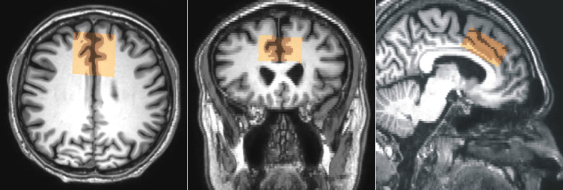
All experiments were performed on 3T scanner (Achieva dStream, Philips Healthcare, Best, The Netherlands). The data were acquired with a 32 channel receiver head coil. Whole brain 3D T1-weighted turbo FFE images (TR/TE 8/3.8 ms, isotropic resolution 1x1x1 mm3) were acquired to guide the positioning of the voxel in the anterior cingulate cortex (ACC) (Fig 1). The GABA spectra were acquired with a MEGA-PRESS sequence using the following parameters: voxel size 4x4x2 cm3, TR/TE 2000/68 ms, 320 alternating ON-OFF spectra, 14 ms GABA-editing RF pulses at 1.9 (ON) and 7.5 (OFF) ppm, spectral bandwidth 2000 Hz, 1024 time domain data points, and 40 blocks. Each block started with the acquisition of one unsuppressed reference water spectral line followed by four pairs of water suppressed ON-OFF spectra acquired with 4-step phase-cycling. The unsuppressed water signal was used as the internal concentration reference, for eddy current corrections, for B0 drift correction and for updating the carrier frequency of RF pulses in real time. Reference metabolite concentrations were measured using the standard PRESS sequence (TR/TE 5000/30 ms, spectral bandwidth 2000 Hz, 1024 data points, 32 averages, 16 phase cycle steps) with the same voxel size and position. Two dummy excitations were followed by 16 non-water-suppressed and 32 water-suppressed scans. MEGA-PRESS and PRESS sequence performed water suppression by two selective RF pulses and spoiler gradients. Suppression was accomplished by adjusting the flip angle of the second RF pulse such that the longitudinal magnetization of the water signal was minimal at the time of the first excitation RF pulse.

Fig 1. Spectroscopy voxel position.

Fig 1

Representative voxel (4x4x2 cm3) placement in anterior cingulate cortex and the results of partial volume segmentation of CSF, GM and WM (brown). Transversal images (reconstructed pixel size 0.47x0.47 mm2, slice thickness 2 mm) were used for segmentation and for reconstruction of coronal and sagittal slices.

Reference frequency of the PRESS pulses was centered at tCr/GABA (~3 ppm) in water-suppressed scans. Frequency offset was centered at water in scans without water suppression, ie, there was no chemical shift displacement between the water and GABA PRESS-boxes. Chemical shift displacement between GABA and tNAA (2 ppm) PRESS boxes was approximately 2.2, 3.7, and 1.9 mm in left-right (90° RF pulse, slice thickness 40 mm), ~feet-head (180° RF pulse, slice thickness 40 mm), and ~anterior-posterior (180° RF pulse, slice thickness 20 mm) directions, respectively. Chemical shift displacement (absolute value) between GABA and tCho (~3.2 ppm) PRESS boxes was lower by factor 5 compare to displacement between tCr/GABA and tNAA PRESS boxes. It should be noted that 90° RF-pulse with broader bandwidth (BW) produces lower chemical shift displacement than 180° RF-pulses with narrow BW.

MEGA-PRESS spectra of GABA, Glu, Gln, and NAA aqueous solutions were measured a few hours after preparation. The voxel size was 3x3x3 cm3 and temperature was kept at 22°C (room temperature) during the acquisition. All other MEGA-PRESS parameters were identical to the in vivo experiments.

Post processing and quantification

Reconstructed brain images (matrix 512x512, pixel size 0.47 mm, slice thickness 2 mm) were used for GM, WM, and CSF segmentation (Fig 1). Segmentation was performed by using the automated segmentation tool (FAST) [19]. A binary mask of the water PRESS box was created using the SVMask tool (Philips Healthcare, Michael Schär).

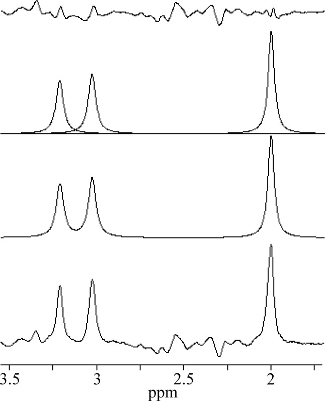
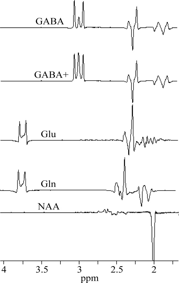
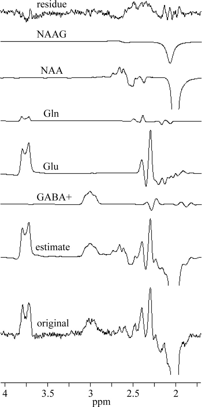
MEGA-PRESS spectra were processed with jMRUI 6.0 software package [17]. Each spectrum was zero filled to 8192 points and the residual water was removed by Hankel-Lanczos Singular Value Decomposition (HLSVD) filter. No apodization of the FIDs was applied in this study. All in vivo OFF spectra were averaged and tCho, tCr, and tNAA singlets were fitted using AMARES algorithm (Fig 2). The zero-order phase correction was estimated by AMARES. The in vivo difference (ON-OFF) spectra were fitted by the QuasarX algorithm (QUEST with new constrains and shape peak selection). This time-domain algorithm uses prior knowledge obtained either theoretically by quantum mechanics, or by measuring in vitro aqueous metabolite solutions (Fig 3). Non-linear least-squares algorithm fits a weighted combination of metabolite FIDs to the considered in vivo FID. Our basis set of FID signals was obtained from MEGA-PRESS spectra of GABA (GABA+), Glu, Gln, NAA and NAAG aqueous solutions (Fig 3). NAAG spectrum was approximated by shifted NAA spectrum, with the main peak shifted to 2.045 ppm from 2.01 ppm [18]. GABA+ spectrum was made by modifying GABA spectrum. Contribution of MM signals to GABA was empirically simulated by adding the Lorentzian line (linewidth 5 Hz) to the central peak of pseudo-triplet at 3 ppm. The amplitude was adjusted to be about ~10% higher compare to outer two peaks (Fig 3). Gaussian line shapes were used to fit GABA+ spectral lines. The in vivo difference spectra were first averaged and then fitted by the QuasarX algorithm (Fig 4). The zero-order phase correction was estimated by QuasarX.

Fig 2. An example of averaged in vivo OFF spectrum.

Fig 2

AMARES fits of the tCho, tCr, and tNAA singlets, and residue.

Fig 3. Basis set of in vitro spectra.

Fig 3

MEGA-PRESS spectra of GABA, Gln, Glu, and NAA aqueous solutions, and simulated GABA+.

Fig 4. Representative in vivo MEGA-PRESS spectrum and fits.

Fig 4

The difference spectra were averaged and fitted by the QuasarX algorithm using the basis set spectra shown in Fig 2.

The unsuppressed water signal was fitted by Hankel-Lanczos Squares Singular Value Decomposition (HLSSVD) algorithm. The AMARES and QuasarX algorithms provide the Cramér-Rao lower bound (CRLB) standard deviation (CRSD). The fitting error was computed as the percentage ratio of CRSD to the FID’s amplitude. Water scaled GABA concentration in relation to wet weight tissue (mol/kg) was computed according to the equation:

CGABA=IGABAIH2O×2NGABA×1RGABA×Wconc×MMcoreffGABA (1)

where IGABA is the GABA+ spectral intensity at ~3 ppm, IH2O is intensity of reference water line, NGABA = 2 is the number of protons contributing to IGABA resonance, RGABA is the GABA attenuation factor, MMcor = 0.5 is a macromolecule correction factor [11,20,21], and effGABA = 0.5 is the editing efficiency [22]. Wconc is the reference water concentration corrected for partial volume and relaxation effects [16,23,24]:

Wconc=WH2O(fGMRH2O-GM+fWMRH2O-WM+fCSFRH2OCSF)(1fCSF) (2)

and

fx=cxϑx0.82ϑGM+0.7ϑWM+0.99ϑCSF (3)

where WH2O is the molal concentration of pure water (55.51 mol/kg), fx is the mole fraction of water in the voxel’s GM, WM and CSF, ϑx is the GM, WM and CSF volume fractions and cx is the relative density of MR visible water in GM (0.82) WM (0.7), and CSF (0.99) [25,26]. RH2O-GM, RH2O-WM, and RH2O-CSF are PRESS relaxation attenuation factors R = exp(-TE/T2)x[1-exp(-TR/T1)] of water in GM, WM, and CSF, respectively. The following relaxation times were used for corrections: GABA (T1 1310 ms, T2 88 ms) [27,28], water in GM (T1 1820 ms, T2 99 ms), WM (T1 1084 ms, T2 69 ms), and CSF (T1 4163 ms, T2 503 ms) [2931].

The GABA concentration was also assessed using tCho, tCr, and tNAA as the internal concentration references. The reference metabolite concentrations CMET, Glu and other metabolites were measured by PRESS sequence with long TR (5000 ms) and short TE (30 ms) to minimize the influence of the water and metabolites relaxation effects. Concentrations were estimated by LCModel [18]. Partial volume and relaxation corrections were performed by adjusting LCModel control parameter WCONC according to the Eq 2, i.e. WCONC = Wconc [24]. It should be noted that the default LCModel control parameter ATTH2O for water attenuation correction was switched off (ATTH2O = 1) because water relaxation corrections were already performed in Eq 2. The absolute GABA concentration (mol/kg) was estimated according to the formula:

CGABA=IGABAIMET×NMETNGABA×RMETRGABA×CMET×MMcoreffGABA (4)

where IMET is the spectral intensity of reference metabolite in OFF spectrum (Fig 3), NMET is the number of protons contributing to IMET resonance (9 for tCho, 3 for tCr and tNAA), RMET is the metabolite attenuation factor. CMET is the reference metabolite concentration (mol/kg) of considered volunteer. Mean relaxation times of tCho (T1 1140 ms, T2 230 ms), tCr (T1 1110 ms, T2 163 ms), and tNAA (T1 1340 ms, T2 260 ms) were used in relaxation corrections [32]. It should be noted that only small differences in metabolite relaxation times were found between GM and WM [32,33].

Statistics

The reported values are given as the mean ± one SD. P < 0.05 of a two-tailed Student’s t-test was considered statistically significant. The relative variances (variance-to-mean ratio) were expressed in %. The two-tailed F-test was performed to compare variances of mean GABA concentrations obtained by different quantification methods.

Results

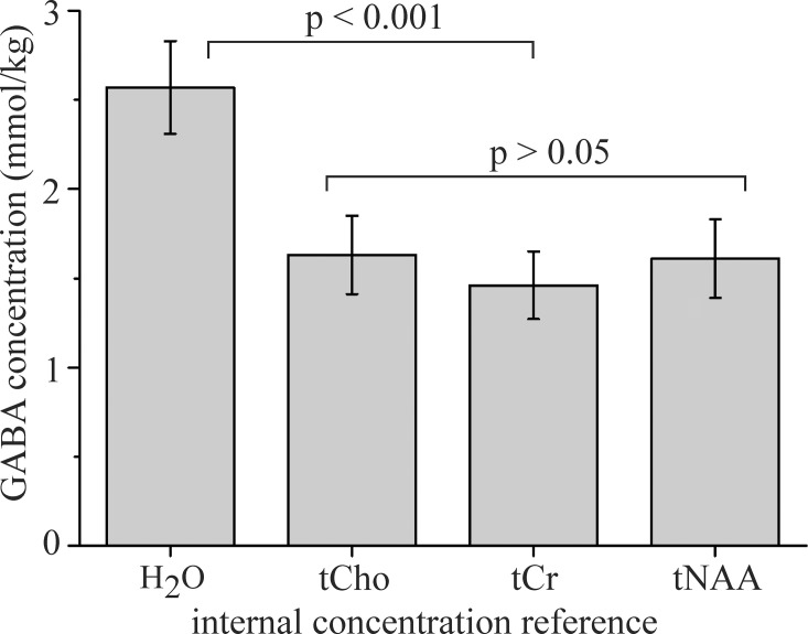
Thirteen volunteers underwent combined PRESS and MEGA-PRESS examinations. All experiments were successful, no spectra had to be discarded. The OFF and difference (ON-OFF) spectra of all volunteers are shown in S1 Fig (Supporting information). Table 1 summarizes the water-scaled metabolite concentrations and CRLBs acquired by the PRESS (TR/TE 5000/30 ms) sequence. The spectra were processed by LCModel. The mean WM, GM, and CSF volume fractions were 52.0 ± 3.5%, 33.2 ± 2.5%, and 14.8 ± 3.5%, respectively. Spectra of GABA, Glu, Gln, and NAA aqueous solutions and simulated GABA+ spectrum are shown in Fig 3. These spectra were used as prior knowledge for fitting the volunteer’s MEGA-PRESS spectra using the QuasarX algorithm. Figs 2 and 4 show representative in vivo results. The mean QuasarX fitting error of GABA+ intensity was 1.5 ± 0.2% (range: 1.2–1.8%). The mean AMARES fitting errors of metabolites were 1.0 ± 0.1%, 0.8% ± 0.1%, and 1.2 ± 0.2% for tCr, tNAA, and tCho, respectively. The mean spectral intensity ratios GABA+/tCr, GABA+/tNAA, and GABA+/tCho were 0.070 ± 0.01, 0.052 ± 0.007, and 0.088 ± 0.013, respectively. The absolute GABA concentrations (mmol/kg) were computed according to Eqs 1 and 4 using tissue water (2.57±0.26 [2.7]), tCho (1.63±0.22 [3.1]), tCr (1.46 ± 0.19 [2.6]), and tNAA (1.61 ± 0.22 [3.1]) as internal concentration references. The square brackets depict relative variances. Concentrations are visualized in Fig 5. Two-tailed F-tests detected no differences in the variances of GABA concentrations obtained by different methods.

Table 1. Water-scaled metabolite concentrations (mmol/kg) and CRLBs (%).

Concentration CRLB
GABA 2.65 ± 0.44 18.62 ± 2.96
Glu 10.84 ± 0.54 5.23 ± 0.44
Glx 13.03 ± 1.03 6.69 ± 0.48
tNAA 12.37 ± 0.68 2.23 ± 0.44
tCr 9.11 ± 0.57 2.0 ± 0.0
tCho 2.42 ± 0.20 2.85 ± 0.38
mI 6.43 ± 0.51 3.92 ± 0.28

Concentrations were estimated from the PRESS spectra (TR/TE 5000/30 ms).

Fig 5. GABA concentrations.

Fig 5

Concentrations (mmol/kg) were estimated using H2O (2.57±0.26), tCho (1.63±0.22), tCr (1.46 ± 0.19), and tNAA (1.6±0.22) as internal concentration references.

Discussion

To the best of our knowledge, this is the first study whereby a PRESS sequence with short TE and long TR together with a MEGA-PRESS sequence were used to estimate the absolute GABA concentration. Applied PRESS method improved the precision of individual reference metabolite concentrations and enabled utilization of tCho, tNAA and tCr as internal concentration references at the expense of a relatively short prolongation of the net measurement time (4 minutes in our case). Spectrum processing approach with QuasarX algorithm and the use of different concentration references have a good potential to improve the reliability of GABA estimation.

The anterior cingulate cortex was chosen because this region acts as a central node in the brain and is important for the regulation of advanced brain functions. The water scaled PRESS spectra were used for the individual reference metabolite quantification. The described approach with short TE and long TR together with the partial volume and relaxation corrections is regarded to be the most accurate. This is because errors due to inaccurate relaxation times were minimized. Low CRLBs of Glu, tCho, tCr, and tNAA LCModel fits (Table 1) indicate very good fitting results of the LCModel algorithm. The fitting errors of GABA signals were at the boundary of acceptable reliability (CRLB ~ 20%). However, it should be noted that average over a group of LCModel results can significantly reduce the uncertainty [18]. The concentration estimates of Glu, Glx, tNAA, tCr and tCho are in line with previous studies performed at 3, 4, and 7 Tesla [5,7].

The relative variances of absolute GABA concentrations estimated using H2O, tCho, tCr, and tNAA concentration references show similar dispersion. The comparison of our GABA concentrations with the literature data is not straightforward due to differences in tissue composition and data processing. Cerebral GABA content from ~1 up to 3.7 mM was previously reported [1115,34,35]. GM/WM ratio is an important issue because GABA content was reported to be from 1.5 to 8.7 times larger in GM relative to WM [14,20,34,36]. Differences in segmentation algorithms, spectrum processing methods, macromolecule correction factor MMcor and accuracy of internal concentration references are also important factors that contribute to the variability of the concentration estimates.

The absolute GABA concentrations estimated using water reference and measured by PRESS and MEGA-PRESS are surprisingly in very good agreement. However, water referenced GABA concentrations were significantly higher than the concentrations estimated with tCho, tCr and tNAA references (Fig 5). The main drawback of water referenced quantification using typical MEGA-PRESS (TR/TE 2000/68 ms) acquisition is the fact that partial volume and relaxation corrections (Eqs 2 and 3) depend on the precision of WM, GM, and CSF segmentation and on the accuracy of nine experimental constants: water fractions and water relaxation times T1, T2 in GM, WM, and CSF. The advantage of GABA quantification using tCr, tCho, and tNAA as the internal concentration references is the fact that partial volume and relaxation corrections are unnecessary because metabolites originate only from GM and WM compartments and the relaxation times of tCho, tCr, and tNAA are approximately equal in both compartments [32,33]. It should be noted, that the described metabolite reference method is still subject to all sources of error as in water referenced MEGA-PRESS approach because tCho, tCr, and tNAA were quantified from water scaled PRESS spectra (TR/TE 5000/30 ms). However, the main difference is in relaxation correction accuracy. Standard water referenced MEGA-PRESS approach with a relatively short TR (2000 ms) and long TE (68 ms) is more susceptible to inaccuracies of relaxation times compare to the proposed metabolite referenced quantification using PRESS with long TR (5000 ms) and short TE (30 ms). It should be noted, that quantification of tCho, tCr, and tNAA can be omitted in comparative studies and the most reliable literature values can be applied instead. Our GABA values can be compared with the concentrations estimated from the water scaled STEAM and SPECIAL spectra measured at 7 T scanners [5,7]. The occipital lobe spectra were measured with long TR and a very short TE (6 ms). High spatial resolution facilitated fitting of the GABA triplet at 2.28 ppm which is uncontaminated by the macromolecules. GABA levels in the range of 1.3–1.6 mmol/kg were reported. These values were slightly underestimated because partial volume and relaxation corrections were not taken into consideration. Nevertheless, we believe that our metabolite referenced results conform to the most reliable literature values such as the GABA values reported by Mekle et al [5] and Tkac et al [7]. We hypothesize that our water referenced GABA values are overestimated due to inaccuracies in partial volume and relaxation corrections.

Conclusion

QuasarX algorithm together with the basis set of in vitro spectra improves reliability of GABA+ fitting. The proposed GABA quantification method with PRESS and MEGA-PRESS acquisitions enables the utilization of tCho, tCr, and tNAA as internal concentration references. Water referenced GABA estimations were significantly higher compared to the values obtained by metabolite references. The use of different concentration references have a good potential to improve the reliability of GABA estimation.

Supporting information

S1 Fig. MEGA-PRESS spectra.

OFF spectra (left) and corresponding difference spectra (right) of all volunteers.

(TIF)

Data Availability

All relevant data are within the manuscript and its Supporting Information files.

Funding Statement

J.P. and A. F. were supported by the Swedish Research Council, the Swedish Brain Foundation, and the Swedish Foundation for Humanities and Social Sciences (Riksbankens jubileumsfond). A.F. was supported by The Kjell and Märta Beijer Foundation and J.P. was supported by a postdoctoral grant from the Swedish Brain Foundation. The funders provided support in the form of salaries for authors [J.P., A.F.], but did not have any additional role in the study design, data collection and analysis, decision to publish, or preparation of the manuscript. The specific roles of these authors are articulated in the ‘author contributions’ section.

References

  • 1.Zhou Y, Danbolt NC. Glutamate as a neurotransmitter in the healthy brain. J Neural Transm 2014; 121:799–817. 10.1007/s00702-014-1180-8 [DOI] [PMC free article] [PubMed] [Google Scholar]
  • 2.McCormic DA. GABA as an inhibitory neurotransmitter in human cerebral cortex. J Neurophysiol 1989; 62:1018–1027. 10.1152/jn.1989.62.5.1018 [DOI] [PubMed] [Google Scholar]
  • 3.Mikkelsen M, Barker PB, Bhattacharyya PK, et al. Big GABA: Edited MR spectroscopy at 24 research sites. Neuroimage 2017; 159: 32–45. 10.1016/j.neuroimage.2017.07.021 [DOI] [PMC free article] [PubMed] [Google Scholar]
  • 4.Harris AD, Saleh MG, Eden RAE. Edited 1H magnetic resonance spectroscopy in vivo: Methods and metabolites. Magn Reson Med 2017; 77:1377–1389. 10.1002/mrm.26619 [DOI] [PMC free article] [PubMed] [Google Scholar]
  • 5.Mekle R, Mlynarik V, Gambarota G, Hergt M, Krueger G, Gruetter R. MR spectroscopy of the human brain with enhanced signal intensity at ultrashort echo times on a clinical platform at 3T and 7T. Magn Reson Med 2009; 61:1279–1285. 10.1002/mrm.21961 [DOI] [PubMed] [Google Scholar]
  • 6.Oeltzschner G, Zöllner HJ, Jonuscheit M, Lanzman RS, Schnitzler A, Wittsack HJ. J-difference-edited MRS measures of γ-aminobutyric acid before and after acute caffeine administration. Magn Reson Med 2018; 80:2356–2365. 10.1002/mrm.27233 [DOI] [PMC free article] [PubMed] [Google Scholar]
  • 7.Tkac I, Öz G, Adriany G, Ugurbil K, Gruetter R. In vivo 1H NMR spectroscopy of the human brain at high magnetic fields: metabolite quantification at 4T vs. 7T. Magn Reson Med 2009; 62:868–879. 10.1002/mrm.22086 [DOI] [PMC free article] [PubMed] [Google Scholar]
  • 8.Mescher M, Merkle H, Kirsch J, Garwood M, Gruetter R. Simultaneous in vivo spectral editing and water suppression. NMR Biomed 1998; 11:266–278. [DOI] [PubMed] [Google Scholar]
  • 9.Öngür D, Prescot AP, Jensen JE, Cohen BM, Renshaw PF. Creatine abnormalities in schizophrenia and bipolar disorder. Psychiatry Res 2009; 172:44–48. 10.1016/j.pscychresns.2008.06.002 [DOI] [PMC free article] [PubMed] [Google Scholar]
  • 10.Pilatus U, Lais C, du Mesnil de Rochmont A, et al. Conversion to dementia in mild cognitive impairment is associated with decline of N-acetylaspartate and creatine as revealed by magnetic resonance spectroscopy. Psychiatry Research: Neuroimaging 2009; 173:1–7. 10.1016/j.pscychresns.2008.07.015 [DOI] [PubMed] [Google Scholar]
  • 11.Harris AD, Puts NAJ, Barker PB, Edden RAE. Spectral-editing measurements of GABA in the human brain with and without macromolecule suppression. Magn Reson Med 2015; 74:1523–1529. 10.1002/mrm.25549 [DOI] [PMC free article] [PubMed] [Google Scholar]
  • 12.Harris A, Puts NAJ, Edden RAE. Tissue correction for GABA-edited MRS: consideration of voxel composition, tissue segmentation, and tissue relaxations. J Magn Reson Imag 2015; 42:1431–1440. [DOI] [PMC free article] [PubMed] [Google Scholar]
  • 13.O’Gorman RL, Michels L, Edden RAE, Murdoch JB, Martin E. In vivo detection of GABA and glutamate with MEGA-PRESS: reproducibility and gender effects. J Magn Reson Imag 2011; 33:1262–1267. 10.1002/jmri.22520 [DOI] [PMC free article] [PubMed] [Google Scholar]
  • 14.Bhattacharyya PK, Phillips MD, Stone LA, Lowe MJ. In vivo magnetic resonance spectroscopy measurement of gray-matter and white-matter gamma-aminobutyric acid concentration in sensorimotor cortex using a motion-controlled MEGA point-resolved spectroscopy sequence. Magn Reson Imag 2011; 29:374–379. 10.1016/j.mri.2010.10.009 [DOI] [PMC free article] [PubMed] [Google Scholar]
  • 15.Grewal M, Dabas A, Saharan S, et al. GABA quantitation using MEGA-PRESS: Regional and hemispheric differences. J Magn Reson Imag 2016; 44:1619–1623. 10.1002/jmri.25324 [DOI] [PMC free article] [PubMed] [Google Scholar]
  • 16.Gasparovic Ch, Song T, Devier D, et al. Use of tissue water as a concentration reference for proton spectroscopic imaging. Magn Reson Med 2006; 55:1219–1226. 10.1002/mrm.20901 [DOI] [PubMed] [Google Scholar]
  • 17.Naressi A, Couturier C, Devos JM, et al. Java-based graphical user interface for the MRUI quantitation package. Magn Reson Mater Phys Biol Med 2001; 12:141–152. 10.1007/BF02668096 [DOI] [PubMed] [Google Scholar]
  • 18.Provencher SW. Estimation of metabolite concentrations from localized in vivo NMR spectra. Magn Reson Med 1993; 30:672–679. 10.1002/mrm.1910300604 [DOI] [PubMed] [Google Scholar]
  • 19.Zhang Y, Brady M, Smith S. Segmentation of brain MR images through a hidden Markov random field model and the expectation-maximization algorithm. IEEE Trans Med Imaging 2001; 20:45–57. 10.1109/42.906424 [DOI] [PubMed] [Google Scholar]
  • 20.Mikkelsen M, Singh KD, Sumner P, Evans CJ. Comparison of the repeatability of GABA-edited magnetic resonance spectroscopy with and without macromolecules suppression. Magn Reson Med 2016; 7:946–953. [DOI] [PubMed] [Google Scholar]
  • 21.Shungu DC, Mao X, Gonzales R, et al. Brain γ-aminobutyric acid (GABA) detection in vivo with the J-editing 1H MRS technique: a comprehensive methodological evaluation of sensitivity enhancement, macromolecule contamination and test-retest reliability. NMR Biomed 2016; 29:932–942. 10.1002/nbm.3539 [DOI] [PMC free article] [PubMed] [Google Scholar]
  • 22.Mullins PG, McConigle DJ, O’Gorman RL, Puts NAJ, Vidyasagar R, Evans CJ. Current practice in the use of MEGA-PRESS spectroscopy for the detection of GABA. Neuroimage 2014; 86:43–52. 10.1016/j.neuroimage.2012.12.004 [DOI] [PMC free article] [PubMed] [Google Scholar]
  • 23.Gasparovic Ch, Chen H, Mullins PG. Errors in 1H-MRS estimates of brain concentrations caused by failing to take into account tissue-specific signal relaxation. NMR Biomed 2018; 31:e3914 10.1002/nbm.3914 [DOI] [PubMed] [Google Scholar]
  • 24.Quadrelli S, Mountford C, Ramadan S. Hitchhiker’s guide to voxel segmentation for partial volume correction of in vivo magnetic resonance spectroscopy. Magn Reson Insights 2016; 9:1–8. 10.4137/MRI.S32903 [DOI] [PMC free article] [PubMed] [Google Scholar]
  • 25.Abbas Z, Gras V, Möllenhoff K, Oros-Peusquens AM, Shah NJ. Quantitative water content mapping at clinically relevant field strengths: A comparative study at 1.5 and 3T. Neuroimage 2015; 106:404–413. 10.1016/j.neuroimage.2014.11.017 [DOI] [PubMed] [Google Scholar]
  • 26.Sabati M and Maudsley AA. Fast and high-resolution quantitative mapping of tissue water content with full brain coverage for clinically-driven studies. Magn Reson Imag 2013; 31:1752–1759. 10.1016/j.mri.2013.08.001 [DOI] [PMC free article] [PubMed] [Google Scholar]
  • 27.Edden RAE, Intrapiromkul J, Zhu H, Cheng Y, Barker PB. Measuring T2 in vivo with J-difference editing: application to GABA at 3 Tesla. J Magn Reson Imag 2012; 35:229–234. 10.1002/jmri.22865 [DOI] [PMC free article] [PubMed] [Google Scholar]
  • 28.Puts NAJ, Barker PB, Edden RAE. Measuring the longitudinal relaxation time of GABA in vivo at 3 Tesla. J Magn Reson Imag 2013; 37:999–1003. 10.1002/jmri.23817 [DOI] [PMC free article] [PubMed] [Google Scholar]
  • 29.Lin C, Bernstein M, Huston J, Fain S. Measurements of T1 relaxation times at 3.0 T: Implication for MRA. In: Proceedings of the 9th Annual Meeting of ISMRM; 2001; Glasgow, 2001:1391.
  • 30.Piechnik SK, Evans J, Bary LH, Wise RG, Jezzard P. Functional changes in CSF volume estimated using measurement of water T2 relaxation. Mag Reson Med 2009; 61,579–586. 10.1002/mrm.21897 [DOI] [PubMed] [Google Scholar]
  • 31.Stanisz GJ, Odrobina EE, Pun J, et al. T1, T2 relaxation and magnetization transfer in tissue at 3 T. Magn Reson Med 2005; 54:507–512. 10.1002/mrm.20605 [DOI] [PubMed] [Google Scholar]
  • 32.Träber F, Block W, Lamerichs R, Gieseke J, Schild HH. 1H metabolite relaxation times at 3.0 Tesla: Measurements of T1 and T2 values in normal brain and determination of regional differences in transverse relaxation. J Magn Reson Imag 2004; 19:537–545. 10.1002/jmri.20053 [DOI] [PubMed] [Google Scholar]
  • 33.Ethofer T, Mader I, Seeger U, et al. Comparison of longitudinal metabolite relaxation times in different regions of the human brain at 1.5 and 3 Tesla. Magn Reson Med 2003; 50:1296–1301. 10.1002/mrm.10640 [DOI] [PubMed] [Google Scholar]
  • 34.Geramita N, van der Veen JW, Barnett AS, et al. Reproducibility of prefrontal γ-aminobutyric acid measurements with J-edited spectroscopy. NMR Biomed 2001; 24:1098–1098. [DOI] [PubMed] [Google Scholar]
  • 35.Mikkelsen M, Rimbault DL, Barker PB, et al. Big GABA II: Water-referenced edited MR spectroscopy at 25 research sites. Neuroimage 2019; 191:537–548. 10.1016/j.neuroimage.2019.02.059 [DOI] [PMC free article] [PubMed] [Google Scholar]
  • 36.Choi C, Bhardwaj PP, Kalra S, et al. Measurement of GABA and contaminants in gray and white matter in human brain in vivo. Magn Reson Med 2007; 58:27–33. 10.1002/mrm.21275 [DOI] [PubMed] [Google Scholar]

Decision Letter 0

Peter Lundberg

30 Nov 2020

PONE-D-20-30419

GABA quantification in human anterior cingulate cortex

PLOS ONE

Dear Dr. Weis,

Thank you for submitting your manuscript to PLOS ONE. After careful consideration, we feel that it has merit but does not fully meet PLOS ONE’s publication criteria as it currently stands. Therefore, we invite you to submit a revised version of the manuscript that addresses the points raised during the review process.

Please update according to reviewers comments. Especially it would be prudent to update on the biological meaning of 'neurotransmitters', also remove some of the more basic explanations of the editing method, while expanding substantially on the experiment details, as suggested by reviewers.

Please submit your revised manuscript by Jan 14 2021 11:59PM. If you will need more time than this to complete your revisions, please reply to this message or contact the journal office at plosone@plos.org. When you're ready to submit your revision, log on to https://www.editorialmanager.com/pone/ and select the 'Submissions Needing Revision' folder to locate your manuscript file.

Please include the following items when submitting your revised manuscript:

  • A rebuttal letter that responds to each point raised by the academic editor and reviewer(s). You should upload this letter as a separate file labeled 'Response to Reviewers'.

  • A marked-up copy of your manuscript that highlights changes made to the original version. You should upload this as a separate file labeled 'Revised Manuscript with Track Changes'.

  • An unmarked version of your revised paper without tracked changes. You should upload this as a separate file labeled 'Manuscript'.

If you would like to make changes to your financial disclosure, please include your updated statement in your cover letter. Guidelines for resubmitting your figure files are available below the reviewer comments at the end of this letter.

If applicable, we recommend that you deposit your laboratory protocols in protocols.io to enhance the reproducibility of your results. Protocols.io assigns your protocol its own identifier (DOI) so that it can be cited independently in the future. For instructions see: http://journals.plos.org/plosone/s/submission-guidelines#loc-laboratory-protocols

We look forward to receiving your revised manuscript.

Kind regards,

Peter Lundberg

Academic Editor

PLOS ONE

Journal Requirements:

When submitting your revision, we need you to address these additional requirements.

1. Please ensure that your manuscript meets PLOS ONE's style requirements, including those for file naming. The PLOS ONE style templates can be found at

https://journals.plos.org/plosone/s/file?id=wjVg/PLOSOne_formatting_sample_main_body.pdf and

https://journals.plos.org/plosone/s/file?id=ba62/PLOSOne_formatting_sample_title_authors_affiliations.pdf

2. We noted that two co-authors are affiliated with Philips healthcare and that you used materials manufactured by Philips healthcare. Please consult our guidelines on declaring competing interests at https://journals.plos.org/plosone/s/competing-interests and update your competing interests declaration in the submission form as appropriate.

  1. Please provide an amended Funding Statement declaring this commercial affiliation, as well as a statement regarding the Role of Funders in your study. If the funding organization did not play a role in the study design, data collection and analysis, decision to publish, or preparation of the manuscript and only provided financial support in the form of authors' salaries and/or research materials, please review your statements relating to the author contributions, and ensure you have specifically and accurately indicated the role(s) that these authors had in your study. You can update author roles in the Author Contributions section of the online submission form.

Please also include the following statement within your amended Funding Statement.

“The funder provided support in the form of salaries for authors [insert relevant initials], but did not have any additional role in the study design, data collection and analysis, decision to publish, or preparation of the manuscript. The specific roles of these authors are articulated in the ‘author contributions’ section.”

If your commercial affiliation did play a role in your study, please state and explain this role within your updated Funding Statement.

         2. Please also provide an updated Competing Interests Statement declaring this commercial affiliation a      long with any other relevant declarations relating to employment, consultancy, patents, products in development, or marketed products, etc.  

Within your Competing Interests Statement, please confirm that this commercial affiliation does not alter your adherence to all PLOS ONE policies on sharing data and materials by including the following statement: "This does not alter our adherence to  PLOS ONE policies on sharing data and materials.” (as detailed online in our guide for authors http://journals.plos.org/plosone/s/competing-interests) . If this adherence statement is not accurate and  there are restrictions on sharing of data and/or materials, please state these. Please note that we cannot proceed with consideration of your article until this information has been declared.

Please include both an updated Funding Statement and Competing Interests Statement in your cover letter. We will change the online submission form on your behalf.

Please know it is PLOS ONE policy for corresponding authors to declare, on behalf of all authors, all potential competing interests for the purposes of transparency. PLOS defines a competing interest as anything that interferes with, or could reasonably be perceived as interfering with, the full and objective presentation, peer review, editorial decision-making, or publication of research or non-research articles submitted to one of the journals. Competing interests can be financial or non-financial, professional, or personal. Competing interests can arise in relationship to an organization or another person. Please follow this link to our website for more details on competing interests: http://journals.plos.org/plosone/s/competing-interests

[Note: HTML markup is below. Please do not edit.]

Reviewers' comments:

Reviewer's Responses to Questions

Comments to the Author

1. Is the manuscript technically sound, and do the data support the conclusions?

The manuscript must describe a technically sound piece of scientific research with data that supports the conclusions. Experiments must have been conducted rigorously, with appropriate controls, replication, and sample sizes. The conclusions must be drawn appropriately based on the data presented.

Reviewer #1: Yes

Reviewer #2: Partly

**********

2. Has the statistical analysis been performed appropriately and rigorously?

Reviewer #1: Yes

Reviewer #2: Yes

**********

3. Have the authors made all data underlying the findings in their manuscript fully available?

The PLOS Data policy requires authors to make all data underlying the findings described in their manuscript fully available without restriction, with rare exception (please refer to the Data Availability Statement in the manuscript PDF file). The data should be provided as part of the manuscript or its supporting information, or deposited to a public repository. For example, in addition to summary statistics, the data points behind means, medians and variance measures should be available. If there are restrictions on publicly sharing data—e.g. participant privacy or use of data from a third party—those must be specified.

Reviewer #1: No

Reviewer #2: Yes

**********

4. Is the manuscript presented in an intelligible fashion and written in standard English?

PLOS ONE does not copyedit accepted manuscripts, so the language in submitted articles must be clear, correct, and unambiguous. Any typographical or grammatical errors should be corrected at revision, so please note any specific errors here.

Reviewer #1: Yes

Reviewer #2: Yes

**********

5. Review Comments to the Author

Please use the space provided to explain your answers to the questions above. You may also include additional comments for the author, including concerns about dual publication, research ethics, or publication ethics. (Please upload your review as an attachment if it exceeds 20,000 characters)

Reviewer #1: Summary:

In this study, the authors propose a combination of PRESS and MEGA-PRESS acquisitions for absolute GABA quantification. In addition, the authors also demonstrate the QuasarX algorithm implemented in jMRUI used for the purpose of fitting the MEGA-PRESS spectra. This proposed GABA quantification method based on utilization of tCho, tCr, and tNAA as internal concentration references showed good potential for improving the reliability of the GABA estimation. The authors give a thorough background of the field ending in well-defined aims of the study. Furthermore, the authors have been very clear by describing every step carefully in the method section and considering potential explanations for the results in the discussion section. However, my main concern about this manuscript is how the results are presented, thus the figures and tables. If this presentation is improved, in combination with some clarifications to the following points, I think this great work could be published.

Major Comments:

1. Please show all spectra and not just representative spectra. It is easier for the reader to see overall quality of the data acquired in this study by looking at all spectra, than just look at a spectrum chosen by the authors. This point is valid for both Figure 3 and 4, but especially Figure 4.

2. Please further clarify how the zero-order correction was performed. Figure 3 is not very well explained. Did you only use OFF data for the correction? (Line 172)

3. Why are not Table 1 and Table 2 in the same table with all the concentration ratios computed for all reference metabolite (as done with the water reference)? Alternatively, remove Table 2 because it contains 3 numbers that easily could be written in the text.

4. Table 3 is completely redundant because it is containing the same information that is described in Figure 5. Consider removing Table 3 and add these numbers in Figure 5 (maybe over each bar?) or simply just include these in the caption and text.

5. All figures (except Figure 5) are very blurry. If this is a consequence of the submission, disregard this comment, otherwise, increasing the figure quality will substantially improve the overall impression of this work.

Minor Comments:

• Methods: What type of water suppression was used?

• Line 165: Consider rewording sentence “This nonlinear least-squares algorithm fits a time-domain model function, made up from a basis set of in vitro spectra, to in vivo data.”

• Line 172: Something is strange: “The zero-order phase correction of in vivo MEGA-PRESS spectrum was estimated by fitting the tCho, tCr, and tNAA singlets in averaged OFF spectrum using AMARES algorithm (Fig 3).”

• Line 248: what were the p-values?

Reviewer #2: This manuscript describes the acquisition and processing of edited spectra obtained from the anterior cingulate cortices of 13 healthy volunteers. It aims to demonstrate a new approach to fitting edited spectra as well as to quantify GABA using both water and other internal metabolites as concentration references. The overarching aim, although not explicitly stated, seems to be to test whether there is superior reliability of the above approaches compared to the published literature.

The authors have obtained some good quality spectra and have used a method for post-processing which is claimed to improve the precision of measurement.

Introduction:

It is stated that glutamate is involved in EVERY major excitatory brain function (referencing a 2000 review by Meldrum). This is a generalization and is not strictly accurate. There are other excitatory neurotransmitters which may act independently of glutamatergic neurotransmission and which also play important roles (without which the organism concerned will die, which suggests that they are probably playing major roles too). If the authors are referencing a review for the role played by glutamate, a more up to date one (e.g. Zhou and Danbolt 2014 or would be more appropriate as the field has moved since 2000.

Several paragraphs are expended describing the basis of the MEGA-PRESS approach, which is already well documented in the literature and could be removed and replaced with suitable references without losing any understanding.

It is important to note that calling both glutamate and GABA “neurotransmitter” is somewhat misleading. Only a very small fraction of the Glu and GABA in the MR spectrum is actually neurotransmitter while the rest of it is metabolic. Glutamate is the major contributor to transaminase reactions in the brain where it is used to maintain equilibria with other amino acids such as aspartate and alanine as well as to buffer the Krebs cycle. While all the metabolic glutamate can potentially become synaptic glutamate (and potentially thereafter a neurotransmitter) by far the biggest impact on glutamate levels is activity based and changes in the levels can be caused by many different factors. Similarly with GABA, it is now fairly well accepted that the GABA measured by MRS is mostly extrasynaptic. While it can serve as a neurotransmitter the mechanism for this is largely through tonic inhibition rather than synaptic. The point is that both of these compounds as not simply “neurotransmitter” and calling them this is oversimplifying, potentially misleading and something that should not be encouraged in the literature.

While the goals of the study are stated they are to demonstrate that a method works and then to measure two other things. While these could be worthy goals it would be useful to state the purpose of doing them – what is the over-arching thing that the authors are attempting to achieve and what is the context? Are they showing that the method they are demonstrating is better than other approaches? Why are they doing the measurements? It is currently not clear.

Methods:

Please include the acquisition frequency (F1 – e.g. was the spectrum acquired at the GABA resonant frequency?) and whether the location of the voxel shown is at the frequency of the water resonance or F1) if this was different to 4.7 ppm? Was the voxel parcellated at the frequency of the water or the GABA?

What was the water suppression method used? This can have some impact on the integral of the creatine and the NAA due to exchange.

What was the rationale for zero filling the spectra to 8k data points? Does this impact the fitting at all? And were the MEGA-PRESS spectra processed offline? Were the blocks added on the spectrometer or in jMRUI (this may impact signal to noise depending on how it was done).

P12 line 286 – it is stated that the size of the CRLBs reflects the accuracy of the concentration estimates. It should be noted that CRLBs, although often used as a proxy for error, actually only represent a measure of the goodness of fit of the algorithm to the data and as such are not predictors of ground truth. In order to be able to make this statement, the authors would need to know the actual brain GABA concentration.

The water reference vs Cho.Cre reference differences may be an artefact of jMRUI, depending on how the postprocessing of the MEGA-PRESS spectra was done. Subtraction of one spectrum from another in jMRUI can give different noise values and interfere with the scaling. The unsuppressed, reference water spectrum would not be impacted by this. It is not possible to know if this is a problem here as the methods section does not explain how the post processing of the spectra was done.

Minor points:

P12 line 275, Applied PRESS method improved the ACCURACY of .. should be PRECISION instead of accuracy. Please be careful about use of words like “reliability” when you may mean “repeatability”. Suggest to refer to Bartlett & Frost Ultrasound Obstet Gynecol 2008; 31: 466–475 for a useful discussion of these terms and their correct usage.

A paper has recently been published online in Magn Reson Med https://doi.org/10.1002/mrm.28587 which examines the repeatability and reliability of GABA measures in the anterior cingulate cortex

For noting:

The T2s of the relevant macromolecules have recently been published doi.org/10.1002/mrm.28282. and could be included in the calculations. Similarly, a method was shown which enabled one to propagate the estimation errors through the calculation; this led to improvements in precision.

**********

6. PLOS authors have the option to publish the peer review history of their article (what does this mean?). If published, this will include your full peer review and any attached files.

If you choose “no”, your identity will remain anonymous but your review may still be made public.

Do you want your identity to be public for this peer review? For information about this choice, including consent withdrawal, please see our Privacy Policy.

Reviewer #1: No

Reviewer #2: No

[NOTE: If reviewer comments were submitted as an attachment file, they will be attached to this email and accessible via the submission site. Please log into your account, locate the manuscript record, and check for the action link "View Attachments". If this link does not appear, there are no attachment files.]

While revising your submission, please upload your figure files to the Preflight Analysis and Conversion Engine (PACE) digital diagnostic tool, https://pacev2.apexcovantage.com/. PACE helps ensure that figures meet PLOS requirements. To use PACE, you must first register as a user. Registration is free. Then, login and navigate to the UPLOAD tab, where you will find detailed instructions on how to use the tool. If you encounter any issues or have any questions when using PACE, please email PLOS at figures@plos.org. Please note that Supporting Information files do not need this step.

PLoS One. 2021 Jan 15;16(1):e0240641. doi: 10.1371/journal.pone.0240641.r002

Author response to Decision Letter 0


21 Dec 2020

Response to reviewers

We thank to the academic editor and reviewers for their professional review and constructive criticism.

Academic editor

Please update according to reviewers comments. Especially it would be prudent to update on the biological meaning of 'neurotransmitters', also remove some of the more basic explanations of the editing method, while expanding substantially on the experiment details, as suggested by reviewers.

Response:

- The first paragraph in “Introduction” (page 3) was rewritten according the hints of the reviewer #2 in point R2.C2.

- Basic explanation of MEGA editing method was removed. Please see point R2.C3.

- The experimental details were expanded following the reviewers comments in points R1.C2, R1.C6, R1.C7, R1.C8, R2.C5, R2.C6, R2.C7

Reviewer #1

R1.C1

1. Please show all spectra and not just representative spectra. It is easier for the reader to see overall quality of the data acquired in this study by looking at all spectra, than just look at a spectrum chosen by the authors. This point is valid for both Figure 3 and 4, but especially Figure 4.

Response: Done. OFF spectra (Fig. 2) and difference spectra (Fig. 4) of all 13 volunteers are in Supporting information, S1 Fig. This information contains 2-nd sentence in “Results”, page 11.

R1.C2

2. Please further clarify how the zero-order correction was performed. Figure 3 is not very well explained. Did you only use OFF data for the correction? (Line 172).

Response: We apologize for unclear text. The text in “Post processing and quantification” section (page 8), was improved and figures 2 and 3 have been replaced. Also the text in Fig. 2 and 4 legends was improved. All (160 in our case) OFF spectra were averaged (Fig. 2) as well as all (160) difference (ON-OFF) spectra (Fig. 4). The zero-order phase correction of averaged OFF spectrum was estimated by AMARES algorithm. The same zero-order phase correction algorithm is a part of QuazarX algorithm as well. Zero-order phase correction of difference in vivo MEGA-PRESS spectrum (Fig. 4) was performed by QuazarX. Please see the new text in “Post processing and quantification” section, page 8.

R1.C3

3. Why are not Table 1 and Table 2 in the same table with all the concentration ratios computed for all reference metabolite (as done with the water reference)? Alternatively, remove Table 2 because it contains 3 numbers that easily could be written in the text.

Response: We agree that Table 2 is redundant. Table 2 was removed and the results were moved to “Results” (page 11) as you suggested.

R1.C4

4. Table 3 is completely redundant because it is containing the same information that is described in Figure 5. Consider removing Table 3 and add these numbers in Figure 5 (maybe over each bar?) or simply just include these in the caption and text.

Response: Thank you for suggestion. Table 3 was removed. GABA concentrations were added to the end of “Results” section (page 11) as well as to Fig. 5 legend.

R1.C5

5. All figures (except Figure 5) are very blurry. If this is a consequence of the submission, disregard this comment, otherwise, increasing the figure quality will substantially improve the overall impression of this work.

Response: Yes, they are blurry. It is the consequence of magnification and by jpg format used by PlosOne. Submitted original images possess a high quality.

Minor Comments:

R1.C6

• Methods: What type of water suppression was used?

Response: We used “excitation” water suppression. This kind of water suppression uses two bandwidth selective RF pulses and spoiler gradients. Suppression was accomplished by adjusting the flip angle of the second RF pulse such that the longitudinal magnetization of the water signal was minimal at the time of the first MEGA-PRESS (PRESS) excitation RF pulse. The “MRI and MRS acquisition protocols” section was completed by this information. Please see page 6.

R1.C7

• Line 165: Consider rewording sentence “This nonlinear least-squares algorithm fits a time-domain model function, made up from a basis set of in vitro spectra, to in vivo data.”

Response: This part of manuscript was rewritten. Please see “Post processing and quantification” section, page 8.

R1.C8

• Line 172: Something is strange: “The zero-order phase correction of in vivo MEGA-PRESS spectrum was estimated by fitting the tCho, tCr, and tNAA singlets in averaged OFF spectrum using AMARES algorithm (Fig 3).”

Response: This problem was already solved in our answer R1.C2.

R1.C9

• Line 248: what were the p-values?

Response: We are not sure what you mean because this line contain concentrations and variations. If you mean GABA concentrations then p-values are in Fig. 5. We performed F-test described here:

https://www.statisticshowto.datasciencecentral.com/probability-and-statistics/hypothesis-testing/f-test/

F_critical_table for alpha 0.025:

https://www.statisticshowto.datasciencecentral.com/tables/f-table/

This F-test of variances does not produce the p-values. F-test evaluates two hypothesis:

Null hypothesis: Ho = the two populations of variances are equal

Alternative hypothesis: H1 = the two populations of variances are unequal

The input values are variances (SD^2) of two concentrations and degree of freedom of two considered data sets (concentrations). F_critical is then computed and with the help of the table of critical values is null hypothesis either rejected or not.

Reviewer #2

R2.C1: It is stated that glutamate is involved in EVERY major excitatory brain function (referencing a 2000 review by Meldrum). This is a generalization and is not strictly accurate. There are other excitatory neurotransmitters which may act independently of glutamatergic neurotransmission and which also play important roles (without which the organism concerned will die, which suggests that they are probably playing major roles too). If the authors are referencing a review for the role played by glutamate, a more up to date one (e.g. Zhou and Danbolt 2014 or would be more appropriate as the field has moved since 2000.

Response: Thank you for explanation. Reference 1 was replaced by the updated review of Zhou Y, Danbolt NC. J Neural Transm 2014; 121:799-817. The word “every” was deleted and this sentence was rewritten. Please see the first paragraph in “Introduction”, page 3.

R2.C2: It is important to note that calling both glutamate and GABA “neurotransmitter” is somewhat misleading. Only a very small fraction of the Glu and GABA in the MR spectrum is actually neurotransmitter while the rest of it is metabolic. Glutamate is the major contributor to transaminase reactions in the brain where it is used to maintain equilibria with other amino acids such as aspartate and alanine as well as to buffer the Krebs cycle. While all the metabolic glutamate can potentially become synaptic glutamate (and potentially thereafter a neurotransmitter) by far the biggest impact on glutamate levels is activity based and changes in the levels can be caused by many different factors. Similarly with GABA, it is now fairly well accepted that the GABA measured by MRS is mostly extrasynaptic. While it can serve as a neurotransmitter the mechanism for this is largely through tonic inhibition rather than synaptic. The point is that both of these compounds as not simply “neurotransmitter” and calling them this is oversimplifying, potentially misleading and something that should not be encouraged in the literature.

Response: We are grateful for these comments. We did not know it. The first paragraph in “Introduction” (page 3) was rewritten.

R2.C3: Several paragraphs are expended describing the basis of the MEGA-PRESS approach, which is already well documented in the literature and could be removed and replaced with suitable references without losing any understanding.

Response: Six lines in second paragraph of “Introduction” (page 3) were removed. We think that ON, OFF, and GABA+ terms need to be introduced in this paragraph, because they are using in “Material and Methods” sections.

R2.C4: While the goals of the study are stated they are to demonstrate that a method works and then to measure two other things. While these could be worthy goals it would be useful to state the purpose of doing them – what is the over-arching thing that the authors are attempting to achieve and what is the context? Are they showing that the method they are demonstrating is better than other approaches? Why are they doing the measurements? It is currently not clear.

Response: The overarching goal of this study is to contribute to the improvement of the GABA quantification methodology. This sentence was added to the end of the “Introduction”, page 5.

R2.C5: Methods:

Please include the acquisition frequency (F1 – e.g. was the spectrum acquired at the GABA resonant frequency?) and whether the location of the voxel shown is at the frequency of the water resonance or F1) if this was different to 4.7 ppm? Was the voxel parcellated at the frequency of the water or the GABA?

Response: Philips scanners visualize two voxels during voxel position planning. The first voxel is “PlanScan metabolite” and the second is “Shifted metabolite”. User defines which spectral line (or position of line in ppm) represents “PlanScan metabolite” voxel and “Shifted metabolite” voxel. We used 3 ppm position, i.e. Cr/GABA line for PlanScan metabolite. This GABA resonance frequency was use as the reference frequency of the PRESS excitation pulses during acquisition with water suppression. Philips approach is that positions of the water voxel (concentration reference, unsuppressed water line) and “PlanScan metabolite” voxel (tCr/GABA at 3 ppm in our case) are identical. It is achieved by changing the reference frequency of the PRESS excitation pulses during acquisition of unsuppressed water line and during acquisition with water suppression. Voxel shown in Fig. 1 represents position of both the water and “PlanScan metabolite” voxel (GABA/tCr, 3 ppm) voxel. There was no chemical shift displacement between GABA (3 ppm) and water PRESS boxes, very low displacement between tCho and water/GABA PRESS boxes, and somewhat higher (but still acceptable low) displacement between tNAA and water/GABA PRESS boxes. SVMask tool (Philips Healthcare, Michael Schär) mentioned in the 1-st paragraph of “Post processing and quantification” section (page 8) computes binary mask for water = Cr = GABA PRESS boxes (Fig. 1). The chemical shift displacement of the Cho, and NAA voxels has very low impact on GABA quantitation with tNAA, tCho, references because of small differences in displacement between tNAA, tCho, and tCr /GABA PRESS boxes. This item is explained in the penultimate paragraph in “MRI and MRS acquisition protocols” section, page 7.

R2.C6: What was the water suppression method used? This can have some impact on the integral of the creatine and the NAA due to exchange.

Response: This question was already answered in point R1.C6.

R2.C7: What was the rationale for zero filling the spectra to 8k data points? Does this impact the fitting at all? And were the MEGA-PRESS spectra processed offline? Were the blocks added on the spectrometer or in jMRUI (this may impact signal to noise depending on how it was done).

Response: The aim of zero filling was to improve digital resolution of the spectra. It improved visualization of the spectra in very short intervals (1.75 and 2.25 ppm) used in Figs. 2-4. It has negligible effect on the fitting. jMRUI can read spectra in Philips SDAT/SPAR format. All spectra (320 in our case) were exported from the scanner. This spectra were already rearranged by the scanner. The first half of the spectra (160 in our case) are ON spectra and second 160 spectra are OFF spectra multiplied by -1. Difference (ON-OFF) spectra (Fig. 3 and 4) are then computed by summation of all 320 spectra. OFF spectra (Fig. 2) were computed by summation of the second half of the spectra. It should be noted that LCModel is unable to read our SDAT/SPAR data. LCModel assumes the alternating spectra ON, OFF, ON, OFF, etc. To circumvent this, we measured our own basis set (Fig. 3) and processed the spectra using jMRUI.

R2.C8: P12 line 286 – it is stated that the size of the CRLBs reflects the accuracy of the concentration estimates. It should be noted that CRLBs, although often used as a proxy for error, actually only represent a measure of the goodness of fit of the algorithm to the data and as such are not predictors of ground truth. In order to be able to make this statement, the authors would need to know the actual brain GABA concentration.

Response: Thank you for this alert. This sentence was rewritten. Please see second paragraph of “Discussion”, page 13.

R2.C9: The water reference vs Cho.Cre reference differences may be an artefact of jMRUI, depending on how the postprocessing of the MEGA-PRESS spectra was done.

Response: We are not sure what artifact you have in mind. Our answer to the point R2.C7 contains an additional description of jMRUI processing. Response R2.C5 deals with possible chemical shift displacement errors. We hope that it is clearer now.

R2.C10: The Subtraction of one spectrum from another in jMRUI can give different noise values and interfere with the scaling. The unsuppressed, reference water spectrum would not be impacted by this. It is not possible to know if this is a problem here as the methods section does not explain how the post processing of the spectra was done.

Response: From S1 Fig (Supporting information) follows that SNR of difference spectra is worse compare to OFF spectrum. It is the consequence of subtraction as you mentioned. However, SNR of difference spectra (Fig. 4, S1 Fig) is still very good taken in to account that no apodization of the FIDs was applied in this study. We think that we used standard description of jMRUI spectrum processing. Our answers to the points R2.C7 and R1.C2 contain an additional description. We hope that it is clearer now. We will be happy to improve spectrum processing section if we know what is unclear.

R2.C11: P12 line 275, Applied PRESS method improved the ACCURACY of .. should be PRECISION instead of accuracy.

Response: Corrected. Please see the first paragraph of Discussion, page 12. Thank you.

Thank you for the recommendation of interesting papers. We will read them.

Attachment

Submitted filename: Response to reviewers.docx

Decision Letter 1

Peter Lundberg

7 Jan 2021

GABA quantification in human anterior cingulate cortex

PONE-D-20-30419R1

Dear Dr. Weis,

We’re pleased to inform you that your manuscript has been judged scientifically suitable for publication and will be formally accepted for publication once it meets all outstanding technical requirements.

Within one week, you’ll receive an e-mail detailing the required amendments. When these have been addressed, you’ll receive a formal acceptance letter and your manuscript will be scheduled for publication.

An invoice for payment will follow shortly after the formal acceptance. To ensure an efficient process, please log into Editorial Manager at http://www.editorialmanager.com/pone/, click the 'Update My Information' link at the top of the page, and double check that your user information is up-to-date. If you have any billing related questions, please contact our Author Billing department directly at authorbilling@plos.org.

If your institution or institutions have a press office, please notify them about your upcoming paper to help maximize its impact. If they’ll be preparing press materials, please inform our press team as soon as possible -- no later than 48 hours after receiving the formal acceptance. Your manuscript will remain under strict press embargo until 2 pm Eastern Time on the date of publication. For more information, please contact onepress@plos.org.

Kind regards,

Peter Lundberg

Academic Editor

PLOS ONE

Additional Editor Comments (optional):

Reviewers' comments:

Acceptance letter

Peter Lundberg

8 Jan 2021

PONE-D-20-30419R1

GABA quantification in human anterior cingulate cortex

Dear Dr. Weis:

I'm pleased to inform you that your manuscript has been deemed suitable for publication in PLOS ONE. Congratulations! Your manuscript is now with our production department.

If your institution or institutions have a press office, please let them know about your upcoming paper now to help maximize its impact. If they'll be preparing press materials, please inform our press team within the next 48 hours. Your manuscript will remain under strict press embargo until 2 pm Eastern Time on the date of publication. For more information please contact onepress@plos.org.

If we can help with anything else, please email us at plosone@plos.org.

Thank you for submitting your work to PLOS ONE and supporting open access.

Kind regards,

PLOS ONE Editorial Office Staff

on behalf of

Professor Peter Lundberg

Academic Editor

PLOS ONE

Associated Data

    This section collects any data citations, data availability statements, or supplementary materials included in this article.

    Supplementary Materials

    S1 Fig. MEGA-PRESS spectra.

    OFF spectra (left) and corresponding difference spectra (right) of all volunteers.

    (TIF)

    Attachment

    Submitted filename: Response to reviewers.docx

    Data Availability Statement

    All relevant data are within the manuscript and its Supporting Information files.


    Articles from PLoS ONE are provided here courtesy of PLOS

    RESOURCES