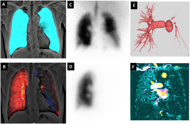
A 74-year-old man with exertional dyspnoea was referred to our hospital. The chest X-ray showed ipsilateral decreased lung vasculature in the left lung field. Pulmonary perfusion and ventilation images were created from dynamic chest radiography (DCR) captured by a flat-panel detector (AeroDR fine, KONICAMINOLTA, Japan) and a conventional X-ray system (RADspeed Pro, SHIMADZU, Japan) using a prototype workstation (KONICAMINOLTA, Japan). These novel images detected normal ventilation dynamics in bilateral lungs (Panel A and Supplementary material online, Video S1) and severely decreased perfusion in the left lung (Panel B and Supplementary material online, Video S2). Lung ventilation–perfusion scintigraphy showed the ventilation–perfusion mismatch of the left lung (Panel C: ventilation, Panel D: perfusion), which is closely similar to the findings of DCR. Afterward, computed tomography (CT) pulmonary angiography (Panel E) and 18F-fluorodeoxyglucose positron emission tomography (FDG-PET) revealed severe stenosis in the left pulmonary artery due to pulmonary arterial wall thickening with high FDG uptake (maximum standardized uptake value = 7.6), which indicated large vessel vasculitis. The iodine mapping acquired with detector-based dual-energy CT (IQon Spectral CT, Philips Healthcare, Netherlands) also showed a similar finding to the pulmonary perfusion image of DCR (Panel F). After careful examinations, the patient was clinically diagnosed with giant cell arteritis, and combination therapy of corticosteroid and tocilizumab started.
Dynamic chest radiography visualizes pulmonary air filling while breathing (ventilation image) and blood supply while breath holding (perfusion image) as slight changes in pixel value even without the use of contrast media. It requires one-fifth as much radiation exposure as lung ventilation–perfusion scintigraphy. This is the first demonstration of pulmonary ventilation–perfusion mismatch by DCR.
The authors would like to thank Kohtaro Abe (Department of Cardiovascular Medicine, Kyushu University, Japan) for his technical advice and Takenori Fukumoto (KONICAMNOLTA, Japan) for technical support in image creation.
This work was supported by the Japan Society for the Promotion of Science (JSPS) KAKENHI (20K16728).
Supplementary material is available at European Heart Journal online.
Supplementary Material
Contributor Information
Yuzo Yamasaki, Department of Clinical Radiology, Graduate School of Medical Sciences, Kyushu University, 3-1-1, Maidashi, Higashi-ku, Fukuoka, 812-8582, Japan.
Kazuya Hosokawa, Department of Cardiovascular Medicine, Graduate School of Medical Sciences, Kyushu University, 3-1-1, Maidashi, Higashi-ku, Fukuoka, 812-8582, Japan.
Hiroyuki Tsutsui, Department of Cardiovascular Medicine, Graduate School of Medical Sciences, Kyushu University, 3-1-1, Maidashi, Higashi-ku, Fukuoka, 812-8582, Japan.
Kousei Ishigami, Department of Clinical Radiology, Graduate School of Medical Sciences, Kyushu University, 3-1-1, Maidashi, Higashi-ku, Fukuoka, 812-8582, Japan.
Associated Data
This section collects any data citations, data availability statements, or supplementary materials included in this article.


