

Abstract
Objective
This study aimed to evaluate the results of surgical treatment of tibial avulsion injuries of the posterior cruciate ligament (PCL) with a 3.5-mm locking compression hook plate (LCHP).
Methods
From June 2012 to June 2015, 16 consecutive patients (10 males and 6 females, mean age: 38 (range: 19–57) years) presented with isolated tibial avulsion injuries of the PCL. We used a 3.5-mm LCHP and lag screws for open reduction and internal fixation (ORIF) through the posterior medial approach. The operation time, quantity of bleeding, visual analog scale (VAS) scores, stability of posterior drawer test (PDT) results, and fracture healing time were studied to assess clinical efficacy. At the 12-month follow-up, a functional evaluation using knee range of motion (ROM) and the Lysholm knee scoring system (LKSS) was performed.
Results
The data from a mean follow-up of 24.1 (range: 14–33) months from 16 patients were recorded. No neurovascular complications, incision infections, or delayed union or nonunion were observed. The mean operation time was 61.4 (range: 45–80) min. The mean quantity of bleeding was 41.6 (range: 25–66) mL. The mean bone healing time was 11.8 (range: 45–80) weeks. The mean VAS score was 1.63 (range: 0–3) after surgery. The average LKSS and ROM of the knee were 51.75±7.67 and 50.94°±10.19° before surgery and 92.75±5.46 and 127.75°±6.13° at 1 year, respectively. The outcomes were judged to be excellent for 11 patients, good for 4, and fair for 1 (excellent and good rates: 93.8% for 15/16). At the final follow-up (≥1 year), the PDT scores returned to normal.
Conclusion
The results showed that 3.5-mm LCHP provided reliable fixation following ORIF of isolated PCL tibial avulsion fractures and was a safe, simple, and effective procedure. This procedure may reduce complications and improve functional recovery relative to those of other procedures.
Trial registration
Chinese Clinical Trial Registry, ChiCTR-1900022920. Registered on 3 May 2019.
Level of Evidence
Level IV, Therapeutic study
Keywords: Avulsion fractures, Knee, Posterior cruciate ligament, Locking compression hook plate
Introduction
Isolated posterior cruciate ligament (PCL) tibial avulsion fractures are unusual injuries that frequently occur in young patients (1, 2). These fractures account for 3% to 38% of all knee injuries (3–5). Femoral detachment, intrasubstance tear, and PCL tibial avulsion injuries have been described in previous studies (6, 7). The global incidence of PCL tibial avulsion fractures is extremely low but higher in developing Asian countries because of frequent motor vehicle accidents (1), especially motorcycle injuries, followed by sports-related injuries (7). PCL injury may lead to instability, abnormal activity, and degeneration of the posterior knee joint (8, 9).
The outcomes and risks of open versus arthroscopic procedures for PCL tibial avulsion fractures reportedly are similar (7). Although the advantages of arthroscopy are well-known, there are some disadvantages, i.e., high requirements for reduction and fixation, long learning curve, expensive arthroscopic system, and occasional difficulty in achieving accurate reduction for severe displaced comminuted fractures (10–12). Therefore, in China, the preferred treatment for avulsion fractures of the PCL tibia is open reduction internal fixation (ORIF) without arthroscopic capabilities.
The optimal fixation method for this kind of injury remains controversial (7, 11, 13–15). The selection of the instrument depends on the size and shape of the fracture. Therefore, an area of interest in clinical research is how to choose a strong and effective instrument so that the fracture can be stabilized and the patient can exercise as early as possible. This retrospective study aimed to assess the effectiveness of ORIF using a 3.5-mm locking compression hook plate (LCHP) to treat isolated avulsion fractures of the tibial PCL attachment, with a 12-month minimum follow-up.
Materials and Methods
Patient eligibility
This was a retrospective study approved by the clinical research ethics committee of our hospital according to the 1964 Declaration of Helsinki principles. Written informed consent from all the participants was obtained.
Twenty-two patients aged between 19 and 57 years (mean age, 38.0 years) with PCL tibial avulsion fractures were recruited between June 2012 and June 2015. The inclusion criteria were acute fracture displacement less than 3 mm on lateral x-ray radiographs and a transverse diameter of the fracture fragments greater than 8 mm on computed tomography (CT) scan; adults between the ages of 18 and 65; duration of injury less than 12 weeks; and treated by a 3.5-mm LCHP (DePuy Synthes, Oberdorf, Switzerland) (Figure 1) fixation with a follow-up period of greater than or equal to 12 months. The exclusion criteria were open knee injuries; pathological fracture or re-fracture; non-displaced fracture fragments or fragment displacement greater than 3 mm; patients with other ligament injuries of knee joint or multiple injuries; contraindication of surgery, and serious complications. Six patients were excluded, and 16 patients with isolated PCL tibial avulsion fractures were enrolled (10 males and 6 females, 9 left and 7 right) (Table 1). The patients had the following injury mechanisms: 8 patients were involved in road traffic accidents, 5 in sports-related trauma, and 3 experienced falls. There were 6 type II and 10 type III fracture types (Meyer-McKeever) (16). All patients underwent thorough clinical examinations (Figure 2. a, b). The posterior drawer test (PDT) results for type III were positive. The visual analog scale (VAS) scores, Lysholm Knee Scoring System (LKSS) scores, and range of motion (ROM) of the knee were recorded before operation. Anteroposterior and lateral knee imaging by x-ray, 3-dimensional CT reconstruction, and magnetic resonance imaging (MRI) were performed before the surgery. All patient eligibility criteria were strictly reviewed by the first author.
Figure 1. a, b.

A 3.5-mm LCHP and lag screws
Table 1.
Characteristics and clinical data of the patients (n=16)
| Sex/age (years) | Side | Duration of injury (days) | Injury mechanism | Surgery time (min) | Loss of bleeding (mL) | Fracture healing (weeks) | Follow-up (months) | Preop VAS score | Postop VAS score | Preop LKSS score | Postop LKSS score | Preop ROM (°) | Postop ROM (°) | Complications | Patient’s satisfaction |
|---|---|---|---|---|---|---|---|---|---|---|---|---|---|---|---|
| male/35 | left | 5 | RTA | 48 | 45 | 11 | 15 | 7 | 3 | 49 | 97 | 43 | 135 | No | excellent |
| male/48 | left | 10 | RTA | 70 | 34 | 13 | 31 | 6 | 1 | 51 | 96 | 47 | 123 | No | excellent |
| female/20 | left | 7 | fall | 45 | 36 | 9 | 29 | 6 | 1 | 63 | 95 | 50 | 133 | No | excellent |
| male/28 | right | 9 | SR | 63 | 50 | 12 | 16 | 7 | 0 | 46 | 96 | 44 | 134 | No | excellent |
| male/31 | right | 3 | SR | 57 | 43 | 11 | 14 | 7 | 3 | 42 | 92 | 55 | 125 | No | Good |
| male/28 | left | 6 | SR | 58 | 46 | 11 | 25 | 5 | 2 | 58 | 95 | 46 | 133 | No | excellent |
| female/19 | left | 3 | SR | 49 | 35 | 9 | 16 | 6 | 1 | 43 | 96 | 51 | 136 | No | excellent |
| male/35 | right | 5 | fall | 68 | 30 | 12 | 20 | 8 | 2 | 54 | 95 | 45 | 128 | No | excellent |
| male/44 | left | 4 | RTA | 71 | 33 | 13 | 29 | 8 | 1 | 61 | 87 | 70 | 120 | No | Good |
| male/47 | left | 11 | RTA | 80 | 45 | 11 | 22 | 6 | 2 | 55 | 88 | 44 | 124 | No | Good |
| female/57 | left | 4 | RTA | 55 | 25 | 14 | 27 | 7 | 3 | 40 | 78 | 46 | 116 | No | fair |
| male/39 | left | 5 | fall | 58 | 45 | 12 | 28 | 6 | 1 | 53 | 85 | 78 | 121 | No | Good |
| male/34 | right | 8 | SR | 61 | 38 | 12 | 24 | 7 | 2 | 56 | 95 | 43 | 128 | No | excellent |
| female/46 | right | 5 | RTA | 76 | 40 | 13 | 27 | 7 | 1 | 47 | 98 | 47 | 135 | No | excellent |
| female/51 | right | 5 | RTA | 65 | 66 | 13 | 29 | 6 | 2 | 45 | 96 | 46 | 125 | No | excellent |
| female/46 | right | 7 | RTA | 58 | 55 | 12 | 33 | 6 | 1 | 65 | 95 | 60 | 128 | No | excellent |
VAS: Visual Analog Scores; ROM: range of motion; RTA: road traffic accident; SR: sports-related; Preop: preoperative; Postop: postoperative
Figure 2. a–f.
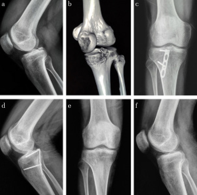
(a) Lateral radiograph of the knee (nonstress view) showing the avulsed fragment from the tibia. (b) Three-dimensional computed tomography scan showing the avulsed fragment of the tibia. (c and d) Radiographs showing bone union 3 months after surgery. (e and f) Photographs of the 3.5-mm LCHP plate removed 1 year after surgery
Surgical technique
After satisfactory anesthetization, the patient was placed in the prone position with mild knee flexion. The approach used an 8–12 cm inverted L-shaped incision that began along the medial border of the gastrocnemius muscle and bent laterally along the flexion crease of the joint. After careful incisions avoiding the small saphenous vein and medial cutaneous nerve to expose the articular capsule, it was incised longitudinally to expose the PCL and avulsion fracture fragments. After clearing blood clots and soft tissue to expose the fracture line, the knee was positioned in flexion. The fracture was reduced, and temporary fixation using two Kirschner wires (<1.5 mm) was applied. After satisfactory reduction under c-arm fluoroscopic viewing, the 3.5-mm LCHP teeth were inserted into the PCL (Figure 2. c, d). Appropriate fixation with lag screws was applied, and the incision was closed if the PDT result was negative.
Postoperative care and follow-up
The injured limb was fixed with an elastic bandage or wrap stretch hose with 15° knee flexion for 2 days postoperatively. Quadriceps exercises and ankle extension training were started initially, and active and passive knee functional exercises with an auxiliary appliance were started 1 week postoperatively with joint activities not exceeding 90° for 4 weeks. Six weeks postoperatively, the patients could walk up and down stairs with crutches. Knee flexion of 110°–120° was required 6–8 weeks postoperatively. The assistive device was removed 8 weeks postoperatively, and the patient was required to bear full weight. Full pre-injury knee function was restored after 3 months (Figures 2. e, f and 3. a–c).
Figure 3. a–c.

Photographs showing a patient with good right knee function 1 year after surgery (a–c)
Clinical and functional outcome assessments
Basic patient information and surgical data were collected. Imaging and functional data were regularly evaluated during follow-up. Outcome measures included VAS, fracture healing time (FHT), PDT, knee ROM, patient satisfaction, and complications. The LKSS score was used to evaluate knee function (17).
Statistical analysis
Statistical analysis was performed by using the Statistical Package for Social Sciences version 23.0 software (IBM SPSS Corp.; Armonk, NY, USA). Data are presented as the mean±standard deviation. The paired t-test with a confidence level of 5% was performed to assess intergroup variability.
Results
The mean follow-up of our 16 patients was 24.1 (range: 14–33) months. The mean surgical time was 61.4 (range: 45–80) min. The mean blood loss was 41.6 (range: 25–66) mL. The mean FHT by radiography was 11.8 (range: 9–14) weeks after surgery.
No deep vein thrombosis, cutaneous numbness, tourniquet palsy, neurovascular complications, incision infection, or delayed union or nonunion were recorded. At the final follow-up, no patients had knee stiffness (Table 2).
Table 2.
Statistical analysis
| Parameter | Preoperative | Postoperative | p |
|---|---|---|---|
| VAS | 6.56±0.81 | 1.63±0.89 | <0.001 |
| LKSS | 51.75±7.67 | 92.75±5.46 | <0.001 |
| ROM (°) | 50.94±10.19 | 127.75±6.13 | <0.001 |
Values are presented as mean±standard deviation
The mean VAS score was 1.63 (range: 0–3) after surgery. The mean LKSS score was 92.75 (range: 78–98). The combined excellent + good rate was 93.8% (15/16) at 12 months. The average knee ROM was 127.75°±6.13° at 1 year.
Postoperatively, there was no laxity in 100% (16/16) of the knees on PDT. The PDT results normalized, and there was no difference in the PDT results and those from the intact knee in all patients at the 1-year follow-up.
Discussion
In this study, the distribution of injuries was similar to those that were previously reported (1, 5, 7, 15, 18). PCL tibial avulsion fractures may lead to knee instability and secondary joint changes resulting in osteoarthritis (8, 9). Delayed union or nonunion also may occur during conservative treatment (19). Therefore, it is generally agreed that surgical intervention is necessary for displaced avulsion fractures of the tibial PCL attachment. Although arthroscopic repair is comparable to ORIF, there are some limitations, such as the long learning curve and high technical requirements. The most common method at present to treat PCL tibial avulsion fractures in hospitals without arthroscopic equipment is ORIF using the posterior approach (11).
Kirschner wires, hollow lag screw, absorbable screw, suture anchor, and straddle screw have been used to treat PCL tibial avulsion fractures (7, 11, 13–15). The challenges of treating these fractures are bone conditions and small fragments. However, there is no consensus regarding which technique will achieve the best functional outcome. A 3.5-mm LCHP designed to use a tension band has been found suitable for upper limb fractures, including ulna aquiline fracture (AO: B1, B3, C1) (20), ulna distal fracture (21, 22), ankle joint avulsion fracture (23), and fifth metatarsal base fractures (24–26). However, there has been no previous study on the clinical results of 3.5-mm LCHP in the treatment of PCL tibial avulsion fractures.
Our results indicate that 3.5-mm LCHP fixation was effective for isolated PCL avulsion fractures in adults without arthroscopy in our 16-patient sample, and the results were excellent + good in 93.8% (15/16) and fair in 6.2% (1/16) of the patients with an average postoperative LKSS of 92.75±5.46. The mean FHT by radiography was 11.8 (range: 9–14) weeks after surgery. In the last follow-up, no laxity in 100% (16/16) of the knees on PDT was observed. No surgical complications occurred in this series, and all patients obtained good clinical and functional prognosis. These results are no worse than previous results. In a study by Chen et al., among 21 patients treated using a tooth plate and hollow lag screw, excellent results were obtained in 90.5%, good results in 9.5% (11). They reported a mean operative time of 65.5±13.7 min, a mean blood loss of 54.3±15.0 mL, a mean knee ROM of 121.9°±10.4°, and a mean LKSS score of 93.6±5.9. The average fracture healing time was 10.2±1.7 (range: 8–12) weeks. Joshi et al. reported 14 patients with PCL avulsion fractures treated with a cancellous screw (1). Their results showed a low incidence of complications and good fracture healing. At the final followup, the average flexion was 121.7°±9.18° with full extension possible in all patients. The functional outcome assessed using the Lysholm scoring system was excellent in 11 patients, good in 2, and fair in 1. The average Lysholm score was 97±7.6 .Instability was clinically tested using the drawer test and radiologically with stress radiography; both tests showed mild intensity (1+) in 4 of the patients, whereas the other patients had no residual instability. Yastrebov et al. in 2010 reported that a 1/3 tubular plate was cut, bent, and shaped into a hook plate for PCL tibial avulsion fractures (27). However, the operation time is obviously shortened by avoiding cutting and shaping a tubular plate during the operation using 3.5-mm LCHP fixation. Li et al. reported the clinical data of 14 patients with an avulsion fracture at the PCL of the tibia who were treated by ORIF with a stellate steel plate (28). The results showed that after 6 months, all fractures had healed, the knee joint displayed normal motion, and there was no stiffness or instability. The LKSS score at the final follow-up was 97.1±1.7 points. In 2019, Yin et al. reported that 19 patients with avulsion fracture of the PCL were treated with a hook plate and achieved good function (29). The preoperative functional score was 50.2±1.4, and the postoperative functional score was 91.4±1.9.
We found that the advantages of a 3.5-mm LCHP in the treatment of Meyer-Mckeever type II and type III fracture fragments were operational simplicity, fixation reliability, and short operative time. The teeth of the LCHP expand the fixed area, whereas the teeth around the plate are embedded into the bone and surface of the ligament to allow the fracture fragments to be fixed by using high torque. The PCL attachment point is directly penetrated through the hook teeth, and the eccentric drilling and sliding pressure of the two nail holes are applied in turn so that the plate has two uniform pressure opportunities and the fracture ends contact more closely. As the teeth of the hook make point contacts, the cutting damage is reduced, hence the larger fracture fragment can be fixed, and smaller or even crushed fractures can be fixed. These characteristics result in stable fixation of the fragments and facilitate healing of bone and ligaments. Finally, the fixation direction of the screw is basically perpendicular to the posterior tibial bone surface. The screw is inserted into the bone in the non-fracture area during the operation, which not only prevents injury of the fracture block, but also avoids loosening and nail removal of the screw along with flexion and extension of the knee joint. The PCL tibial attachment bone is mainly cancellous with an abundant blood supply, which leads to excellent reduction and firm fixation of fractures that heal successfully within 3 months postoperatively. Our patients began functional exercise at an early stage after surgery and ultimately achieved pre-injury knee joint function.
However, one of the surgical pitfalls that must be mentioned is that the 3.5-mm LCHP is not suitable for some comminuted fractures. Moreover, in some cases, the hooks cannot adapt to the proximal fracture part.
The limitations of this study were the small sample size, relatively short follow-up time, retrospective analysis, and lack of comparison with other fixation techniques. Therefore, to confirm our positive knee function and biomechanical results in this small uncontrolled study, a large-sample prospective randomized controlled study would be required. In addition, biomechanical experimental evidence is needed to provide additional mechanistic support.
In conclusion, we found that the use of a 3.5-mm LCHP provided reliable fixation following ORIF of isolated PCL tibial avulsion fractures and was a safe, simple, and effective procedure. Because satisfactory fixation was achieved, complications were minimized and recovery was better than that observed using many other procedures. Furthermore, this procedure could potentially be used at hospitals without arthroscopic capabilities.
HIGHLIGHTS
We would like to highlight that to our knowledge, this is the first study to investigate the clinical and functional outcomes of treatment with a 3.5-mm LCP hook plate for PCL tibial avulsion fractures.
As per our literature review, we have demonstrated, for the first time, the treatment efficacy of this method in this injury in adults without any arthroscopic conditions.
By directly tapping into the PCL attachment point with the teeth of the hook, the two nails were drilled, and sliding pressure was successively applied to ensure that the plate had two uniform pressure spots and the fracture ends were closer. The teeth of the hook belong to the point of contact; therefore, damage from cutting is minimized such that large, small, or even crushed fractures can be fixed.
Footnotes
Ethics Committee Approval: Ethics committee approval was received for this study from the Biomedical Research Ethic Committee of West China Hospital of Sichuan University (file version number: 1.0; file number: 2019-220).
Informed Consent: Written informed consent from all the participants was obtained in the study.
Author Contributions: Concept - H.Z.; Design - W.D., H.Z.; Supervision - F.H, H.Z.; Materials - W.D., Y.L.; Data Collection and/or Processing - Y.L., S.W.; Analysis and/or Interpretation - W.D., X.L.; Literature Search - W.D., Y.L.; Writing Manuscript - W.D.; Critical Review - X.L., F.H.
Conflict of Interest: The authors have no conflicts of interest to declare.
Financial Disclosure: The authors declared that this study has received no financial support.
References
- 1.Joshi S, Bhatia C, Gondane A, Rai A, Singh S, Gupta S. Open reduction and internal fixation of isolated posterior cruciate ligament avulsion fractures: Clinical and functional outcome. Knee Surg Relat Res. 2017;29:210–6. doi: 10.5792/ksrr.17.022. [DOI] [PMC free article] [PubMed] [Google Scholar]
- 2.Katsman A, Strauss EJ, Campbell KA, Alaia MJ. Posterior cruciate ligament avulsion fractures. Curr Rev Musculoskelet Med. 2018;11:503–9. doi: 10.1007/s12178-018-9491-2. [DOI] [PMC free article] [PubMed] [Google Scholar]
- 3.Barros MA, Cervone GL, Costa AL. Surgical treatment of avulsion fractures at the tibial insertion of the posterior cruciate ligament: functional result. Rev Bras Ortop. 2015;50:631–7. doi: 10.1016/j.rbo.2015.04.012. [DOI] [PMC free article] [PubMed] [Google Scholar]
- 4.Fanelli GC, Edson CJ. Posterior cruciate ligament injuries in trauma patients: Part II. Arthroscopy. 1995;11:526–9. doi: 10.1016/0749-8063(95)90127-2. [DOI] [PubMed] [Google Scholar]
- 5.Janousek AT, Jones DG, Clatworthy M, Higgins LD, Fu FH. Posterior cruciate ligament injuries of the knee joint. Sports Med. 1999;28:429–41. doi: 10.2165/00007256-199928060-00005. [DOI] [PubMed] [Google Scholar]
- 6.Giordano BD, Dehaven KE, Maloney MD. Acute femoral “peel-off” tears of the posterior cruciate ligament: technique for arthroscopic anatomical repair. Am J Orthop (Belle Mead NJ) 2011;40:226–32. [PubMed] [Google Scholar]
- 7.Hooper PO, 3rd, Silko C, Malcolm TL, Farrow LD. Management of posterior cruciate ligament tibial avulsion injuries: A systematic review. Am J Sports Med. 2018;46:734–42. doi: 10.1177/0363546517701911. [DOI] [PubMed] [Google Scholar]
- 8.Zhang X, Cai G, Xu J, Wang K. A minimally invasive postero-medial approach with suture anchors for isolated tibial avulsion fracture of the posterior cruciate ligament. Knee. 2013;20:96–9. doi: 10.1016/j.knee.2012.10.016. [DOI] [PubMed] [Google Scholar]
- 9.Strobel MJ, Weiler A, Schulz MS, Russe K, Eichhorn HJ. Arthroscopic evaluation of articular cartilage lesions in posterior-cruciate-ligament-deficient knees. Arthroscopy. 2003;19:262–8. doi: 10.1053/jars.2003.50037. [DOI] [PubMed] [Google Scholar]
- 10.Abdallah AA, Arafa MS. Treatment of posterior cruciate ligament tibial avulsion by a minimally-invasive open posterior approach. Injury. 2017;48:1644–9. doi: 10.1016/j.injury.2017.05.032. [DOI] [PubMed] [Google Scholar]
- 11.Chen W, Luo W, Chen Z, Jiang Y. Treatment of posterior cruciate ligament avulsion fractures of the tibia using a toothed plate and hollow lag screw. Singapore Med J. 2016;57:39–44. doi: 10.11622/smedj.2016010. [DOI] [PMC free article] [PubMed] [Google Scholar]
- 12.Gavaskar AS, Karthik B, Gopalan H, Srinivasan P, Tummala NC. A novel MIS technique for posterior cruciate ligament avulsion fractures. Knee. 2017;24:890–6. doi: 10.1016/j.knee.2017.03.014. [DOI] [PubMed] [Google Scholar]
- 13.Seitz H, Schlenz I, Pajenda G, Vecsei V. Tibial avulsion fracture of the posterior cruciate ligament: K-wire or screw fixation? A retrospective study of 26 patients. Arch Orthop Trauma Surg. 1997;116:275–8. doi: 10.1007/BF00390052. [DOI] [PubMed] [Google Scholar]
- 14.Torisu T. Isolated avulsion fracture of the tibial attachment of the posterior cruciate ligament. J Bone Joint Surg Am. 1977;59:68–72. doi: 10.2106/00004623-197759010-00011. [DOI] [PubMed] [Google Scholar]
- 15.Bali K, Prabhakar S, Saini U, Dhillon MS. Open reduction and internal fixation of isolated PCL fossa avulsion fractures. Knee Surg Sports Traumatol Arthrosc. 2012;20:315–21. doi: 10.1007/s00167-011-1618-6. [DOI] [PubMed] [Google Scholar]
- 16.Huang TW, Hsu KY, Cheng CY, et al. Arthroscopic suture fixation of tibial eminence avulsion fractures. Arthroscopy. 2008;24:1232–8. doi: 10.1016/j.arthro.2008.07.008. [DOI] [PubMed] [Google Scholar]
- 17.Lysholm J, Gillquist J. Evaluation of knee ligament surgery results with special emphasis on use of a scoring scale. Am J Sports Med. 1982;10:150–4. doi: 10.1177/036354658201000306. [DOI] [PubMed] [Google Scholar]
- 18.Schulz MS, Russe K, Weiler A, Eichhorn HJ, Strobel MJ. Epidemiology of posterior cruciate ligament injuries. Arch Orthop Trauma Surg. 2003;123:186–91. doi: 10.1007/s00402-002-0471-y. [DOI] [PubMed] [Google Scholar]
- 19.Singla R, Devgan A, Gogna P, Batra A. Fixation of delayed union or non-union posterior cruciate ligament avulsion fractures. J Orthop Surg (Hong Kong) 2014;22:70–4. doi: 10.1177/230949901402200118. [DOI] [PubMed] [Google Scholar]
- 20.Wagner FC, Konstantinidis L, Hohloch N, Hohloch L, Suedkamp NP, Reising K. Biomechanical evaluation of two innovative locking implants for comminuted olecranon fractures under high-cycle loading conditions. Injury. 2015;46:985–9. doi: 10.1016/j.injury.2015.02.010. [DOI] [PubMed] [Google Scholar]
- 21.Nunez FA, Jr, Li Z, Campbell D, Nunez FA., Sr Distal ulna hook plate: Angular stable implant for fixation of distal ulna. J Wrist Surg. 2013;2:87–92. doi: 10.1055/s-0032-1333427. [DOI] [PMC free article] [PubMed] [Google Scholar]
- 22.Nunez FA, Jr, Barnwell J, Li Z, Nunez FA., Sr Metaphyseal ulnar shortening osteotomy for the treatment of ulnocarpal abutment syndrome using distal ulna hook plate: case series. J Hand Surg Am. 2012;37:1574–9. doi: 10.1016/j.jhsa.2012.04.034. [DOI] [PubMed] [Google Scholar]
- 23.Zhenhua F, Waizy H, Ming X, Wusheng K. Lateral malleolus hook plate for comminuted Weber A and B fractures: A retrospective study. Indian J Orthop. 2013;47:364–9. doi: 10.4103/0019-5413.114918. [DOI] [PMC free article] [PubMed] [Google Scholar]
- 24.Xie L, Guo X, Zhang SJ, Fang ZH. Locking compression plate distal ulna hook plate fixation versus intramedullary screw fixation for displaced avulsion fifth Metatarsal Base fractures: A comparative retrospective cohort study. BMC Musculoskelet Disord. 2017;18:405. doi: 10.1186/s12891-017-1766-z. [DOI] [PMC free article] [PubMed] [Google Scholar]
- 25.Kim JB, Song IS, Park BS, Ahn CH, Kim CU. Comparison of the outcomes between headless cannulated screw fixation and fixation using a locking compression distal ulna hook plate in fracture of fifth metatarsal base. J Foot Ankle Surg. 2017;56:713–7. doi: 10.1053/j.jfas.2017.01.048. [DOI] [PubMed] [Google Scholar]
- 26.Lee SK, Park JS, Choy WS. Locking compression plate distal ulna hook plate as alternative fixation for fifth metatarsal base fracture. J Foot Ankle Surg. 2014;53:522–8. doi: 10.1053/j.jfas.2014.02.021. [DOI] [PubMed] [Google Scholar]
- 27.Yastrebov O, Lobenhoffer P. Refixation of tibial bony avulsions of the posterior cruciate ligament with a hook plate. Oper Orthop Traumatol. 2010;22:347–53. doi: 10.1007/s00064-010-9021-x. [DOI] [PubMed] [Google Scholar]
- 28.Li L, Tian W. Displaced avulsion fractures of the posterior cruciate ligament: Treated by stellate steel plate fixation. Indian J Orthop. 2015;49:171–5. doi: 10.4103/0019-5413.152454. [DOI] [PMC free article] [PubMed] [Google Scholar]
- 29.Yin QD, Rui YJ, Wu YW, et al. Surgical treatment of avulsion fracture around joints of extremities using hook plate fixation. BMC Musculoskelet Disord. 2019;20:200. doi: 10.1186/s12891-019-2585-1. [DOI] [PMC free article] [PubMed] [Google Scholar]