PLOS ONE logoLink to PLOS ONE
. 2021 Jan 20;16(1):e0242067. doi: 10.1371/journal.pone.0242067

A randomized clinical trial of a new anti–cervical stenosis device after conization by loop electrosurgical excision

Marcelo de Andrade Vieira 1,*,#, Raphael Leonardo Cunha de Araújo 2,, Carlos Eduardo Mattos da Cunha Andrade 1,, Ronaldo Luis Schmidt 1,, Agnaldo Lopes Filho 3,, Ricardo dos Reis 1,#
Editor: Emily Chenette4
PMCID: PMC7816995  PMID: 33471788

Abstract

Background

The complications inherent to conization include vaginal bleeding, cervical stenosis, amenorrhea, dysmenorrhea, and deep dyspareunia. Cervical stenosis is the most important complication due to the clinical repercussions. Studies show rates of cervical stenosis ranging from 1.3 to 19% after the Loop Electrosurgical Excision Procedure (LEEP).

Objective

Our primary outcome was to compare the role of a new endocervical device to prevent cervical stenosis after LEEP in patients with high-grade squamous intraepithelial lesions (HSILs).

Methods

A randomized clinical trial was performed including phases II and III for evaluation of a new device for cervical stenosis prevention. In Phase II, we included 25 patients who underwent LEEP and placement of the device to assess its toxicity and efficacy. In phase III, we compared two groups (with and without the use of an anti-stenosis device) to evaluate its efficacy and safety.

Results

From August 2015 to June 2018, 265 participants were randomized (Phase II: 25, Phase III: 120 with DUDA and 120 without DUDA). The toxicity during phase II was observed in only one patient (4%) with pain grade > 7. There were 7 cases of toxicity during Phase III, 2 in the DUDA group (1.8%), and 5 in the No DUDA group (4.5%). The complications rate was numerically higher in the No DUDA group (2.5x higher) than the DUDA group, but this difference did not reach statistical significance (p = 0.52). The rate of cervical stenosis in DUDA group was (4–7,3%), and in No DUDA group was (4.3–5.8%) (p = 0.5). We did not find a significant difference when comparing the evolution at 3, 6, and 12 months in terms of cervical patency and visualization of the squamocolumnar junction (SCJ) during colposcopy. The DUDA group exhibited 15% to 19% nonvisualization of the SCJ, whereas that rate ranged from 10 to 12% in the No DUDA group.

Conclusions

The rate of cervical stenosis was not different comparing the use of a new device, specifically produced to prevent cervical stenosis, compared to no use after LEEP procedure. This clinical trial opens up space for a discussion of the utility of using cervical stenosis devices after LEEP. Perhaps in another type of conization it can be evaluated to avoid cervical stenosis.

Introduction

Infection with the human papillomavirus (HPV) virus is common among women. In most cases, after 6 to 12 months of infection, the immune system eliminates the virus. If the lesion persists, women may develop cancer precursor lesions in the cervix and, if not treated, cervical cancer [14]. The term used for cervical precursor lesions or high-grade cervical intraepithelial neoplasia (CIN) is used to define lesions previously reported as moderate dysplasia (CIN II) and severe dysplasia/carcinoma in situ (CIN III) [5]. The natural history of CIN II indicates that 32% to 43% of untreated lesions will regress without treatment, while 35% to 56% will persist and 14% to 22% will progress to carcinoma in situ or invasive carcinoma [57]. When assessing CIN III lesions, studies show that within 30 years after infection, 31% of patients will develop cervical cancer [2]. When we group the premalignant lesions into CIN II and III, studies show that in 10 to 20 years, the progression to cancer will occur in some cases if they are left.

Untreated [8]. Progression depends on factors such as the health system, socioeconomic status, HPV status, smoking and awareness of the importance of Pap smears [9].

Treatment of high-grade CIN (CIN II and III) is chosen according to the patient’s colposcopic assessment. In cases of satisfactory colposcopy (visible squamocolumnar junction), both ablative and excisional methods are adequate, whereas in cases of the nonvisible squamocolumnar junction, only excisional treatment is accepted [10, 11]. The complications inherent to conization include vaginal bleeding, cervical stenosis, amenorrhea, dysmenorrhea and deep dyspareunia [12, 13]. Cervical stenosis is the most important complication due to the clinical repercussions, which may range from menstrual cramps to hematometra, infertility, and the impossibility of early detection of relapse and or recurrence of the premalignant lesion due to the difficulty of follow-up [12, 1417]. Studies show rates of cervical stenosis ranging from 3 to 25% after laser conization and from 1.3 to 19% after LEEP [12, 14, 1821]. Also besides, the diagnosis can be made up to 28 months after conization [14]. At present, there is no consensus standard procedure for the prevention and treatment of cervical stenosis after conization. The clinical studies evaluating anti–cervical stenosis measures have all been nonrandomized, and they have used temporary devices in an attempt to avoid this type of complication [22, 23]. In addition, studies published to date have used several different types of devices without adequate standardization [17, 24, 25]. We propose the first randomized, prospective clinical trial evaluating the role of a new endocervical device called DUDA (Uterine Device to Dilate the Endocervical Canal) developed with characteristics proportional to the size of the cervical and endocervical canal to prevent cervical stenosis after LEEP in patients with high-grade squamous intraepithelial lesions (HSILs).

Materials and methods

The study was conducted at the Barretos Cancer Hospital from August 2015 to June 2018 (first randomization and the last study follow up). It was started after approval by the Research Ethics Committee (CEP–Local IRB) from Barretos and National Committee in Research Ethics (CONEP–National Committee) because it was on a new medical device (CAAE 36018714.4.0000.5437). It is registered at ClinicalTrials.gov (NCT02500966). After checking the inclusion criteria in the study, the patients were informed about the study and, in accordance with the research terms, signed the Free and Informed Consent Form (ICF). After this step, online randomization was carried out on the website. A randomization list was generated randomly using the R. statistical software. This list was a sequence with dichotomous values divided into even-sized blocks to ensure a balance between patients who would or would not use the DUDA device. Subsequently, this list was attached to a project made on the RedCap Platform to guarantee the accuracy and reliability of data and results [26]. This randomization was blinded, but due to the nature of the study, the patients and physicians were not blinded to the intervention after the outcome of the randomization. The gynecologist who performed the colposcopy during the follow up was blinded to the groups. Furthermore, the patients were allocated to groups with (DUDA Group) or without placement of the DUDA device (No DUDA Group). It was a prospective and randomized manner 1:1. The sample estimate ranged from 104 to 203 in each group. The sample size calculation assumed a rate of cervical stenosis of approximately 15% to 20% after LEEP, as described in the literature, and a reduction of this rate to 6% with the use of an endocervical device, according to the uncontrolled trial of Luesley et al. [22]. Considering this difference in stenosis rates (15–20% vs. 6%), an alpha error of 5%, a beta error of 20%, and balanced allocation of 1:1. All patients signed an informed consent form before joining the study. Patients aged 18 to 65 years with a histopathological diagnosis of CIN2/3 in cervical biopsy and indication for LEEP were included, whereas women were pregnant before LEEP, women with antecedent of LEEP, women who did not understand or did not agree to participate in the study, and women under conditions of high vulnerability (e.g., prisoners, indigenous, etc.) were excluded.

Study design

We performed phases II and III on the new cervical device according to the standards of randomized clinical trials. In stage II, we included 25 patients who underwent conization and placement of the device to assess its toxicity. In this stage, all patients underwent conization and placement of the device.

In phase III (evaluation of safety and efficacy), the patients were allocated into two groups: 120 patients in the DUDA group, who underwent conization and placement of DUDA, and 120 patients in the No DUDA group who underwent conization but without placement of DUDA.

Surgical procedure

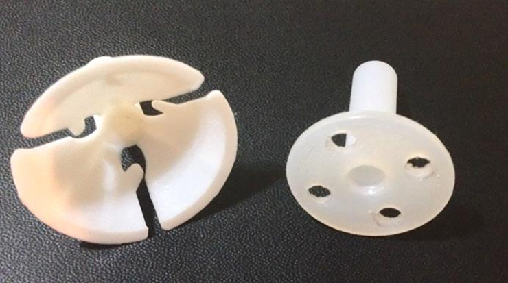
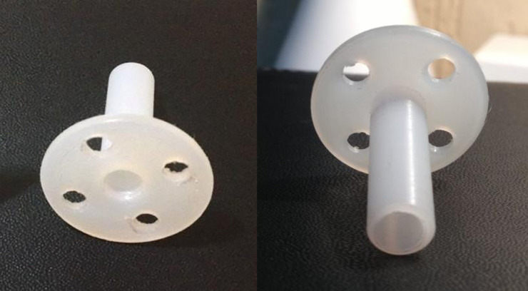
After intravenous anesthesia, the patients in the lithotomy position received local anesthesia in the cervix at four time-points (3, 6, 9, and 12 hours) with 2% xylocaine without vasoconstrictor using 2 ml per point. Then the LEEP was performed, hemostasis was performed with a roller-ball electrode, the cervix was dilated to Hegar number 8, and DUDA was placed (Figs 1 and 2) on the cervix and secured with four Prolene 2–0 stitches in the DUDA group. Patients in the No DUDA group underwent the LEEP procedure and hemostasis without endocervical canal dilation. Patients were observed for 3 hours and then discharged.

Fig 1. The balloon holder on the left, the DUDA device on the right.

Fig 1

The DUDA device was created based on the balloon holder.

Fig 2. Device DUDA.

Fig 2

Anterior and posterior views of DUDA.

Follow-up

The patients in phase 1 and phase 3 in the DUDA group removed the DUDA when they returned for a follow-up visit on day 30. The follow-ups were done in phase II at 15 and 30 days and 3, 6, and 12 months. In phase III, they were done at 30 days and 3, 6, and 12 months. The following-up, the squamocolumnar junction (SCJ) and the transformation zone (TZ) were assessed by colposcopy. The gynecologist who performed the colposcopy was blinded to the groups. Both were classified according to international colposcopy standards [27]. During the physical examination, the external visualization of the external cervical orifice (visible or nonvisible/stenosis), menstrual flow (days), menstrual cramps after surgery (mild: spontaneous improvement; moderate: improvement after the use of oral medications; and severe: requiring intravenous analgesia) and patency of the endocervical canal were assessed through inserting the hysterometer through the canal, classifying the resistance to insertion as none, little or moderate, or failed insertion (complete resistance to the insertion of the hysterometer). Moreover, oncotic cytology was also collected in all follow-ups. The data were stored on the RedCap platform (Research Electronic Data Capture) [26].

Characteristics of the device

DUDA consists of a thermoplastic device of the polyoxymethylene (polyacetal). It was designed by the researcher responsible for the study, made by hand in a lathe by machining a cylindrical piece which it was vacuum-packed and sterilized in hydrogen peroxide (Figs 1 and 2). DUDA is cylindrical at one of its ends, with four holes (where the stitches are anchored), is 2.5 cm in diameter and 2 cm long, accompanied by a cylindrical rod with a 4-mm diameter and a central lumen. The rod is introduced into the endocervical canal, and the more substantial cylindrical portion makes contact with the external surface of the uterine cervix (Fig 2). DUDA was designed and constructed to adjust to the anatomy and functionality of the uterine cervix in terms of length, thickness, the diameter of the fenestration and base of fixation in the uterine cervix (Fig 2). The device remains in the endocervical canal for 30 days and causes a mechanical obstruction to central healing in an attempt to prevent its stenosis.

Local tolerance assessment

The adverse events were related to the procedure during the first 30 days after surgery, and the main safety events considered in this analysis were vaginal bleeding and uterine infection which were recorded according to the criteria of CTCAE version 4.0 [28]. If there were a single adverse event grade 3 or higher, the device would be considered unsafe, and consequently, the trial would be discontinued. After evaluation of the first 25 cases by an independent data monitoring committee and after analysis and approval of the partial (safety) report by the CEP/CONEP System, the study moved into phase III.

Objectives

The primary objective of the present study was to evaluate the efficacy of a new cervical device called DUDA in preventing late stenosis of the cervical canal in women undergoing LEEP conization. The secondary objectives were to evaluate the safety of the device, to compare the SCJ visualization rate and the rate of patients classified in each type of transformation zone (ZT1, ZT2, and ZT3) between women who did and did not use the device.

Statistical analysis

Statistical analysis was performed using the chi-squared test or Fisher’s exact test. Normality was assessed using the Kolmogorov-Smirnov normality test, and when necessary, a parametric test (t-test), with a description of the mean (m) and standard deviation (SD), and nonparametric test (Mann-Whitney), with a description of the median (md) and interquartile range (IQR), were used. To calculate the sample size according to the difference between the groups for the proportions of stenosis, we used the study by Luesley et al. [26]. To calculate the power of the test according to the sample size and the difference observed between proportions, we used the software GPower v.3.1.9.2. Fisher's exact test was used to calculate the significance of differences in proportions between two independent groups, assuming significance level of 0.05 and a test power of 0.80. For the symptoms of menstruation and dysmenorrhea, we excluded the menopausal women from the analysis.

Results

Baseline covariates

A total of 265 patients were selected to participate in the Phase III of the study. However, 20 patients were excluded from randomization and did not enter the study (18 not meeting inclusion criteria and 02 declined to participate in the study) as noted in the CONSORT attachment. All samples underwent LEEP conization.

Patients in phase II had a median age of 34 years old (22–68), had on average 2 children (0–6), were mostly white (60%) and crossbred (32%), and mostly had HSIL (52%) in the initial Pap smear and cervical biopsy with CIN III (44%).

The average duration of placement of the device was 17.7 min (10–30, SD = 6.02 min) (Table 1). All devices were removed within 30 days after the procedure. All patients underwent general and intravenous anesthesia.

Table 1. Demographic characteristics—phases II and III.

Phase II Phase III Phase III p-value*
N = 25 DUDA No DUDA
N = 111 N = 110
Duration of Surgery Median (minutes) (min-max)a 17 (10–30)
Median (minutes) (IQR)b 15 (6) 10 (5)
Pregnancy N (%) 6 (24) 1 (0.9) 3 (2.7) 0.36

* p-value comparisons across treatment groups for categorical variables based on Fisher’s exact test. P-values for quantitative variables based on Wilcoxon rank-sum test. Normality was assessed using the Kolmogorov-Smirnov normality test, and when necessary, a parametric test (t-test), with a description of the mean (m) and standard deviation (SD), and nonparametric test (Mann-Whitney), with a description of the median (md) and inter-quartile range (IQR), were used.

a- Median (min = minimum, max = maximum)

b Median (inter-quartile range).

The toxicity during phase II was observed in only one patient (4%) with pain grade > 7, who required intravenous analgesia on the 23rd postoperative day. It was resolved with a single dose of intravenous analgesia without the need to remove the device. No other cases of toxicity were observed (Table 2). Three devices were removed before the expected date during Phase II (3/25 = 12%). There were two loose devices at 15 days of follow-up that were removed on the 15th postoperative day, one due to a fever, which was resolved with antipyretic alone within 48 hours, and the other after a gynecologic examination. Another device was loose on the 30th postoperative day and was removed.

Table 2. Evaluation of the toxicity of the antistenosis device.

Phase II Phase III Phase III
DUDA No DUDA p-value*
N(%) N(%) N(%)
TOXICITY 0.52
Pain grade II 1 (4) 0 2 (1.8)
Leucorrhea 0 2 (1.8) 3 (2.7)
None 24 (96) 109 (98.2) 105 (95.5)

* p-value comparisons across treatment groups for categorical variables based on the Fisher’s exact test.

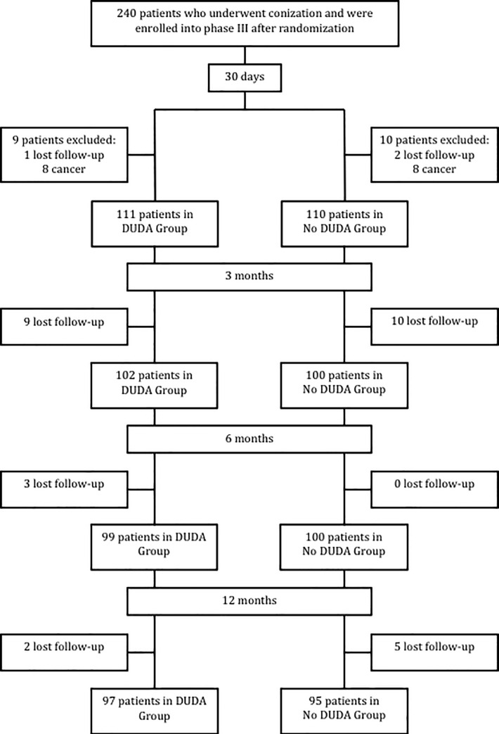
During phase III, in the first 30 days after surgery, the results of 221 patients from the 240 randomized ones were analyzed, with 111 patients (50.2%) in the DUDA group and 110 (49.8%) in the No DUDA group. A total of 19 patients were excluded from the surgical analysis (after conization surgery): 16 presented with invasive squamous cell carcinoma on final histology examination, and 3 were lost to follow-up. For the 3-month analysis, 202 patients were evaluated, 199 patients at the 6-month evaluation, and 192 patients at the 12-month evaluation (Fig 3). All primary and secondary outcomes were evaluated in all phase III patients. The demographic characteristics of phase III patients showed a homogeneous group (Table 3).

Fig 3. Study population enrollment.

Fig 3

Table 3. Demographic characteristics—phases II and III.

Phase II Phase III Phase III
N = 25 DUDA No DUDA
N = 111 N = 110
Age (median) 35.16 (22–68) 34 (21–64) 34 (23–63)
Race n (%) White 15 (60) 75 (68) 75 (68)
Black 2 (8) 9 (8) 6 (6)
Mixed 8 (32) 27 (24) 29 (26)
Pap smear n (%) Minor findings 1 (4) 5 (9) 4 (4)
Major findings 13 (52) 45 (41) 44 (40)
Negative/Benign 11 (44) 61 (55) 52 (57)
Colposcopy n (%) Negative/Benign 0 3 (3) 0
Minor findings 2 (8) 2(2) 28 (26)
Major findings 23 (92) 75 (68) 76 (69)
Suggestive of invasion 0 0 0
SCJ visualization n (%) Visible 23 (92) 104 (94) 105 (96)
Partially visible 2 (8) 0 0
Nonvisible 0 7 (6) 5 (5)
Cervical biopsy n (%) CIN IIc 8 (32) 19 (17) 27 (25)
CIN IIId 11 (44) 58 (52) 54 (49)
HILs 6 (24) 34 (31) 29 (26)
Intraoperative complications n (%) Yes 0 0 0
No 25 (100) 111 (100) 110 (100)

Normality was assessed using the Kolmogorov-Smirnov normality test, and when necessary, a parametric test (t-test), with a description of the mean (m) and standard deviation (SD), and nonparametric test (Mann-Whitney), with a description of the median (md) and inter-quartile range (IQR), were used.

a- Median (min = minimum, max = maximum)

b–Squamocolumnar junction

c- Cervical Intraepithelial neoplasia grade 2

d- Cervical Intraepithelial neoplasia grade 3.

Regarding the final histology examination, we observed 142 cases of CIN III (64.3%), 58 cases of cervicitis (26.2%), 20 cases of CIN II (9%), and 1 case of adenocarcinoma in situ (0.5%). We had four pregnancies during follow-up, one in the DUDA group and three in the No DUDA group (p = 0.36) (Table 1). The patient chose the anesthesia for the procedure in phase III: local in 106 patients (48%) and sedation plus local in 115 patients (52%). The principal investigator of the study performed all surgeries. The median duration of surgery was 15 min (IQR = 6) for the DUDA group and 10 min (IQR = 5) for the No DUDA group (p<0.001) (Table 1).

There were 7 cases of toxicity, 2 in the DUDA group (1.8%) and 5 in the No DUDA group (4.5%). In the DUDA group, we had two cases of grade 1 leukorrhea that were treated clinically without antibiotics. No grade 3 or 4 toxicity was detected. In the No DUDA group, we had two cases of grade II pelvic pain that were clinically resolved, and three cases of grade 1 leukorrhea all resolved clinically without antibiotics. No grade 3 or 4 toxicity was detected. Although there was no statistically significant difference between the groups (p = 0.52), the No DUDA group had a 2.5x higher toxicity rate than the DUDA group (Table 2).

During the evaluation at 30 postoperative days in phase III, three devices (2.7%) had loosened. The first case loosed 19 days after surgery and was removed, and the other two were removed on the 30th postoperative day because they were inside of the vagina, not attached to the cervix. The stenosis was evaluated over time, and there was no statistically significant difference, ranging from 4 to 7.3% and 5.8 to 4.3% in the groups with and without DUDA, respectively (Table 4).

Table 4. Cervical patency—cervical stenosis (%) and SCJ visualization (%).

DUDA NO DUDA p-value*
Cervical stenosis N (%) N (%)
3 months (N = 202) 4 (4) 6 (5.8) 0.5
6 months (N = 199) 5 (5.1) 9 (9.1) 0.4
12 months (N = 192) 7 (7.3) 4 (4.3) 0.5
Nonvisualization of SCJa (TZ3)b N (%) N (%)
3 months (N = 202) 14 (14.1) 11 (10.8) 0.45
6 months (N = 199) 17 (17.3) 12 (11.9) 0.5
12 months (N = 192) 19 (19.4) 11 (11.7) 0.2

*- P-value comparisons across treatment groups for categorical variables based on the chi-squared test.

a–Squamocolumnar junction

b- TZ3—transformation zone type 3 (unsatisfactory colposcopy).

When comparing the evolution at 3, 6, and 12 months in terms of cervical patency (stenosis after insertion of the hysterometer) and visualization of the squamocolumnar junction during colposcopy, we did not find a significant difference. The DUDA group exhibited 14.1% to 19.4% nonvisualization of the SCJ, whereas that rate ranged from 10.8 to 11.7% in the No DUDA group (Table 4).

Discussion

Even though this results in our clinical trial showed no difference regarding the anti–cervical stenosis potential after LEEP with the placement of DUDA, in the group in which the device was not placed, there was an evolution over time from a totally visible SCJ to a partially visible SCJ. This finding was owed to the greater difficulty of colposcopic follow-up and thus may be related to greater difficulty for early detection of high-grade lesion recurrence.

Regarding research implications, although the findings of nonrandomized studies are favorable toward the use of an anti–cervical stenosis device, there is still a lack of robust evidence on the safety and efficacy of these methods. It is necessary to explore the application of devices in the cervical canal with greater scientific rigor to prevent cervical stenosis. Studies have shown a high risk of cervical stenosis after cervical conization when there is a larger excision of the entire endocervix (RR 5.07) and when the excision of the cervical canal is > 2 cm (RR 1.95) [14]. It is important to emphasize that excisional procedures allow a detailed evaluation of the entire excised tissue, reducing the risk of inadvertently treating a microinvasive or occult invasive carcinoma as a preinvasive lesion. When the colposcopic evaluation is unsatisfactory (nonvisible squamocolumnar junction), there is an up to 7% chance of a diagnosis of occult invasive carcinoma in the conization specimen by biopsy showing CIN II/III. Thus, diagnostic conization procedures that allow histological examination of the endocervix are usually employed for women with biopsies confirming CIN II/III who have unsatisfactory colposcopic evaluation.

While many studies report cervical stenosis after conization, few studies have described techniques for treating the endocervical canal's stenosis. Those that described some type of intervention were not randomized and had few patients. In addition, they used different types of devices to prevent cervical stenosis. A significant strength of this study is the randomization of the treatment and it is the first prospective randomized clinical trial in a large sample of patients evaluating the antistenosis role of an endocervical device. In our study, we observed a maximum cervical stenosis rate of 8% in the DUDA group and 9% in the No DUDA group. Luesley et al., based on their initial experience with 40% cervical stenosis after conization, conducted a pilot study in 1990 with 33 patients who underwent cold knife conization and placement of an antistenosis device that was secured in the cervix and maintained for 14 days. None of the patients exhibited discomfort during the removal of the device. The second evaluation, in this study, of patency of the endocervical canal was performed on the 6th month, and stenosis was observed in 6% of the DUDA group, though the transformation zone was not fully visible in 36% of them. In contrast to the cold knife conization done by Luesley at el., our study performed LEEP, which has been shown to have lower stenosis rates [22] Given the stenosis and SCJ visualization rates found in our study, considering a power of 80% and a significance level of 5%, the required sample sizes would be at least 3194 patients to find an anti-stenosis difference between the groups.

Studies with anti-stenosis devices have been done in an attempt to reduce cervical stenosis rates. Nasu K and Narahara H reported on four patients subjected to cold knife conization with intrauterine device placement. In the reassessment at 6 months, no recurrence of stenosis was observed in any case [23]. Motegi E et al. reported on two cases and found that the IUD was effective for stenosis after an unsuccessful attempt at endocervical canal dilation alone [29]. This study is interesting because it shows that stenosis can occur after 6 months of manipulation of the cervix, so we analyzed it 6 months after conization. Dilation alone did not solve the problem of cervical stenosis, since stenosis relapse occurred early and frequently. Grund et al. described a case report using a vascular stent that was secured in the cervix after dilation and left for 9 months after laser conization. After removal, the patient became pregnant and had a successful pregnancy, demonstrating the efficacy of the device in maintaining patency of the endocervical canal [24]. The choice of material to be used in the study was due it is unique and pure (no additives), with excellent dimensional stability, low coefficient of friction, low moisture absorption, and high resistance to abrasion. It maintains its characteristics when immersed in hot water. It exhibits high durability, is resistant to biological attacks and impacts, and can even hold weight for an indefinite period of time. It also has low weight, high melting point, good impact strength, high lubricant ability, thermal insulation, low coefficient of friction, and high resistance to wear and chemical agents, so it is superior to bronze, brass, aluminum, cast iron, and steel for our purposes. In addition to the advantages above, this material is inexpensive. It has a nonporous surface, which prevents 380 the accumulation of substances. consequently, it does not form a biofilm, which reduces the risk of infections associated with it. For the above properties, along with the fact that polyacetal is used in the manufacturing of disposable surgical materials and has very low allergenicity, we choose this material to make the device.

For developing countries or those with low socioeconomic levels, these alternatives are not feasible due to the high cost of the devices and the instruments used for the laser procedure. Our study proposes the use of a device developed specifically to avoid the cervix’s stenosis, which is fully anatomically adapted to the size of the endocervical canal and the ectocervix. Besides that, it has low toxicity, safety, and low cost. Another alternative already described in the literature by Yasmin Tan et al. is the use of a urinary catheter stent without a cuff on the distal end. It was evaluated in a case report with five patients to describe the safety, ease of use, and effectiveness of this device for cervical stenosis. That study did not show relapse after use, and of the five patients in the study, four of them had a successful pregnancy without presenting adverse effects [25]. In phase 1 of our study, we had six pregnancies, and during phase 2, we had one pregnancy in the DUDA group and three in the No DUDA group. Although the No DUDA group had a 3x higher rate of pregnancies (2.7% x 0.9%), this difference was not statistically significant. We propose a simple placement technique for a single-use device that can be removed in the doctor’s office without anesthesia.

The device’s loss rate (2.7%) or infection (4.5%) related to the device was very low, which does not make its use unfeasible. There are reports of the use of devices that maintain cervical patency for a short period time to avoid re-stenosis of the cervical canal and maintain patient fertility or to facilitate follow-up in women of non-fertile age [2224].

Thus study has limitations. The fact that the surgery was performed by the same surgeon and all patient follow-ups were performed by a single physician could have generated a bias in the study. However, the colposcopic analysis was performed by a second researcher who was blinded to the groups, which provides greater credibility to the data. We believe that although this study did not present results outwardly favorable to the use of an anti–cervical stenosis device, we found no significant toxicity from the device secured in the cervix and kept there for up to 30 days. For this, perhaps a multicenter study could be useful, to combine a larger number of cases in a short period of time, and the final evaluation of the stenosis could be performed with a longer follow-up period to assess whether there is also a long-term impact on cervical stenosis. Another question that still needs to be answered is whether the use of DUDA would change the stenosis rates in laser and cold knife conization. New studies with more patients are needed because cervical stenosis is widespread and there is nothing that considerably decreases its rate.

Conclusions

This randomized clinical trial that evaluated the role of a new device to prevent cervical stenosis after LEEP conization demonstrated the absence of severe adverse effects that would preclude placement of the device. There was no statistically significant difference between the use or non-use of the anti-stenosis device to prevent cervical stenosis. Studies with more patients and with standardization of the type of conization to be performed are necessary to draw more robust conclusions. DUDA’s use opens up a discussion of cervical stenosis related to the type of conization performed and the use of anti-stenosis devices to lower the stenosis rate.

Supporting information

S1 Checklist. CONSORT 2010 checklist.

(DOC)

S1 File. CONSORT 2010 flow diagram.

(DOC)

S2 File. Study protocol.

(DOCX)

Acknowledgments

The authors will like to thank the Barretos Cancer Hospital (BCH), São Paulo, SP, Brazil, Department of Pathology, Prevention and all team from the Teaching and Resource Institute from BCH.

Data Availability

All relevant data are within the manuscript and its Supporting Information files.

Funding Statement

Hospital de Amor provided material and financial support for the study, but had no role in study design, data collection and analysis, decision to publish, or preparation of the manuscript. The authors did not receive any additional funding for this work.

References

  • 1.Doorbar J., et al. , The biology and life-cycle of human papillomaviruses. Vaccine. 2012;30(5):55–70. 10.1016/j.vaccine.2012.06.083 [DOI] [PubMed] [Google Scholar]
  • 2.McCredie M.R., et al. , Natural history of cervical neoplasia and risk of invasive cancer in women with cervical intraepithelial neoplasia 3: a retrospective cohort study. Lancet Oncol. 2008;9(5):425–34. 10.1016/S1470-2045(08)70103-7 [DOI] [PubMed] [Google Scholar]
  • 3.Hamkar R., et al. , Type-Specific Human Papillomavirus Prevalence in Iranian Women with Normal Cervical Cytology: The Impact of Current HPV Vaccines. Intervirology. 2017;60(4):125–130. 10.1159/000485898 [DOI] [PubMed] [Google Scholar]
  • 4.zur Hausen H., Papillomaviruses in the causation of human cancers–a brief historical account. Virology. 2009;384(2):260–5. 10.1016/j.virol.2008.11.046 [DOI] [PubMed] [Google Scholar]
  • 5.Wright TC, F.A., Kurman RJ., Precancerous lesions of the cervix In: Kurman RJ. In: Kurman RJ, editor. Blaustein's pathology of the female genital tract.New York: Springer-Verlag, 2002. [Google Scholar]
  • 6.Genest D.R., et al. , A binary (Bethesda) system for classifying cervical cancer precursors: criteria, reproducibility, and viral correlates. Hum Pathol. 1993;24(7):730–6. 10.1016/0046-8177(93)90009-6 [DOI] [PubMed] [Google Scholar]
  • 7.Mitchell M.F., et al. , Cervical human papillomavirus infection and intraepithelial neoplasia: a review. J Natl Cancer Inst Monogr. 1996;(21):17–25. [PubMed] [Google Scholar]
  • 8.Santesso N., et al. , Systematic reviews and meta-analyses of benefits and harms of cryotherapy, LEEP, and cold knife conization to treat cervical intraepithelial neoplasia. Int J Gynaecol Obstet. 2016;132(3):266–71. 10.1016/j.ijgo.2015.07.026 [DOI] [PubMed] [Google Scholar]
  • 9.Pontes V.B., et al. , Factors associated 478 with cervical intraepithelial neoplasia (CIN2/CIN3), early stage and advanced stage of cervical cancer diagnosis in the Brazilian Amazonian region. Eur J Cancer Prev. 2019. [DOI] [PubMed] [Google Scholar]
  • 10.Martin-Hirsch P.P., et al. , Surgery for cervical intraepithelial neoplasia. Cochrane Database Syst Rev. 2010;(6): CD001318 10.1002/14651858.CD001318.pub2 [DOI] [PubMed] [Google Scholar]
  • 11.Wright T.C. Jr., et al. , 2001 Consensus guidelines for the management of women with cervical intraepithelial neoplasia. J Low Genit Tract Dis. 2003;7(3):154–67. 10.1097/00128360-200307000-00002 [DOI] [PubMed] [Google Scholar]
  • 12.Monteiro A.C., et al. , Cervical stenosis following electrosurgical conization. Sao Paulo Med J.2008;126(4):209–14. 10.1590/s1516-31802008000400002 [DOI] [PMC free article] [PubMed] [Google Scholar]
  • 13.Holmskov A., Qvist N., and Moller A., Conization of the cervix uteri. Complications in connection with plain catgut or silk suturing. Acta Obstet Gynecol Scand. 1984;63(7): 587–9. 10.3109/00016348409155542 [DOI] [PubMed] [Google Scholar]
  • 14.Baldauf J.J., et al. , Risk of cervical stenosis after large loop excision or laser conization. Obstet Gynecol. 1996;88(6):933–8. 10.1016/S0029-7844(96)00331-6 [DOI] [PubMed] [Google Scholar]
  • 15.Puzey M.S. and Wright M., A case of haematocervix and hematometra. S Afr Med J. 1990;78(1):46 [PubMed] [Google Scholar]
  • 16.Pschera H. and Kjaeldgaard A., Haematocervix after conization diagnosed by ultrasonography. Gynecol Obstet Invest. 1990;29(4):309–10. 10.1159/000293343 [DOI] [PubMed] [Google Scholar]
  • 17.English N., Harker E., and Epee-Bekima M., Cervical stenosis causing haematocervix and haematometra in a postmenopausal woman. BMJ Case Rep. 2016. [DOI] [PMC free article] [PubMed] [Google Scholar]
  • 18.Brun J.L., Youbi A., and Hocke C., [Complications, sequellae and outcome of cervical conizations: evaluation of three surgical technics]. J Gynecol Obstet Biol Reprod (Paris). 2002;31(6):558–64. [PubMed] [Google Scholar]
  • 19.Ferris D.G., et al. , Electrosurgical loop 503 excision of the cervical transformation zone: the experience of family physicians. J Fam Pract. 1995;41(4):337–44. [PubMed] [Google Scholar]
  • 20.Mathevet P., et al. , Long-term outcome of a randomized study comparing three techniques of conization: cold knife, laser, and LEEP. Eur J Obstet Gynecol Reprod Biol. 2003;106(2):214–8. 10.1016/s0301-2115(02)00245-2 [DOI] [PubMed] [Google Scholar]
  • 21.Mathevet P., et al. , A randomized prospective study comparing three techniques of conization: cold knife, laser, and LEEP. Gynecol Oncol. 1994;54(2):175–9. 10.1006/gyno.1994.1189 [DOI] [PubMed] [Google Scholar]
  • 22.Luesley D.M., et al. , Prevention of post-cone biopsy cervical stenosis using a temporary cervical stent. Br J Obstet Gynaecol. 1990;97(4):334–7. 10.1111/j.1471-0528.1990.tb01811.x [DOI] [PubMed] [Google Scholar]
  • 23.Nasu K. and Narahara H., Management of severe cervical stenosis after conization by detention of nylon threads tied up to intrauterine contraceptive device. Arch Gynecol Obstet. 2010; 281(5):887–9. 10.1007/s00404-009-1205-y [DOI] [PubMed] [Google Scholar]
  • 24.Grund D., et al. , A new approach to preserve fertility by using a coated nitinol stent in a patient with recurrent cervical stenosis. Fertil Steril. 2007;87(5):1212–6. [DOI] [PubMed] [Google Scholar]
  • 25.Tan Y. and Bennett M.J., Urinary catheter stent placement for treatment of cervical stenosis. Aust N Z J Obstet Gynaecol. 2007;47(5):406–9. 10.1111/j.1479-828X.2007.00766.x [DOI] [PubMed] [Google Scholar]
  • 26.Harris P.A., et al. , Research electronic data capture (RedCap)—a metadata-driven methodology and workflow process for providing translational research informatics support. J Biomed Inform. 2009;42(2):377–81. 10.1016/j.jbi.2008.08.010 [DOI] [PMC free article] [PubMed] [Google Scholar]
  • 27.Khan M.J., et al. , ASCCP Colposcopy Standards: Role of Colposcopy, Benefits, Potential Harms, and Terminology for Colposcopic Practice. J Low Genit Tract Dis. 2017;21(4):223–229. 10.1097/LGT.0000000000000338 [DOI] [PubMed] [Google Scholar]
  • 28.Atkinson T.M., et al. , The association between 528 clinician-based common terminology criteria for adverse events (CTCAE) and patient-reported outcomes (PRO): a systematic review. Support Care Cancer. 2016; 24(8):3669–76. 10.1007/s00520-016-3297-9 [DOI] [PMC free article] [PubMed] [Google Scholar]
  • 29.Motegi E., et al. , Levonorgestrel-releasing intrauterine system placement for severe uterine cervical stenosis after conization: two case reports. J Med Case Rep. 2016; 10:56 10.1186/s13256-016-0831-9 [DOI] [PMC free article] [PubMed] [Google Scholar]

Decision Letter 0

Richard Hodge

7 Apr 2020

PONE-D-20-04004

Randomized Clinical Trial of a new Anti–Cervical Stenosis Device after Conization by Loop Electrosurgical Excision

PLOS ONE

Dear MD vieira,

Thank you for submitting your manuscript to PLOS ONE. After careful consideration, we feel that it has merit but does not fully meet PLOS ONE’s publication criteria as it currently stands. Therefore, we invite you to submit a revised version of the manuscript that addresses the points raised during the review process.

Specifically, the reviewers raised overlapping concerns with the reporting of the definitions, demographic information and methodology of the Clinical Trial protocol. In addition, one reviewer raised concerns about the statistical reporting in the manuscript. 

We would appreciate receiving your revised manuscript by May 21 2020 11:59PM. When you are ready to submit your revision, log on to https://www.editorialmanager.com/pone/ and select the 'Submissions Needing Revision' folder to locate your manuscript file.

If you would like to make changes to your financial disclosure, please include your updated statement in your cover letter.

To enhance the reproducibility of your results, we recommend that if applicable you deposit your laboratory protocols in protocols.io, where a protocol can be assigned its own identifier (DOI) such that it can be cited independently in the future. For instructions see: http://journals.plos.org/plosone/s/submission-guidelines#loc-laboratory-protocols

Please include the following items when submitting your revised manuscript:

  • A rebuttal letter that responds to each point raised by the academic editor and reviewer(s). This letter should be uploaded as separate file and labeled 'Response to Reviewers'.

  • A marked-up copy of your manuscript that highlights changes made to the original version. This file should be uploaded as separate file and labeled 'Revised Manuscript with Track Changes'.

  • An unmarked version of your revised paper without tracked changes. This file should be uploaded as separate file and labeled 'Manuscript'.

Please note while forming your response, if your article is accepted, you may have the opportunity to make the peer review history publicly available. The record will include editor decision letters (with reviews) and your responses to reviewer comments. If eligible, we will contact you to opt in or out.

We look forward to receiving your revised manuscript.

Kind regards,

Richard Hodge

Associate Editor

PLOS ONE

Journal Requirements:

When submitting your revision, we need you to address these additional requirements.

1. Please ensure that your manuscript meets PLOS ONE's style requirements, including those for file naming. The PLOS ONE style templates can be found at

http://www.journals.plos.org/plosone/s/file?id=wjVg/PLOSOne_formatting_sample_main_body.pdf and http://www.journals.plos.org/plosone/s/file?id=ba62/PLOSOne_formatting_sample_title_authors_affiliations.pdf

2. Thank you for your ethics statement: 'The study was conducted at the Barretos Cancer Hospital from August 2015 to June 2018. It was started after approval by the hospital and national ethics committees'.

(a)Please amend your current ethics statement to include the full name of the ethics committee/institutional review board(s) that approved your specific study.

(b) Once you have amended this/these statement(s) in the Methods section of the manuscript, please add the same text to the “Ethics Statement” field of the submission form (via “Edit Submission”).

For additional information about PLOS ONE ethical requirements for human subjects research, please refer to " ext-link-type="uri" xlink:type="simple">http://journals.plos.org/plosone/s/submission-guidelines#loc-human-subjects-research."

3. In your Methods section, please provide additional information about the participant recruitment method and the demographic details of your participants. Please ensure you have provided sufficient details to replicate the analyses such as: a) the recruitment date range (month and year) and b) a description of how participants were recruited."

4. During our internal evaluation, the on-house editorial staff noted that the image presented in Figure 3 may be unsuitable for publication. At this time, we ask that you remove it or replace it with a suitable diagram instead. Thank you for your attention to this request.

5. Thank you for stating the following financial disclosure: 'NO'

At this time, please address the following queries:

a)    Please clarify the sources of funding (financial or material support) for your study. List the grants or organizations that supported your study, including funding received from your institution.

b)    State what role the funders took in the study. If the funders had no role in your study, please state: “The funders had no role in study design, data collection and analysis, decision to publish, or preparation of the manuscript.”

c)    If any authors received a salary from any of your funders, please state which authors and which funders.

d)    If you did not receive any funding for this study, please state: “The authors received no specific funding for this work.”

Please include your amended statements within your cover letter; we will change the online submission form on your behalf.

[Note: HTML markup is below. Please do not edit.]

Reviewers' comments:

Reviewer's Responses to Questions

Comments to the Author

1. Is the manuscript technically sound, and do the data support the conclusions?

The manuscript must describe a technically sound piece of scientific research with data that supports the conclusions. Experiments must have been conducted rigorously, with appropriate controls, replication, and sample sizes. The conclusions must be drawn appropriately based on the data presented.

Reviewer #1: No

Reviewer #2: Yes

Reviewer #3: Yes

**********

2. Has the statistical analysis been performed appropriately and rigorously?

Reviewer #1: Yes

Reviewer #2: I Don't Know

Reviewer #3: Yes

**********

3. Have the authors made all data underlying the findings in their manuscript fully available?

The PLOS Data policy requires authors to make all data underlying the findings described in their manuscript fully available without restriction, with rare exception (please refer to the Data Availability Statement in the manuscript PDF file). The data should be provided as part of the manuscript or its supporting information, or deposited to a public repository. For example, in addition to summary statistics, the data points behind means, medians and variance measures should be available. If there are restrictions on publicly sharing data—e.g. participant privacy or use of data from a third party—those must be specified.

Reviewer #1: Yes

Reviewer #2: No

Reviewer #3: Yes

**********

4. Is the manuscript presented in an intelligible fashion and written in standard English?

PLOS ONE does not copyedit accepted manuscripts, so the language in submitted articles must be clear, correct, and unambiguous. Any typographical or grammatical errors should be corrected at revision, so please note any specific errors here.

Reviewer #1: No

Reviewer #2: Yes

Reviewer #3: Yes

**********

5. Review Comments to the Author

Please use the space provided to explain your answers to the questions above. You may also include additional comments for the author, including concerns about dual publication, research ethics, or publication ethics. (Please upload your review as an attachment if it exceeds 20,000 characters)

Reviewer #1: The paper is on an interesting subject: prevention of cervical stenosis following conization.

But I would not recommend this paper for publication in its current form as there are too many problems with this study.

. The main problem is that the primary objective of the study is the prevention of cervical stenosis and the authors don?t give any precision how stenosis is evaluated. Is it with visual inspection or colposcopy ? Is it when the all junction zone is not seen ? Is it when the patency of the os is not obtained with heggar dilatator...?

. Also the way the study was realized in 2 phases is not well defined. And in my opinion it is a phase 2 and a phase 3 study (and not phase 1 and 2).

. The way, the device used should prevent stenosis is not clearly described. Is it mechanical or medical (product used for the device...). Also we have some doubt concerning the pre-study evaluation of the device. Does it has been tested in animal models ? Do we kmow if there are no potential dangerous side effects of the device ?

. Last, an english revision should be performed on the text and mainly the abstract.

Reviewer #2: This study sets out to evaluate the safety of DUDA in a Phase 1 study. Followed by evaluation of efficacy and safety in Phase II study.

The manuscript reads well, however could benefit from a few suggestions below.

1) Under materials and methods- there is the first mention of randomisation process which is in relation to presumably phase II, however randomisation process is also mentioned again under study design Better to mention in one place. Also, although randomisation was done by an online computer program, more detail is required, e.g simple randomisation or was there some element of block randomisation.

2) It’s understood that patients and physicians were not blinded to the allocation group. It would be good to know if there was allocation concealment for those that were recruiting and randomising patients.

3) Also was the statistical analysis carried in unblended or blinded fashion?

4) Authors should explicitly state reason for choosing a sample size of 25 in Phase I. For phase II sample size calculation, range between 104 to 203 per group and in the end 120 per groups were recruited, was this the max number reached in the recruitment period or was this decided as priory since it was in the range of sample size. Was attrition rate into account?

5) The sentence re sample size under statistical analysis (line 101) should really be moved to line 100.

6) Also the sentence in 143 re the blinded gynaecologist, this could to move earlier section in manuscript (i.e. after line 124).

7) According to ICH E9 , its suggested to define populations included in analyses.

8) Recruitment only mentions people randomised, did the authors collect information relating to number screened (also people excluded pre-randomisation), if so it would be good include this information as well.

9) Figure 1 shows all randomised including Phase 1. In my opinion the CONSORT should be focused on Phase II study. Also there should be another level (110 vs 110), followed by the n=19 excluded (indicating number in each group).

10) Table 1, its recommended not to carry out statistical tests at baseline and also Table 1 should only include variables measured at baseline. For an example duration of surgery and pregnancy should not be Table 1.

11) Table 2- is this fishers exact test as opposed to chi-squared and also the numbers are so few to carry formal statistical tests, and chi2 test not replicable, please check –value.

12) Line 247 mentioning range 5 – 8%, please check rounded numbers as table 3 shows 4 – 7%. Same comment for line 252, table 3 shows 14 – 19% and 11- 12%.

13) Again statistical tests in Table 3, ideally should be fishers exact test due to small numbers.

14) Result for physical examinations are not presented as part of the results although these data were collected.

Reviewer #3: Review PONE-D-20-04004:

Randomized Clinical Trial of a new Anti-Cervical Stenosis Device after Conization by Loop Electrosurgical Excision.

Dear Editor,

thank you for the opportunity to read above mentioned manuscript in advance. With pleasure I’ve read this randomized trial. The question asked is relevant since especially in postmenopausal patients cervical stenosis post conization is a problem. The device used was not beneficial. Thus, this is a study with negative findings.

Studies with negative findings have the same importance for the medical community compared to studies with positive findings

However, I do have considerable questions and remarks with respect to current version of the manuscript.

1) The definition of cervical stenosis is not clear and possibly wrong: non-visualization of the SCJ does not imply cervical stenosis! Non-passage of a hysterometer does not imply cervical stenosis!!! A hysterometer usually has a diameter of Hegar 2 or 3, which is not cervical stenosis.

Cervical stenosis post conization is either dysmenorrhea associated with hematometra, complete occlusion of the cervical os on colposcopy or impossibility of taking a follow-up cervical smear using a cytobrush. This was not measured in the study.

2) In addition development of stenosis is highly dependent on menstruation or non-menstruation: if regular menses is present stenosis is rare since blood flow keeps the canal open, whereas in menopause the incidence of stenosis is as high as 20%.

We do not get information how many patients were postmenopausal which is the most important demographic parameter.

3) There is no information about depth of the conisation or volumetric measurement of resected volume, a fact that can directly attribute to later development of cervical stenosis.

4) Additional comments: Line 56 to 70 should be skipped since common knowledge and do not adhere to the topic of stenosis

5) How can the gynecologic be blinded if the patient knows if she had DUDA or not: "The gynecologist who performed the colposcopy was blinded to the groups."

6) Was there any PID between surgery and post day 30 (one patient developed fever, why?)

7) Why did 29 patients not come to all follow-up visits? Could this be explained by morbidity associated with the DUDA device?

8) Why general and local anaesthesia and not either one?

In conclusion, it is a huge merit of Brazilian colleagues having designed and finished this interesting trail. Due to its negative result proposed anti-stenosis device probably will not be implemented in gynecologic practice. Therefore the weaknesses of the trial carry no so much weight in interpretation. Thank you for considering me as a reviewer of this manuscript.

**********

6. PLOS authors have the option to publish the peer review history of their article (what does this mean?). If published, this will include your full peer review and any attached files.

If you choose “no”, your identity will remain anonymous but your review may still be made public.

Do you want your identity to be public for this peer review? For information about this choice, including consent withdrawal, please see our Privacy Policy.

Reviewer #1: Yes: Patrice Mathevet

Reviewer #2: No

Reviewer #3: No

[NOTE: If reviewer comments were submitted as an attachment file, they will be attached to this email and accessible via the submission site. Please log into your account, locate the manuscript record, and check for the action link "View Attachments". If this link does not appear, there are no attachment files to be viewed.]

While revising your submission, please upload your figure files to the Preflight Analysis and Conversion Engine (PACE) digital diagnostic tool, https://pacev2.apexcovantage.com/. PACE helps ensure that figures meet PLOS requirements. To use PACE, you must first register as a user. Registration is free. Then, login and navigate to the UPLOAD tab, where you will find detailed instructions on how to use the tool. If you encounter any issues or have any questions when using PACE, please email us at figures@plos.org. Please note that Supporting Information files do not need this step.

PLoS One. 2021 Jan 20;16(1):e0242067. doi: 10.1371/journal.pone.0242067.r002

Author response to Decision Letter 0


2 Jun 2020

Response to Reviewers

PONE-D-20-04004

Reviewer #1

The paper is on an interesting subject: prevention of cervical stenosis following conization.

But I would not recommend this paper for publication in its current form as there are too many problems with this study.

Comment:

1. The main problem is that the primary objective of the study is the prevention of cervical stenosis and the authors don’t give any precision how stenosis is evaluated. Is it with visual inspection or colposcopy ? Is it when the all junction zone is not seen? Is it when the patency of the os is not obtained with heggar dilatator...?

Response

We provide the information requested by the Reviewer in the Follow Up

During the physical examination, the external visualization of the external cervical orifice (visible or nonvisible/stenosis), menstrual flow (days), menstrual cramps after surgery (mild: spontaneous improvement; moderate: improvement after the use of oral medications; and severe: requiring intravenous analgesia) and patency of the endocervical canal were assessed through inserting the hysterometer through the canal, classifying the resistance to insertion as none, little or moderate, or failed insertion (complete resistance to the insertion of the hysterometer).

Comment:

2. Also the way the study was realized in 2 phases is not well defined. And in my opinion, it is a phase 2 and a phase 3 study (and not phase 1 and 2).

Response

We agree with your comment. The Phase II evaluated the toxicity and safety and the Phase III was done to investigate the efficacy of device compared to standard treatment. We have made the suggested changes. The sentence now reads in Study Design as follows:

We performed phases II and III on the new cervical device according to the standards of randomized clinical trials.

Comment:

3. The way, the device used should prevent stenosis is not clearly described. Is it mechanical or medical (product used for the device...). Also we have some doubt concerning the pre-study evaluation of the device. Does it has been tested in animal models? Do we know if there are no potential dangerous side effects of the device?

Response

We have provided clarification it. The following has been added to the Materials and Methods / Characteristics of device and sentence now reads as follows:

The device remains in the endocervical canal for 30 days and causes a mechanical obstruction to central healing in an attempt to prevent its stenosis.

Comment:

4. Last, an english revision should be performed on the text and mainly the abstract.

Response

With regard to “english revision” this manuscript was reviewed and edited by American Journal Experts again.

Reviewer #2

This study sets out to evaluate the safety of DUDA in a Phase 1 study. Followed by evaluation of efficacy and safety in Phase II study.

The manuscript reads well, however could benefit from a few suggestions below.

Comment:

1. Under materials and methods- there is the first mention of randomization process which is in relation to presumably phase II, however randomisation process is also mentioned again under study design Better to mention in one place. Also, although randomisation was done by an online computer program, more detail is required, e.g simple randomisation or was there some element of block randomisation.

Response

We appreciate the observation and we have made the suggested changes removing the part of randomization from study design and added this information about randomization process. The sentence now reads as follows in Materials and Methods

After this step an online randomization was carried out on the website. A randomization list was generated randomly using the R. statistical software. This list was a sequence with dichotomous values divided into even-sized blocks in order to ensure balance between patients who would or would not use the DUDA device. Subsequently, this list was attached to a project made on the RedCap Platform to guarantee the fidelity and security of data and results. [26]. This randomization was blinded, but due to the nature of the study, the patients and physicians were not blinded to the intervention after the outcome of the randomization. And the patients were allocated to groups with (DUDA Group) or without placement of the DUDA device (No DUDA Group). It was a prospective and randomized manner 1:1.

Comment:

2. It’s understood that patients and physicians were not blinded to the allocation group. It would be good to know if there was allocation concealment for those that were recruiting and randomising patients.

Response

We have provided clarification in the Materials and Methods. But both were blinded until randomization. Only after the randomization process they knew in which group they were allocated (with or without DUDA). Obviously, the doctor who was going to place the device should know in which arm of the study the patient was allocated. But the entire randomization process was done blindly. Characterizing the allocation as concealment. It was impossible blind patients and doctors after randomization.

The statement in the Materials and Methods reads as follows:

This randomization was blinded, but due to the nature of the study, the patients and physicians were not blinded to the intervention after the outcome of the randomization. And the patients were allocated to groups with (DUDA Group) or without placement of the DUDA device (No DUDA Group).

Comment:

3. Also was the statistical analysis carried in unblended or blinded fashion?

Response

We added the information requested by the Reviewer in the Materials and Methods.

The sentence now reads as follows:

A randomization list was generated randomly using the R. statistical software. This list was a sequence with dichotomous values divided into even-sized blocks in order to ensure balance between patients who would or would not use the DUDA device. Subsequently, this list was attached to a project made on the RedCap Platform to guarantee the fidelity and security of data and results. [26]. This randomization was blinded, but due to the nature of the study, the patients and physicians were not blinded to the intervention after the outcome of the randomization. And the patients were allocated to groups with (DUDA Group) or without placement of the DUDA device (No DUDA Group). It was a prospective and randomized manner 1:1.

Comment:

4. Authors should explicitly state reason for choosing a sample size of 25 in Phase I. For phase II sample size calculation, range between 104 to 203 per group and in the end 120 per groups were recruited, was this the max number reached in the recruitment period or was this decided as priory since it was in the range of sample size. Was attrition rate into account?

Response

In the statistical analysis demonstrated in Materials and Methods we reach in the minimum and maximum number of patients that should be allocated in the study (104-203) during phase II, as mentioned by the reviewer. Therefore, we defined a value of 120 for each arm, considering the attrition rate (loss of follow-up or exit from the study) of 20%, so we would reach in the minimum of 104 patients in each arm of the study.

After calculating the sample size for phase II, considering endocervical canal stenosis rates of 1.3 to 19%, according to the literature, we calculated 19% of 240 patients (total number that would be randomized). Therefore, we reached in the value of 45, being divided into two groups (each group around 23 patients with or without the device). Thus, we considered the ideal number of 25 women (maximum number of stenosis that could be found when compared to the literature without the use of any device) for the assessment of toxicity during phase I.

Comment:

5. The sentence re sample size under statistical analysis (line 101) should really be moved to line 100.

Response

We appreciate the observation and we have made the suggested changes. The sentence now reads as follows:

It was a prospective and randomized manner 1:1. The sample estimate ranged from 104 to 203 in each group.

Comment:

6. Also the sentence in 143 re the blinded gynaecologist, this could to move earlier section in manuscript (i.e. after line 124).

Response

We appreciate the observation and we have made the suggested changes. The sentence now reads as follows:

This randomization was blinded, but due to the nature of the study, the patients and physicians were not blinded to the intervention after the outcome of the randomization. The gynecologist who performed the colposcopy during the follow up was blinded to the groups.

Comment:

7. According to ICH E9, its suggested to define populations included in analyses.

8. Recruitment only mentions people randomised, did the authors collect information relating to number screened (also people excluded pre-randomisation), if so it would be good include this information as well.

Response

We agree with your comment. The inclusion criteria of the study were described at Materials and methods. We have added the following statement to the manuscript:

A total of 265 patients were selected to participate in the Phase III of the study. However, 20 patients were excluded from randomization and did not enter in the study (18 not meeting inclusion criteria and 02 declined to participate in the study) as noted in the CONSORT attachment. All samples underwent LEEP conization.

Comment:

9. Figure 1 shows all randomised including Phase 1. In my opinion the CONSORT should be focused on Phase II study. Also there should be another level (110 vs 110), followed by the n=19 excluded (indicating number in each group).

Response

We appreciate the observation and we have made the suggested changes. The Phase I has been removed from CONSORT as suggested by the Reviewer and the modifications in the Figure 4 was performed. As Figure 3 was excluded by request of PlosOne, we renamed figure 4 now for Figure 3. This modification can be seen in the in the figure 3 that it was modified according to your suggestions.

Comment:

10. Table 1, its recommended not to carry out statistical tests at baseline and also Table 1 should only include variables measured at baseline. For an example duration of surgery and pregnancy should not be Table 1.

Response

We agree and we have made the suggested changes. The Table 1 was divided in two Tables now. Table 1 with variable measured at baseline and Table 2 with duration of the surgery and pregnancy.

Comment:

11. Table 2- is this fishers exact test as opposed to chi-squared and also the numbers are so few to carry formal statistical tests, and chi2 test not replicable, please check –value.

Response

We agree with the Reviewer and we have made the suggested changes in Table 2.

Comment:

12. Line 247 mentioning range 5 – 8%, please check rounded numbers as table 3 shows 4 – 7%. Same comment for line 252, table 3 shows 14 – 19% and 11- 12%.

Response

The sentence has been corrected and now reads as follows:

The stenosis was evaluated over time, and there was no statistically significant difference, ranging from 4 to 7.3% and 5.8 to 4.3% in the groups with and without DUDA, respectively (Table 4).

The DUDA group exhibited 14.1% to 19.4% nonvisualization of the SCJ, whereas that rate ranged from 10.8 to 11.7% in the No DUDA group (Table 4).

Comment:

13. Again statistical tests in Table 3, ideally should be fishers exact test due to small numbers.

Response

We agree with the Reviewer and we have made the suggested changes in Table 3.

Comment:

14. Result for physical examinations are not presented as part of the results although these data were collected.

Response

We appreciate the observation regarding the physical examinations however during the gynecological physical examination, as described in the study, it was performed in all follow-up returns as subjective variables. These variables were

categorized to facilitate the interpretation and described in the tables as toxicity (Table 3): degree of pain, leukorrhea, proportion of endocervical stenosis through hysterometry and visualization of SCJ during the colposcopy exam (Table 4). We believe that categorized analysis reduces the chance of bias in the study.

Reviewer #3

Randomized Clinical Trial of a new Anti-Cervical Stenosis Device after Conization by Loop Electrosurgical Excision.

Dear Editor,

thank you for the opportunity to read above mentioned manuscript in advance. With pleasure I’ve read this randomized trial. The question asked is relevant since especially in postmenopausal patients cervical stenosis post conization is a problem. The device used was not beneficial. Thus, this is a study with negative findings.

Studies with negative findings have the same importance for the medical community compared to studies with positive findings

However, I do have considerable questions and remarks with respect to current version of the manuscript.

Comment:

1. The definition of cervical stenosis is not clear and possibly wrong: non-visualization of the SCJ does not imply cervical stenosis! Non-passage of a hysterometer does not imply cervical stenosis!!! A hysterometer usually has a diameter of Hegar 2 or 3, which is not cervical stenosis. Cervical stenosis post conization is either dysmenorrhea associated with hematometra, complete occlusion of the cervical os on colposcopy or impossibility of taking a follow-up cervical smear using a cytobrush. This was not measured in the study.

Response

First of all we know that our study had negative findings but as we know, this the first clinical trial evaluating the antistenosis capacity of a device specificaly built to avoid this complication. Other studies have shown different results but with small number of patients and different devices. We appreciate your observation regarding the definition of cervical stenosis. However, the concept reported by the reviewer refers to the degree of complete stenosis. As can be seen by the reviewer, we described the main complications and symptoms of partial or total cervical stenosis. The statement in the Introduction reads as follows:

The complications inherent to conization include vaginal bleeding, cervical stenosis, amenorrhea, dysmenorrhea and deep dyspareunia [12, 13]. Cervical stenosis is the most important complication due to the clinical repercussions, which may range from menstrual cramps to hematometra, infertility, and the impossibility of early detection of relapse and / or recurrence of the premalignant lesion due to the difficulty of follow-up [12 , 14-17].

And, we have observed in our patients an evolution in the degree of stenosis with progressive symptoms until the complete closure of the endocervical canal followed by intense dysmenorrhea and amenorrhea that can take months to occur. The statement in the Introduction reads as follows:

In addition, the diagnosis can be made up to 28 months after conization [14].

For these reasons and also we believe that there are different degrees of cervical stenosis of the endocervical canal. We respectfully request to the Editor and the reviewer to allow the manuscript remains in its current structure.

Comment:

2. In addition development of stenosis is highly dependent on menstruation or non-menstruation: if regular menses is present stenosis is rare since blood flow keeps the canal open, whereas in menopause the incidence of stenosis is as high as 20%. We do not get information how many patients were postmenopausal which is the most important demographic parameter.

Response

We agree with your comment that cervical stenosis development is highly dependent on menstruation or non-menstruation. This is the reason why we have included in our inclusion criterias only patients aged 18 to 65 years. Some points we took into consideration for the elaboration of this trial. For evaluation of menstrual symptoms, we excluded menopausal patients from the analysis according to your comment. It can be seen in the last paragraph of the Statistical Analysis:

For the symptoms of menstruation and dysmenorrhea, we excluded the menopausal women from the analysis.

Another point that must be taken into account is that when we applied statistical tests comparing non-menopausal and menopausal women, no difference was observed between the groups. As the result of the study was negative, we evaluated that the information would only generate confusion without adding data to the study. In Table 1, we can see that menopausal patients were included, evaluating the age variation of the groups that were part of the study. And, all the studies that we are aware of to date have evaluated both age groups together due to the chance of stenosis in both.

Comment:

3. There is no information about depth of the conisation or volumetric measurement of resected volume, a fact that can directly attribute to later development of cervical stenosis.

Response

We agree with this comment. After the first 30 days, in the first follow-up, the size of the specimen of conization in 3 dimensions (height, depth and width) were analyzed and there was no statistically significant difference between the groups. For this reason, we believe that it would be a confusing criterion for the study without adding relevant information.

Comment:

4) Additional comments: Line 56 to 70 should be skipped since common knowledge and do not adhere to the topic of stenosis

Response

We appreciate and agree with this observation regarding the Additional Comments and these lines (56 to 70) has been removed. However, we request that the introduction remains in this form, because although it is information already known to everyone, for medical students and residents of this area, it would be very interesting to keep this information. Otherwise, we are open to exclude this part if the reviewers and editor will keep this suggestion.

Comment:

5. How can the gynecologic be blinded if the patient knows if she had DUDA or not: "The gynecologist who performed the colposcopy was blinded to the groups."

Response

We have provided the clarification in the Follow up. as follows:

The gynecologist who performed the colposcopy was blinded to the groups.

The colposcopy exam was performed prior to all follow-ups by the same gynecologist in all patients and was not considered an appointment, as there was an appointment only for this exam. Even the gynecologist only had access to the patient's medical record after the examination. As it was a routine exam, he only knew the follow-up of the patient. After the patient was referred to the oncology surgeon (Study PI) who continued the follow-up during the follow-up. Each visit was preceded by a colposcopy on different days, hence the blindness of the Gynecologist who performed the colposcopies.

Comment:

6. Was there any PID between surgery and post day 30 (one patient developed fever, why?)

Response

The patient who had a fever belonged to the group with DUDA and the report of having a fever at home around the 15th day after surgery, however there was no hemodynamic instability, not reported in the discharge and with normal gynecological examination and the DUDA was well positioned. No other symptoms were reported. When in doubt, the device was removed and as there was no identified infectious focus, only anti-pyretic medication was prescribed. As the clinical situation was resolved in 48 hours, the adverse event was ended. Therefore, DIP was not considered because treatment with antibiotic therapy was not necessary.

Comment:

7. Why did 29 patients not come to all follow-up visits? Could this be explained by morbidity associated with the DUDA device?

Response

We work in a tertiary hospital and patients are referred to us from all over Brazil. We are a country, as you know, with a very large territory and the majority ( 95%) of our patients are from other cities and depend on locomotion offered by the government to come to the hospital. Thus, the loss of follow-up during follow-ups is frequent. However, we have a Research Support Center that makes phone calls to inform a new appointment and / or find out the reason for the loss of appointment. We consider this number as low due to the screening carried out by the hospital's ethics and research department.

Comment:

8. Why general and local anaesthesia and not either one?

Response

As a routine protocol of our institution, we perform local anesthesia in the cervix, in 04 points with Lidocaine 2% as described in Surgical Procedure. However, we observed that some patients in our hospital feel very uncomfortable and request general anesthesia to aid in the procedure. As anesthesia does not influence the outcome of the study, we leave it to patients as an option. And, we have the availability of the anesthetic team to perform this procedure. We believe that in some conizations, more deeply, the general anesthesia is necessary because the patient can bleed and without general anesthesia, the control can be difficult. In small conizations as excision of the transformation zone only, we believe that only local anesthesia is suficient.

Decision Letter 1

Emily Chenette

27 Oct 2020

A Randomized Clinical Trial of a new Anti–Cervical Stenosis Device after Conization by Loop Electrosurgical Excision

PONE-D-20-04004R1

Dear Dr. vieira,

We’re pleased to inform you that your manuscript has been judged scientifically suitable for publication and will be formally accepted for publication once it meets all outstanding technical requirements.

Within one week, you’ll receive an e-mail detailing the required amendments; in addition to these requirements, please amend the Methods to include the dimensions of the DUDA so that others can reproduce this work. When these have been addressed, you’ll receive a formal acceptance letter and your manuscript will be scheduled for publication.

An invoice for payment will follow shortly after the formal acceptance. To ensure an efficient process, please log into Editorial Manager at http://www.editorialmanager.com/pone/, click the 'Update My Information' link at the top of the page, and double check that your user information is up-to-date. If you have any billing related questions, please contact our Author Billing department directly at authorbilling@plos.org.

If your institution or institutions have a press office, please notify them about your upcoming paper to help maximize its impact. If they’ll be preparing press materials, please inform our press team as soon as possible -- no later than 48 hours after receiving the formal acceptance. Your manuscript will remain under strict press embargo until 2 pm Eastern Time on the date of publication. For more information, please contact onepress@plos.org.

Kind regards,

Emily Chenette

Deputy Editor-in-Chief

PLOS ONE

Additional Editor Comments (optional):

Reviewers' comments:

Reviewer's Responses to Questions

Comments to the Author

1. If the authors have adequately addressed your comments raised in a previous round of review and you feel that this manuscript is now acceptable for publication, you may indicate that here to bypass the “Comments to the Author” section, enter your conflict of interest statement in the “Confidential to Editor” section, and submit your "Accept" recommendation.

Reviewer #1: All comments have been addressed

Reviewer #2: All comments have been addressed

**********

2. Is the manuscript technically sound, and do the data support the conclusions?

The manuscript must describe a technically sound piece of scientific research with data that supports the conclusions. Experiments must have been conducted rigorously, with appropriate controls, replication, and sample sizes. The conclusions must be drawn appropriately based on the data presented.

Reviewer #1: Yes

Reviewer #2: Yes

**********

3. Has the statistical analysis been performed appropriately and rigorously?

Reviewer #1: Yes

Reviewer #2: Yes

**********

4. Have the authors made all data underlying the findings in their manuscript fully available?

The PLOS Data policy requires authors to make all data underlying the findings described in their manuscript fully available without restriction, with rare exception (please refer to the Data Availability Statement in the manuscript PDF file). The data should be provided as part of the manuscript or its supporting information, or deposited to a public repository. For example, in addition to summary statistics, the data points behind means, medians and variance measures should be available. If there are restrictions on publicly sharing data—e.g. participant privacy or use of data from a third party—those must be specified.

Reviewer #1: Yes

Reviewer #2: Yes

**********

5. Is the manuscript presented in an intelligible fashion and written in standard English?

PLOS ONE does not copyedit accepted manuscripts, so the language in submitted articles must be clear, correct, and unambiguous. Any typographical or grammatical errors should be corrected at revision, so please note any specific errors here.

Reviewer #1: Yes

Reviewer #2: Yes

**********

6. Review Comments to the Author

Please use the space provided to explain your answers to the questions above. You may also include additional comments for the author, including concerns about dual publication, research ethics, or publication ethics. (Please upload your review as an attachment if it exceeds 20,000 characters)

Reviewer #1: Authors responded clearly to the different problems assessed during the revision. This paper can be published in its current form.

Reviewer #2: (No Response)

**********

7. PLOS authors have the option to publish the peer review history of their article (what does this mean?). If published, this will include your full peer review and any attached files.

If you choose “no”, your identity will remain anonymous but your review may still be made public.

Do you want your identity to be public for this peer review? For information about this choice, including consent withdrawal, please see our Privacy Policy.

Reviewer #1: Yes: Patrice Mathevet

Reviewer #2: No

Acceptance letter

Emily Chenette

16 Nov 2020

PONE-D-20-04004R1

  A Randomized clinical trial of a new anti–cervical stenosis device after conization by loopelectrosurgical excision

Dear Dr. vieira:

I'm pleased to inform you that your manuscript has been deemed suitable for publication in PLOS ONE. Congratulations! Your manuscript is now with our production department.

If your institution or institutions have a press office, please let them know about your upcoming paper now to help maximize its impact. If they'll be preparing press materials, please inform our press team within the next 48 hours. Your manuscript will remain under strict press embargo until 2 pm Eastern Time on the date of publication. For more information please contact onepress@plos.org.

If we can help with anything else, please email us at plosone@plos.org.

Thank you for submitting your work to PLOS ONE and supporting open access.

Kind regards,

PLOS ONE Editorial Office Staff

on behalf of

Dr Emily Chenette

Staff Editor

PLOS ONE

Associated Data

    This section collects any data citations, data availability statements, or supplementary materials included in this article.

    Supplementary Materials

    S1 Checklist. CONSORT 2010 checklist.

    (DOC)

    S1 File. CONSORT 2010 flow diagram.

    (DOC)

    S2 File. Study protocol.

    (DOCX)

    Data Availability Statement

    All relevant data are within the manuscript and its Supporting Information files.


    Articles from PLoS ONE are provided here courtesy of PLOS

    RESOURCES