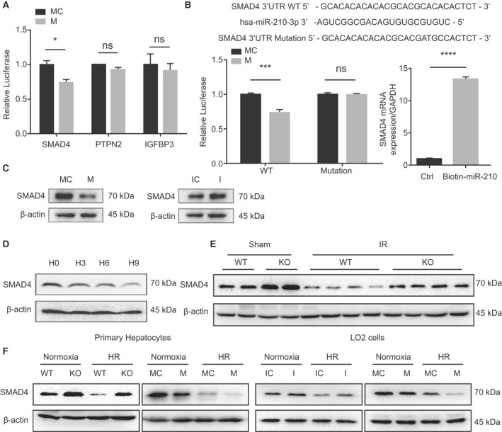
Fig. 4.

miR‐210 targets SMAD4. (A) luciferase analysis of SMAD4, PTPN2, and IGFBP3 3′UTR reporter activities (n = 3, *P < 0.05). (B) Predicted binding site of miR‐210 in the SMAD4 3′UTR and luciferase analysis of SMAD4 3′UTR reporter activities with WT or mutated binding seed regions in LO2 transfected with miR‐210 mimic or mimic control (left, n = 3, ***P < 0.001) and biotinylated miRNA pull‐down assay (right, n = 3, ****P < 0.0001). (C) Western blot analysis of SMAD4 levels in LO2 cells transfected with the indicated oligos under normoxia condition. (D) Western blot analysis of SMAD4 levels in primary hepatocytes with gradient hypoxia. (E) Western blot analysis of SMAD4 in hepatocytes isolated from WT and miR‐210 KO mice liver with indicated stimulation. (F) Western blot analysis of SMAD4 in primary hepatocytes and LO2 cells transfected with indicated oligos under normoxia or HR condition. Abbreviation: Ctrl, control.
