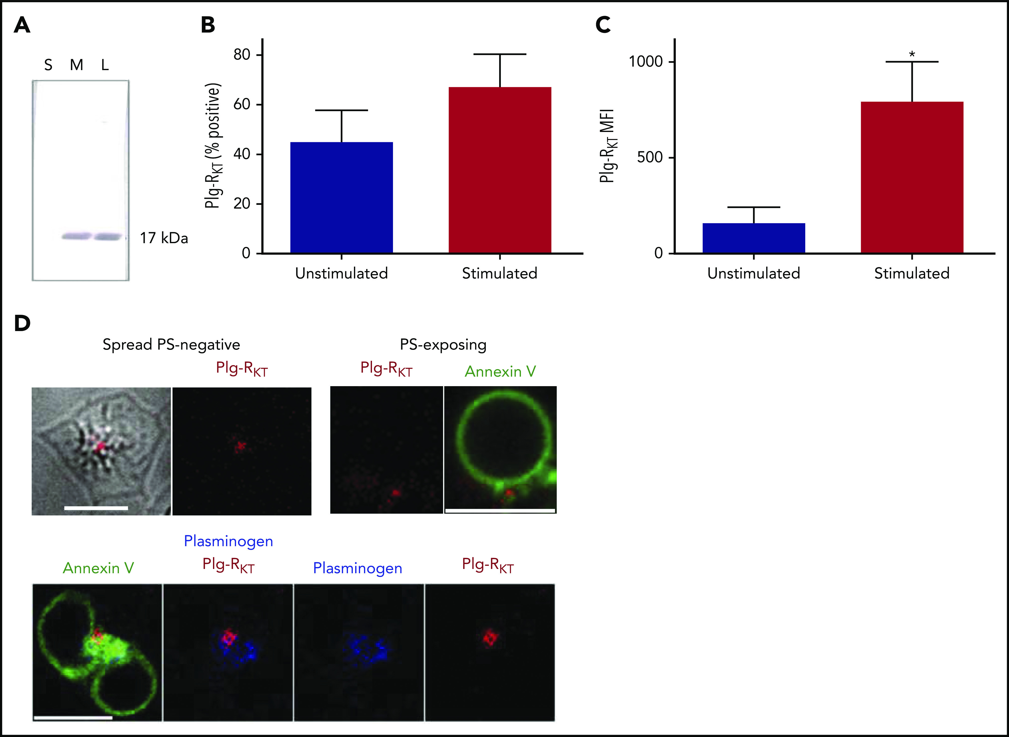
Figure 4.

Platelets express the transmembrane Plg-RKT. (A) Fractions of platelets (1 × 10⁷), lysate (L), soluble (S), and membrane (M) were run on 4% to 12% NuPAGE gels followed by western blotting with anti–Plg-RKT mAb (7H1) fractions. Representative image of n = 4. (B-C) Platelets (2 × 10⁸ platelets/mL) were either unstimulated or stimulated with CVX (100 ng/mL) plus thrombin (100 nM) in the presence of anti–Plg-RKT mAb-DL550. Data are presented as mean ± SEM for (B) percentage platelets positive for Plg-RKT and (C) MFI. *P < .05 vs unstimulated, n = 3. (D) Platelets (0.5 × 10⁸ platelets/mL) stimulated on a collagen (0.6 µg) and thrombin (3 pmol) coated surface for 45 minutes were stained with Annexin V-AF488, anti-plasminogen mAb-DL633, and anti-Plg-RKT mAb-DL550. Scale bars, 5 μm; representative images of n = 4.
