PLOS ONE logoLink to PLOS ONE
. 2021 Jan 22;16(1):e0245369. doi: 10.1371/journal.pone.0245369

Fluorescent detection of PARP activity in unfixed tissue

Soumaya Belhadj 1, Andreas Rentsch 2, Frank Schwede 2, François Paquet-Durand 1,*
Editor: Alfred S Lewin3
PMCID: PMC7822349  PMID: 33481867

Abstract

Poly-ADP-ribose-polymerase (PARP) relates to a family of enzymes that can detect DNA breaks and initiate DNA repair. While this activity is generally seen as promoting cell survival, PARP enzymes are also known to be involved in cell death in numerous pathologies, including in inherited retinal degeneration. This ambiguous role of PARP makes it attractive to have a simple and fast enzyme activity assay, that allows resolving its enzymatic activity in situ, in individual cells, within complex tissues. A previously published two-step PARP activity assay uses biotinylated NAD+ and streptavidin labelling for this purpose. Here, we used the fluorescent NAD+ analogues ε-NAD+ and 6-Fluo-10-NAD+ to assess PARP activity directly on unfixed tissue sections obtained from wild-type and retinal degeneration-1 (rd1) mutant retina. In standard UV microscopy ε-NAD+ incubation did not reveal PARP specific signal. In contrast, 6-Fluo-10-NAD+ resulted in reliable detection of in situ PARP activity in rd1 retina, especially in the degenerating photoreceptor cells. When the 6-Fluo-10-NAD+ based PARP activity assay was performed in the presence of the PARP specific inhibitor olaparib, the activity signal was completely abolished, attesting to the specificity of the assay. The incubation of live organotypic retinal explant cultures with 6-Fluo-10-NAD+, did not produce PARP specific signal, indicating that the fluorescent marker may not be sufficiently membrane-permeable to label living cells. In summary, we present a new, rapid, and simple to use fluorescence assay for the cellular resolution of PARP activity on unfixed tissue, for instance in complex neuronal tissues such as the retina.

Introduction

Poly (ADP-ribose) polymerase (PARP) is a family of enzymes involved in numerous cellular processes [1, 2]. An important function of PARP is to detect DNA damage and to activate the enzymatic machinery involved in the DNA repair process. To this effect, PARP uses NAD+ as a substrate to build poly-ADP ribose polymers (PAR) [3]. While this activity of PARP is generally seen as beneficial and cell-survival promoting, paradoxically, PARP enzymes are also known to be involved in cell death [4]. Notably, a specific type of caspase-independent programmed cell death, called PARthanatos, appears to be triggered by the accumulation of PAR polymers [5, 6]. These polymers may translocate to the mitochondria to cause the release of apoptosis-inducing factor (AIF) and its subsequent translocation to the nucleus, where AIF induces DNA fragmentation. PARthanatos is caspase-independent, as caspases would cleave and inactivate PARP [7]. Since PARP remains intact and active during the process, PAR synthesis is strongly increased, leading to the depletion of NAD+ and ATP, further precipitating cell death [8]. This phenomenon may be relevant in a situation where PARP and its functional antagonist poly-ADP-ribose-glycohydrolase (PARG) [9] are activated simultaneously. While PARP can in principle self-regulate its activity via inhibitory auto-PARylation, the removal of this self-inhibition by PARG allows for continued and unabated activity of PARP.

PARP overactivation associated with AIF release has been shown to be involved in acute neuronal and myocardial cell death following ischemia reperfusion [10]. PARP is also known to have a role in various inflammatory disorders. For example, PARP-1 deficient mice, or mice that have been treated with PARP inhibitors, are resistant to various types of inflammation, including streptozotocin-induced diabetes and lipopolysaccharide-induced sceptic shock [10]. Moreover, in January 2018, olaparib (trade name Lynparza) became the first PARP inhibitor to be approved by the FDA for the treatment of metastatic breast cancer [11]. Altogether, these findings suggest that while PARP is already a target for therapies in oncology, it may also be of interest for therapy development in inflammatory, cardiac, and neurological disorders, including such that affect the neuroretina. In this context it then becomes highly interesting to be able to study PARP activity in an easy and rapid assay that enables the detection in individual cells. Previously, we have adapted and established such an in situ PARP activity assay, using the retina as a model for neurodegeneration [12, 13].

The retina is affected by a group of genetic diseases collectively referred to as inherited retinal degeneration (IRD) and characterized by a progressive degeneration of photoreceptors, leading to vision impairment and ultimately blindness. The pathological mechanisms behind IRD are still unresolved and effective therapy is lacking. The rd1 mouse is to date one of the best characterized animal models for IRD. This animal suffers from a mutation in the beta subunit of the rod photoreceptor Pde6 gene, producing a non-functional phosphodiesterase-6 (PDE6) and leading to an accumulation of cGMP [14]. Various studies [13, 15, 16] showed that inherited photoreceptor cell death involves PARP activity. Indeed, PARP activity, as well as its product PAR, are dramatically increased in the outer nuclear layer (ONL) of rd1 mice at postnatal day (PN) 11, a time-point which corresponds to the onset of cell death in this animal model. Even further, PARP inhibition, in vitro and in vivo, resulted in increased photoreceptor survival [13, 17].

In the present study, we tested three different analogues of the PARP substrate NAD+ to assess and compare their capacity to detect PARP activity in retinal tissue sections derived from wild-type and rd1 mice. While our previously established in situ PARP activity assay required two separate steps for positive detection, the use of a fluorescent NAD+ analogue allowed the detection of PARP activity in a single-step assay, with essentially the same specificity and detection rate, and ease of use in standard microscopic equipment as in the two-step assay. This single-step procedure is faster and easier to perform and would, in principle, also lend itself to in vivo biomarker development.

Materials & methods

Animals

C3H rd1 and wild type (WT) mice were used [14]. Animals were housed under standard white cyclic lighting, had free access to food and water, and were used irrespective of gender. Animal protocols compliant with §4 of the German law of animal protection were reviewed and approved by the Tübingen University committee on animal protection (Einrichtung für Tierschutz, Tierärztlichen Dienst und Labortierkunde; Registration No. AK02/19M).

Histology

For histology, two different types of retinal tissue preparations were collected: 1) Retinal tissue sections fixed with 4% paraformaldehyde (PFA), to be used for PAR staining, and 2) retinal tissue that was directly subjected to cryosectioning without fixation, to be used for PARP activity assay.

Fixed sections (for PAR DAB staining)

Animals were sacrificed in the morning (10–11 am), their eyes enucleated and fixed in PFA in 0.1 M phosphate buffer (pH 7.4) for 45 min at 4°C. After fixation, tissues were washed for 10 min in PBS. For cryoprotection, they were incubated in 10% sucrose solution for 10 min, 20% sucrose solution for 20 min, and 30% sucrose solution for at least 30 min. The retinas were frozen in Tissue-Tek O.C.T. compound (Sakura Finetek Europe, Alphen aan den Rijn, Netherlands)-filled boxes. 12 μm tissue sections were prepared on a Thermo Scientific NX50 microtome (Thermo Scientific, Waltham, MA), thaw-mounted onto Superfrost Plus object slides (R. Langenbrinck, Emmendingen, Germany). Tissue sections were stored at -20°C.

Unfixed sections (for PARP in situ activity assay)

Animals were sacrificed in the morning (10-11am), their eyes enucleated and snap frozen on liquid nitrogen. Then, the eyes were embedded in Tissue-Tek (Sakura Finetek). Sagittal 12 μm sections were obtained as above and stored at -20°C.

PARP in situ activity assays

For the detection of PARP activity in situ, we used unfixed sections. For the PARP activity assay using biotinylated NAD+, the reaction mixture (10 mM MgCl2, 1 mM dithiothreitol, 5 μm 6-Biotin-17-NAD+ (Biolog, Bremen, Germany, Cat. Nr.: N 012) in 100 mM Tris buffer with 0.2% Triton X100, pH 8.0) was applied to the sections for 2 h 30 min at 37°C. After three 5 min washes in PBS, incorporated biotin was detected by avidin-Alexa fluor488 conjugate (Invitrogen, Carlsbad, CA, dilution 1:800 in PBS, incubation 1 h at room temperature). After three 5 min washes in phosphate buffered saline (PBS), the sections were mounted in Vectashield (Vector, Burlingame, CA). For controls, the biotinylated-NAD+ was omitted from the reaction mixture, resulting in absence of detectable reaction product.

For the PARP activity assay using fluorescent NAD+, the reaction mixture (10 mM MgCl2, 1 mM dithiothreitol, 50 μM 6-Fluo-10-NAD+ (Biolog, Cat. Nr.: N 023) or 50 μM ε-NAD+ (Biolog, Cat. Nr.: N 024), in 100 mM Tris buffer with 0.2% Triton X100, pH 8.0) was applied to the sections for 2 h 30 min at 37°C. After three 5 min washes in PBS the sections were mounted in Vectashield (Vector). For controls, the fluorescent-NAD+ was omitted from the reaction mixture, resulting in absence of detectable reaction product.

PAR DAB staining

For the detection of PAR DAB staining, we used fixed sections. 3,3′-diaminobenzidine (DAB) staining commenced with quenching of endogenous peroxidase activity using 40% MeOH and 10% H2O2 in PBS with 0.3% Triton X-100 (PBST) and 20 min incubation. The sections were further incubated with 10% normal goat serum (NGS) in PBST for 30 min followed by anti-PAR antibody (1:200; Enzo Life Sciences, Farmingdale, New-York) incubation overnight, at 4°C. Incubation with the biotinylated secondary antibody (1:150, Vector; in 5% NGS in PBST) for 1 h was followed by application of Vector ABC-Kit (Vector Laboratories, solution A and solution B in PBS, 1:150 each) for 1 h. DAB staining solution (0.05 mg/ml NH4Cl, 200 mg/ml glucose, 0.8 mg/ml nickel ammonium sulphate, 1 mg/ml DAB, 0.1 vol. % glucose oxidase in phosphate buffer) was applied evenly, incubated for exactly 2 min and immediately rinsed with phosphate buffer to stop the reaction. The sections were mounted in Aquatex (Merck, Darmstadt, Germany).

Inhibition of PARP activity using olaparib

The tissue sections were pre-incubated with olaparib (Biomol, Hamburg, Germany, 100 nM in PARP reaction buffer) for 30 min. The PARP activity assay was then conducted as described above with olaparib added to the reaction mixture at a concentration of 100 nM. Olaparib was omitted from the positive control.

Testing fluorescent NAD+ on live retina

At post-natal (P) day 5 rd1 animals were killed and the eyes rapidly enucleated in an aseptic environment. The entire eyes were incubated in R16 serum-free culture medium (Gibco, Carlsbad, CA), with 0.12% proteinase K (MP Biomedicals, Illkirch-Grafenstaden, France), at 37°C for 15 min, to allow preparation of retinal cultures with retinal pigment epithelium (RPE) attached. The proteinase K was inactivated with 10% FCS (Gibco) in R16 medium, and thereafter the eyes were dissected aseptically in a Petri dish containing fresh R16 medium. The anterior segment, lens, vitreous, sclera, and choroid were carefully removed by fine scissors, and the retina was cut perpendicular to its edges, giving a cloverleaf-like shape. Subsequently, the retina was transferred to a culture dish filter insert (COSTAR, NY) with the RPE layer facing the membrane. The insert was put into a six-well culture plate and incubated in R16 medium with supplements [18] at 37°C. The full volume of nutrient medium, 1 ml per dish, was replaced with fresh R16 medium every second day.

At P11, the cultures were incubated for 24 h with 6-Fluo-10-NAD+ at a concentration of 100 μM. At P12, the cultures were stopped by 45 min fixation in 4% PFA. This was followed by graded sucrose cryoprotection, embedding, and collecting of 12-μm-thick retinal cross-sections on a Thermo Scientific NX50 cryotome.

Microscopy, cell counting, and statistical analysis

Light and fluorescence microscopy were performed at room temperature on an Axio Imager Z.1 ApoTome Microscope, equipped with a Zeiss Axiocam MRm digital camera (for more details about the channels used and fluorescence properties of the probes see Table 1). Images were captured using the Zeiss Zen software; representative pictures were taken from central areas of the retina using a 20x / 0.8 Zeiss Plan-APOCHROMAT objective. For quantifications, pictures were captured on three entire sagittal sections from at least three different animals. The average area occupied by a photoreceptor cell (i.e. cell size) was determined by counting 4′,6-diamidino-2-phenylindole (DAPI) stained nuclei in nine different areas of the retina. The total number of photoreceptor cells was estimated by dividing the outer nuclear layer (ONL) area by this average cell size. The number of positively labelled cells in the ONL was counted manually. Errors in graphs and text are given as standard deviation (STD).

Table 1. Fluorescence of NAD+ analogues and microscope filters.

Excitation (exc.) and emission (em.) characteristics of the two fluorescent probes and of the microscope filter sets used to visualize them.

NAD+ analogue exc. max. / em. max. (nm) microscope chan. exc. filter / em. filter (nm)
6-Fluo-10-NAD+ 494 / 517 AF488 450–490 /500-550
ε-NAD+ 300/410 DAPI 335–383 /420-470

For statistical comparisons, the unpaired Student t-test (PAR staining) and a two-way ANOVA with multiple comparison analysis (PARP activity assay) as implemented in Prism 8 for Windows (GraphPad Software, San Diego, CA) were employed. Adobe Photoshop (CS5Adobe Systems Incorporated, San Jose, CA) was used for image processing and figure preparation.

Results

Using NAD+ analogues to probe for in situ PARP activity

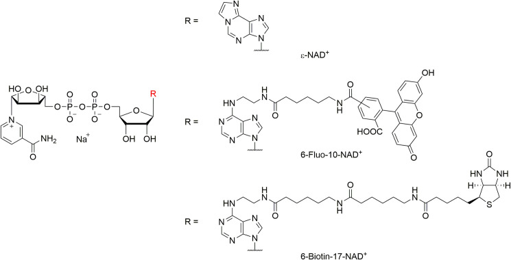
To detect the activity of PARP in unfixed tissue sections, we used three different analogues of NAD+ that were differently substituted at the N6 position of the molecule (Fig 1). Of these three analogues ε-NAD+ and 6-Fluo-10-NAD+ exhibited fluorescence properties (cf. Table 1), while the biotinylated 6-Biotin-17-NAD+ was not by itself fluorescent.

Fig 1. Molecular structures of NAD+-analogues used in this study.

Fig 1

The fluorescent properties of the NAD+ analogues employed here are established via the additional etheno-function bridging the N6- and 1-position (ε-NAD+) or via the fluorescent dye fluorescein attached via a spacer of 10 bond lengths at the N6-position (6-Fluo-10-NAD+). 6-Biotin-17-NAD+ is not fluorescent itself, but features a biotin moiety, which is also attached to the N6-position (via a spacer of 17 bond lengths) and can be addressed by a fluorescent dye such as avidin-Alexa fluor488.

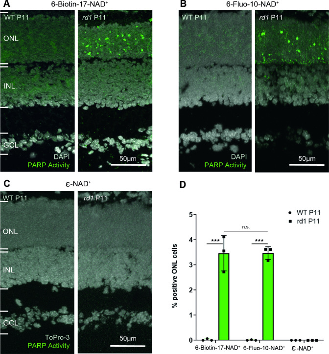
The current assay to evaluate PARP activity ex vivo on tissue sections is using biotinylated NAD+, which in a second step is recognized by fluorescently labelled streptavidin [12]. When this two-step assay was performed on rd1 P11 retinal sections, a large number of positive cells was seen in the ONL, while essentially no positive cells were seen in the WT condition at the same age (Fig 2A).

Fig 2. PARP activity detection with three different NAD+ analogues, in wild-type and rd1 retina.

Fig 2

Retinal tissue sections derived from either wild-type (WT) or rd1 animals, at post-natal (P) day 11, were incubated with different NAD+ analogues. (A) In the two-step assay, employing 6-Biotin-17-NAD+, PARP activity positive cells were rarely seen in the WT retina but readily detected in the rd1 outer nuclear layer (ONL). (B) The single-step assay with 6-Fluo-10-NAD+ detected similar numbers of PARP activity positive cells in the rd1 ONL. (C) No PARP activity was observable with the assay employing ε-NAD+ and a standard UV band-pass filter intended for DAPI visualization. (D) Quantification of PARP activity positive cells in WT and rd1 ONL in assays using the three different NAD+ analogues. DAPI was used as nuclear counterstaining in A and B; To-Pro-3 was used in C. PARP assays performed on tissue sections derived from at least three different WT and rd1 animals (n = 3); error bars indicate STD; statistical analysis: two-way ANOVA with multiple comparison. INL = inner nuclear layer, GCL = ganglion cell layer.

The aim of the present study was to develop a faster and easier to use single-step PARP activity assay, that might potentially be suitable also for in vivo application. The two-step assay would indeed not be applicable in vivo. To this end, the two fluorescent analogues of NAD+ were tested on ex vivo retinal tissue for their capacity to reliably detect PARP activity using standard microscopic equipment.

When 6-Fluo-10-NAD+ was used as a substrate for PARP, a high amount of PARP positive cells was observed in the ONL of rd1 P11 sections. Some signal was also seen at the lower border of the ONL, close to the outer plexiform layer (OPL) (Fig 2B). In contrast, ε-NAD+ did not reveal any PARP specific signal, neither in rd1 tissue sections, nor in WT ones, when visualized with a standard UV band-pass filter for DAPI (Fig 2C, cf. Table 1).

When the single-step 6-Fluo-10-NAD+ based assay was compared to the two-step 6-Biotin-17-NAD+ assay, no significant difference was seen in the rate of PARP activity detection. In the rd1 mouse model ONL at P11, both probes detected on average 3.47 ± 0.25 STD and 3.46 ± 0.70 STD PARP activity positive cells, respectively (Fig 2D).

To validate the results obtained for PARP activity with an independent method, we used an immunostaining for PAR to detect the cellular products of PARP activity and to thereby assess PARP activity indirectly. While some positive cells were observed in the ONL of the rd1 mouse model at P11 (Fig 3B), none were seen in the WT condition at the same age (Fig 3A). In average, 0.99 ± 0.08 STD PAR positive cells were detected in the ONL of the rd1 mouse model at P11 (Fig 3C).

Fig 3. Immunostaining for poly-ADP-ribosylation in wild-type and rd1 retina.
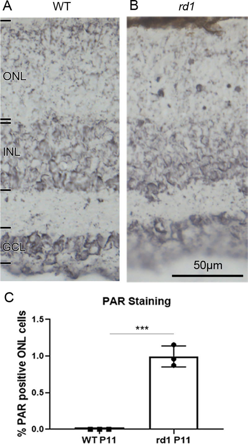
Fig 3

To indirectly assess PARP activity an immunostaining for poly-ADP-ribosylation (PAR) was employed. While post-natal (P) day 11 wild-type (WT) outer nuclear layer (ONL) is essentially devoid of nuclear PAR staining (A), in the rd1 situation (B) numerous photoreceptor nuclei show a marked PAR label. (C) Quantification of PAR positive nuclei in the ONL, with n = 3. error bars indicate STD; statistical analysis: Student´s t-test. INL = inner nuclear layer, GCL = ganglion cell layer. Images representative for PAR immunostaining performed on tissue sections derived from at least three different WT and rd1 animals.

Inhibition of PARP activity: Effect on detection rate

To assess the specificity of the newly set up activity assay, the PARP inhibitor olaparib was used. Olaparib is inhibiting PARP1, PARP2, and PARP3 and is used as a therapy for cancer in people with hereditary BRCA1 or BRCA2 mutations, which include some ovarian, breast and prostate cancers [19]. This inhibitor has multiple modes of action. First, it competes with the binding of NAD+, thus inhibiting PARylation. Additionally, olaparib traps PARP1 and PARP2 on the DNA strand, therefore interfering with DNA damage repair [11]. It has been reported that the IC50 of olaparib for PARP1 is 5 nM, while it is 1 nM for PARP2 [20].

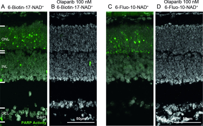
In the positive control condition, for which the PARP activity assay was conducted normally on rd1 P11 retinal sections, a high amount of PARP positive cells was observed in the ONL (Fig 4A and 4C). This signal was extinguished when the activity assay was carried out on retinal sections from the same animal model, at the same age, with olaparib present in the reaction mixture at a concentration of 100 nM (Fig 4B and 4D). These results suggest that both activity assays are specific of PARP activity.

Fig 4. The PARP inhibitor olaparib abolishes PARP activity signals.

Fig 4

Retinal sections derived from post-natal (P) day 11 rd1 animals were incubated with NAD+ analogues, together with the selective PARP inhibitor olaparib. In both the two-step assay with 6-Biotin-17-NAD+ (A, B) and the single-step assay using 6-Fluo-10-NAD+ (C, D), olaparib, at 100 nM, completely abolished positive PARP activity detection. DAPI was used as nuclear counterstaining. ONL = outer nuclear layer, INL = inner nuclear layer, GCL = ganglion cell layer. Images representative for assays performed on tissue sections derived for at least three different rd1 animals per assay condition.

Testing NAD+ analogues on live retina in vitro

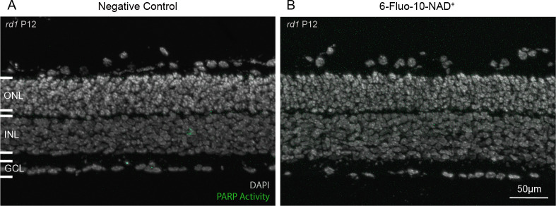
To assess whether 6-Fluo-10-NAD+ could be used to assess PARP activity in live tissue, with intact cell membranes, organotypic retinal explant cultures [2123] were treated with this probe at a concentration of 100 μM for 24h. No significant difference was observed between the negative control condition (not treated with 6-Fluo-10-NAD+) and the treated condition, and no PARP specific signal could be seen (Fig 5A and 5B). These negative results indicate that 6-Fluo-10-NAD+ may not be able to penetrate the membranes of living cells.

Fig 5. The 6-Fluo-10-NAD+ analogue does not allow live tissue detection of PARP activity.

Fig 5

Live, organotypic retinal explant cultures were incubated with 100 μM 6-Fluo-10-NAD+ from P11 to P12 (24h). No fluorescent signal was observed within the retina, indicating that the NAD+ analogue may not have penetrated live cells. DAPI was used as nuclear counterstaining. ONL = outer nuclear layer, INL = inner nuclear layer, GCL = ganglion cell layer. Images representative for results obtained from 5 independent retinal explant cultures.

Discussion

The detection and quantification of cellular PARP activity is highly desirable in the context of many diseases, including in neurodegenerative diseases of the retina. Here, we identified a fluorescent analogue of NAD+ that enables the rapid in situ detection of PARP activity in a simple, single-step assay procedure. This assay could potentially facilitate the development of drugs targeting PARP, and, because of PARPs connection to cell death, could be used to improve our understanding of cell death mechanisms. Moreover, PARP activity detection could also serve as a surrogate biomarker for the study and diagnosis of degenerative diseases.

Methods for the detection of PARP activity

PARP activity can be assessed by a number of commercially available assay kits, which detect PARP activity in tissue lysates or cell cultures [24]. The drawback of these assays is that they do not offer cellular resolution, something that is highly desirable in the context of studies into specific disease pathomechanisms, for instance. Another way to assess PARP activity in whole tissue lysates is to perform an activity blot. Using whole tissue samples, the technique combines western blotting followed by a protein renaturation and activity assay. A labelling will be seen in protein bands with molecular weights corresponding to PARP enzymes [25]. While this technique may allow attributing activity to specific protein bands or isoforms, again, the drawback is that it does not offer cellular resolution. PARP activity can also be assessed indirectly at the cellular level by immunohistology with antibodies against PAR. The staining can be performed on cell cultures, sectioned tissue, or after western blotting [13]. The disadvantage of this method is that PARP activity and PAR product accumulation may not necessarily correspond. PAR may, for instance, be hydrolyzed secondarily by the specific glycohydrolase PARG, potentially reducing detection sensitivity.

Using NAD+ analogues for the in situ detection of PARP activity

The above-mentioned methods do not allow for the detection of PARP activity within individual cells. This, however, is highly relevant in complex tissues, such as when they occur in the central nervous system where only some of the many different cell types may be affected by a disease condition or where only a specific cell type may be amenable to PARP inhibition treatment. As mentioned previously, PARP inhibition is a therapeutic strategy for some cancers [11]. To help the diagnosis with PARP inhibitor therapy, Shuhendler et al. developed a novel NAD+ analogue radioactive probe ([18F]-SuPAR) for noninvasive imaging of PARP activity using positron emission tomography. The probe was tested in breast and cervical cancer xenograft mouse models [26]. Recently, Wigle et al. developed a set of NAD+ competitors using FRET/BRET for in vitro and cellular high-throughput biophysical assays to study PARP activity and inhibition [27]. In this study, to assess PARP activity in situ, a new single-step assay, based on the use of the fluorescent NAD+ analogue 6-Fluo-10-NAD+, was set up. This new assay had a similar detection rate compared to the established two-step assay using biotinylated NAD+ (6-Biotin-17-NAD+). However, the sensitivity appeared to be higher for the two-step activity assay using biotinylated NAD+, since the concentration of 6-Fluo-10-NAD+ (50 μM) used to perform the assay was ten times higher than the one of 6-Biotin-17-NAD+ (5 μM). This can be explained by the specific amplification of the signal provided by the fluorescently labelled avidin used in the second detection step.

When the activity assay was performed with ε-NAD, and with the limitations of standard microscopic equipment and filter sets, no PARP specific signal could be observed. This is, to some extent, in contrast to a previous study, which reported the use of ε-NAD+ to assess PARP activity in cultured cells lines and rat brain slices [28]. However, the NAD+ analogue concentration used by Davis et al. was 400 μM (8X compared to the concentration used here) and even then, the PARP-specific signal was detectable only after incubation with an anti-ethenoadenosine antibody as a second step, to detect ε-NAD+ containing PAR polymers. Combined with the fact that 300 nm UV excitation is required, these limitations make ε-NAD+ appear poorly suited for in situ detection assays, especially so if live cell detection was required, as the high energy UV irradiation would like result in significant cell damage.

Specificity of in situ PARP activity detection

NAD+ is very widely used by enzymes as a cofactor or substrate. This fact may raise questions about the specificity of the assay as the fluorescence observed could originate from the activity of other enzymes as well. To address this potential issue, we used PAR immunostaining as an indirect validation of the activity assay. While the PARP activity assay detected approximately 3.5% positive cells on average, in the rd1 mouse model at postnatal day 11, the PAR immunostaining detected around 1% positive cells on average in the same model, at the same age. This corresponds very well with previous observations on the relation of PARP activity positive to PAR positive cells [13]. Nevertheless, this difference between PARP activity and PAR detection might suggest that some cells detected as PARP positive were, in fact, positive for another enzyme that used NAD+. This could, for instance, be the case for the signal observed at the ONL to OPL border when the assay was performed with 6-Fluo-10-NAD+. This OPL signal was not revealed by the PAR immunostaining. Several reasons could explain this discrepancy: 1) the PAR polymers produced in the photoreceptor synapses may be not long enough to be detected by the PAR antibody. 2) the activity of an enzyme hydrolyzing PAR polymers in the synapse (such as PARG) may degrade the polymers at this location only. 3) the PAR antibody may not reach into the synapse as well as in the nucleus or the cytoplasm. 4) the signal may have been produced by an isoform of PARP present in the synapses only. On the other hand, when the single-step assay using 6-Fluo-10-NAD+ was performed in the presence of PARP inhibitor olaparib, the staining produced in both photoreceptor nuclei and along the OPL disappeared. Altogether, these findings strongly suggest that the staining produced by 6-Fluo-10-NAD+ incubation was indeed specific for the activity of PARP.

Tissue penetration and permeability of 6-Fluo-10-NAD+

Once the assay and its specificity for PARP was established, we wanted to see whether the single-step PARP activity detection could also be applied to live cells and tissues, and potentially even in vivo. In the retina such a method could potentially enable a direct, non-invasive detection of PARP activity in vivo, with single-cell resolution, using techniques such adaptive optics scanning laser ophthalmoscopy (AO-SLO) [2931].

As opposed to what we had observed in unfixed retinal tissue sections, in living organotypic retinal explant cultures incubation 6-Fluo-10-NAD+ did not reveal any PARP specific signal. This could indicate that the probe was either not able to penetrate into the tissue or could not reach across the intact cell membranes of live cells. Such a lack of permeability may be explained by the negative net charge of the 6-Fluo-10-NAD+ molecule (Fig 1). It is likely that this property limits any in vivo application of the fluorescent probe. A possible solution might be to change the molecule in way that would reduce its net charge. This, however, could also affect its capacity to serve as substrate for PARP. An alternative solution might be to encapsulate the molecule in a delivery system such as liposomes or nanoparticles [32] that would release the molecule only intracellularly. In summary, while the 6-Fluo-10-NAD+ compound appears well suited for PARP activity detection on unfixed ex vivo tissue preparations, it cannot readily be used for such purpose in live tissue.

Conclusion

In this study, we set up a new, simple to perform, one-step assay to assess PARP activity on unfixed tissue, using the NAD+ analogue 6-Fluo-10-NAD+. The apparent sensitivity of this single-step assay, as determined by the positive cell detection rate, is similar to that of the previously established two-step assay that used 6-Biotin-17-NAD+. The specificity of the new PARP activity assay was confirmed on the one hand by comparison with PAR immunostaining and on the other hand by inhibition of PARP activity with olaparib. Altogether, the new PARP activity assay is well suited for ex vivo applications on unfixed, native tissue. Moreover, further development and combination with a suitable delivery system could potentially enable future use as an in vivo biomarker for PARP activity [33, 34].

Acknowledgments

We thank Norman Rieger for excellent technical assistance and Mathias Seeliger, Timm Schubert, Valeria Marigo, Per Ekström, and John Groten for helpful discussions. We thank Ayse Sahaboglu for kindly providing olaparib, Yiyi Chen for help with figure making, and Torsten Strasser for help with statistical analysis.

Data Availability

All relevant data are within the manuscript.

Funding Statement

This research was funded by grants from the European Union (transMed; H2020-MSCA-765441), the Charlotte and Tistou Kerstan Foundation, and the German research council (DFG; PA1751/8-1, 10-1). The funders provided support in the form of salaries for authors SB and FPD, but did not have any additional role in the study design, data collection and analysis, decision to publish, or preparation of the manuscript. Biolog Life Science Institute GmbH & Co. KG provided support in the form of salary for authors AR and FS, but had no role in study design, data collection and analysis, decision to publish, or preparation of the manuscript. The specific roles of these authors are articulated in the ‘author contributions’ section.

References

  • 1.Schreiber V, Dantzer F, Ame JC, de Murcia G. Poly(ADP-ribose): novel functions for an old molecule. Nat Rev Mol Cell Biol. 2006;7(7):517–28. 10.1038/nrm1963 [DOI] [PubMed] [Google Scholar]
  • 2.Vyas S, Chesarone-Cataldo M, Todorova T, Huang YH, Chang P. A systematic analysis of the PARP protein family identifies new functions critical for cell physiology. Nat Commun. 2013;4:2240 10.1038/ncomms3240 [DOI] [PMC free article] [PubMed] [Google Scholar]
  • 3.Pascal JM. The comings and goings of PARP-1 in response to DNA damage. DNA Repair (Amst). 2018;71:177–82. 10.1016/j.dnarep.2018.08.022 [DOI] [PMC free article] [PubMed] [Google Scholar]
  • 4.Wang Y, Dawson VL, Dawson TM. Poly(ADP-ribose) signals to mitochondrial AIF: a key event in parthanatos. Exp Neurol. 2009;218(2):193–202. 10.1016/j.expneurol.2009.03.020 [DOI] [PMC free article] [PubMed] [Google Scholar]
  • 5.Andrabi SA, Kim NS, Yu SW, Wang H, Koh DW, Sasaki M, et al. Poly(ADP-ribose) (PAR) polymer is a death signal. Proc Natl Acad Sci U S A. 2006;103(48):18308–13. 10.1073/pnas.0606526103 [DOI] [PMC free article] [PubMed] [Google Scholar]
  • 6.Yu SW, Wang H, Poitras MF, Coombs C, Bowers WJ, Federoff HJ, et al. Mediation of poly(ADP-ribose) polymerase-1-dependent cell death by apoptosis-inducing factor. Science. 2002;297(5579):259–63. 10.1126/science.1072221 [DOI] [PubMed] [Google Scholar]
  • 7.Soldani C, Scovassi AI. Poly(ADP-ribose) polymerase-1 cleavage during apoptosis: an update. Apoptosis. 2002;7(4):321–8. 10.1023/a:1016119328968 [DOI] [PubMed] [Google Scholar]
  • 8.David KK, Andrabi SA, Dawson TM, Dawson VL. Parthanatos, a messenger of death. Front Biosci (Landmark Ed). 2009;14:1116–28. 10.2741/3297 [DOI] [PMC free article] [PubMed] [Google Scholar]
  • 9.Sahaboglu A, Bolz S, Lowenheim H, Paquet-Durand F. Expression of poly(ADP-ribose) glycohydrolase in wild-type and PARG-110 knock-out retina. Adv Exp Med Biol. 2014;801:463–9. 10.1007/978-1-4614-3209-8_59 [DOI] [PubMed] [Google Scholar]
  • 10.Jagtap P, Szabo C. Poly(ADP-ribose) polymerase and the therapeutic effects of its inhibitors. Nat Rev Drug Discov. 2005;4(5):421–40. 10.1038/nrd1718 [DOI] [PubMed] [Google Scholar]
  • 11.Bixel K, Hays JL. Olaparib in the management of ovarian cancer. Pharmgenomics Pers Med. 2015;8:127–35. 10.2147/PGPM.S62809 [DOI] [PMC free article] [PubMed] [Google Scholar]
  • 12.Ekstrom PA, Ueffing M, Zrenner E, Paquet-Durand F. Novel in situ activity assays for the quantitative molecular analysis of neurodegenerative processes in the retina. Curr Med Chem. 2014;21(30):3478–93. 10.2174/0929867321666140601201337 [DOI] [PubMed] [Google Scholar]
  • 13.Paquet-Durand F, Silva J, Talukdar T, Johnson LE, Azadi S, van Veen T, et al. Excessive activation of poly(ADP-ribose) polymerase contributes to inherited photoreceptor degeneration in the retinal degeneration 1 mouse. J Neurosci. 2007;27(38):10311–9. 10.1523/JNEUROSCI.1514-07.2007 [DOI] [PMC free article] [PubMed] [Google Scholar]
  • 14.Keeler CE. The Inheritance of a Retinal Abnormality in White Mice. Proc Natl Acad Sci U S A. 1924;10(7):329–33. 10.1073/pnas.10.7.329 [DOI] [PMC free article] [PubMed] [Google Scholar]
  • 15.Arango-Gonzalez B, Trifunovic D, Sahaboglu A, Kranz K, Michalakis S, Farinelli P, et al. Identification of a common non-apoptotic cell death mechanism in hereditary retinal degeneration. PLoS One. 2014;9(11):e112142 10.1371/journal.pone.0112142 [DOI] [PMC free article] [PubMed] [Google Scholar]
  • 16.Kaur J, Mencl S, Sahaboglu A, Farinelli P, van Veen T, Zrenner E, et al. Calpain and PARP activation during photoreceptor cell death in P23H and S334ter rhodopsin mutant rats. PLoS One. 2011;6(7):e22181 10.1371/journal.pone.0022181 [DOI] [PMC free article] [PubMed] [Google Scholar]
  • 17.Sahaboglu A, Barth M, Secer E, Amo EM, Urtti A, Arsenijevic Y, et al. Olaparib significantly delays photoreceptor loss in a model for hereditary retinal degeneration. Sci Rep. 2016;6:39537 10.1038/srep39537 [DOI] [PMC free article] [PubMed] [Google Scholar]
  • 18.Romijn HJ. Development and advantages of serum-free, chemically defined nutrient media for culturing of nerve tissue. Biology of the Cell. 1988;63(3):263–8. 10.1016/0248-4900(88)90116-5 [DOI] [PubMed] [Google Scholar]
  • 19.Fong PC, Boss DS, Yap TA, Tutt A, Wu P, Mergui-Roelvink M, et al. Inhibition of poly(ADP-ribose) polymerase in tumors from BRCA mutation carriers. N Engl J Med. 2009;361(2):123–34. 10.1056/NEJMoa0900212 [DOI] [PubMed] [Google Scholar]
  • 20.Menear KA, Adcock C, Boulter R, Cockcroft XL, Copsey L, Cranston A, et al. 4-[3-(4-cyclopropanecarbonylpiperazine-1-carbonyl)-4-fluorobenzyl]-2H-phthalazin- 1-one: a novel bioavailable inhibitor of poly(ADP-ribose) polymerase-1. J Med Chem. 2008;51(20):6581–91. 10.1021/jm8001263 [DOI] [PubMed] [Google Scholar]
  • 21.Caffe AR, Soderpalm AK, Holmqvist I, van Veen T. A combination of CNTF and BDNF rescues rd photoreceptors but changes rod differentiation in the presence of RPE in retinal explants. Invest Ophthalmol Vis Sci. 2001;42(1):275–82. [PubMed] [Google Scholar]
  • 22.Vighi E, Trifunović D, Veiga-Crespo P, Rentsch A, Hoffmann D, Sahaboglu A, et al. Combination of cGMP analogue and drug delivery system provides functional protection in hereditary retinal degeneration. Proc Natl Acad Sci U S A. 2018;115(13):E2997–e3006. 10.1073/pnas.1718792115 [DOI] [PMC free article] [PubMed] [Google Scholar]
  • 23.Belhadj S, Tolone A, Christensen G, Das S, Chen Y, Paquet-Durand F. Long-Term, Serum-Free Cultivation of Organotypic Mouse Retina Explants with Intact Retinal Pigment Epithelium. J Vis Exp. 2020(165). 10.3791/61868 [DOI] [PubMed] [Google Scholar]
  • 24.Yuan Z, Chen S, Sun Q, Wang N, Li D, Miao S, et al. Olaparib hydroxamic acid derivatives as dual PARP and HDAC inhibitors for cancer therapy. Bioorg Med Chem. 2017;25(15):4100–9. 10.1016/j.bmc.2017.05.058 [DOI] [PubMed] [Google Scholar]
  • 25.Desnoyers S, Shah GM, Brochu G, Poirier GG. Erasable blot of poly(ADP-ribose) polymerase. Anal Biochem. 1994;218(2):470–3. 10.1006/abio.1994.1212 [DOI] [PubMed] [Google Scholar]
  • 26.Shuhendler AJ, Cui L, Chen Z, Shen B, Chen M, James ML, et al. [(18)F]-SuPAR: A Radiofluorinated Probe for Noninvasive Imaging of DNA Damage-Dependent Poly(ADP-ribose) Polymerase Activity. Bioconjug Chem. 2019;30(5):1331–42. 10.1021/acs.bioconjchem.9b00089 [DOI] [PubMed] [Google Scholar]
  • 27.Wigle TJ, Blackwell DJ, Schenkel LB, Ren Y, Church WD, Desai HJ, et al. In Vitro and Cellular Probes to Study PARP Enzyme Target Engagement. Cell Chem Biol. 2020;27(7):877–87.e14. 10.1016/j.chembiol.2020.06.009 [DOI] [PubMed] [Google Scholar]
  • 28.Davis RE, Mysore V, Browning JC, Hsieh JC, Lu QA, Katsikis PD. In situ staining for poly(ADP-ribose) polymerase activity using an NAD analogue. J Histochem Cytochem. 1998;46(11):1279–89. 10.1177/002215549804601108 [DOI] [PubMed] [Google Scholar]
  • 29.Cordeiro MF, Guo L, Coxon KM, Duggan J, Nizari S, Normando EM, et al. Imaging multiple phases of neurodegeneration: a novel approach to assessing cell death in vivo. Cell Death Dis. 2010;1:e3 10.1038/cddis.2009.3 [DOI] [PMC free article] [PubMed] [Google Scholar]
  • 30.Cordeiro MF, Normando EM, Cardoso MJ, Miodragovic S, Jeylani S, Davis BM, et al. Real-time imaging of single neuronal cell apoptosis in patients with glaucoma. Brain. 2017;140(6):1757–67. 10.1093/brain/awx088 [DOI] [PMC free article] [PubMed] [Google Scholar]
  • 31.Sharma R, Schwarz C, Hunter JJ, Palczewska G, Palczewski K, Williams DR. Formation and Clearance of All-Trans-Retinol in Rods Investigated in the Living Primate Eye With Two-Photon Ophthalmoscopy. Invest Ophthalmol Vis Sci. 2017;58(1):604–13. 10.1167/iovs.16-20061 [DOI] [PMC free article] [PubMed] [Google Scholar]
  • 32.Himawan E, Ekstrom P, Buzgo M, Gaillard P, Stefansson E, Marigo V, et al. Drug delivery to retinal photoreceptors. Drug Discov Today. 2019;24(8):1637–43. 10.1016/j.drudis.2019.03.004 [DOI] [PMC free article] [PubMed] [Google Scholar]
  • 33.Power M, Das S, Schutze K, Marigo V, Ekstrom P, Paquet-Durand F. Cellular mechanisms of hereditary photoreceptor degeneration—Focus on cGMP. Prog Retin Eye Res. 2020;74:100772 10.1016/j.preteyeres.2019.07.005 [DOI] [PubMed] [Google Scholar]
  • 34.Tolone A, Belhadj S, Rentsch A, Schwede F, Paquet-Durand F. The cGMP Pathway and Inherited Photoreceptor Degeneration: Targets, Compounds, and Biomarkers. Genes (Basel). 2019;10(6). [DOI] [PMC free article] [PubMed] [Google Scholar]

Decision Letter 0

Alfred S Lewin

15 Jul 2020

PONE-D-20-16995

Fluorescent detection of PARP activity in unfixed tissue

PLOS ONE

Dear Dr. Belhadj,

Thank you for submitting your manuscript to PLOS ONE. After careful consideration, we feel that it has merit but does not fully meet PLOS ONE’s publication criteria as it currently stands. Therefore, we invite you to submit a revised version of the manuscript that addresses the points raised during the review process.

You will see that there is disagreement between the expert reviewers concerning the merit of this submission. At a minimum, you should fully cite and acknowledge alternative methods for labeling PARP activity in tissue, and immunocytochemistry to illustrate PAR staining should be performed on the same sections as those stained with 6-Fluo-10-NAD. Because there is an apparent conflict of interest involving two of the authors, the novelty  and relative value of your method should be explained.

Please submit your revised manuscript by Aug 29 2020 11:59PM. If you will need more time than this to complete your revisions, please reply to this message or contact the journal office at plosone@plos.org. When you're ready to submit your revision, log on to https://www.editorialmanager.com/pone/ and select the 'Submissions Needing Revision' folder to locate your manuscript file.

Please include the following items when submitting your revised manuscript:

  • A rebuttal letter that responds to each point raised by the academic editor and reviewer(s). You should upload this letter as a separate file labeled 'Response to Reviewers'.

  • A marked-up copy of your manuscript that highlights changes made to the original version. You should upload this as a separate file labeled 'Revised Manuscript with Track Changes'.

  • An unmarked version of your revised paper without tracked changes. You should upload this as a separate file labeled 'Manuscript'.

If you would like to make changes to your financial disclosure, please include your updated statement in your cover letter. Guidelines for resubmitting your figure files are available below the reviewer comments at the end of this letter.

If applicable, we recommend that you deposit your laboratory protocols in protocols.io to enhance the reproducibility of your results. Protocols.io assigns your protocol its own identifier (DOI) so that it can be cited independently in the future. For instructions see: http://journals.plos.org/plosone/s/submission-guidelines#loc-laboratory-protocols

We look forward to receiving your revised manuscript.

Kind regards,

Alfred S Lewin, Ph.D.

Academic Editor

PLOS ONE

Journal Requirements:

When submitting your revision, we need you to address these additional requirements.

1. Please ensure that your manuscript meets PLOS ONE's style requirements, including those for file naming. The PLOS ONE style templates can be found at

https://journals.plos.org/plosone/s/file?id=wjVg/PLOSOne_formatting_sample_main_body.pdf and

https://journals.plos.org/plosone/s/file?id=ba62/PLOSOne_formatting_sample_title_authors_affiliations.pdf

2. Thank you for including your competing interests statement; "I have read the journal's policy and the authors of this manuscript have the following competing interests: AR and FS are employees of Biolog Life Science Institute GmbH & Co. KG, which is selling NAD+ analogues."

We note that one or more of the authors are employed by a commercial company: Biolog Life Science Institute GmbH & Co. KG

1. Please provide an amended Funding Statement declaring this commercial affiliation, as well as a statement regarding the Role of Funders in your study. If the funding organization did not play a role in the study design, data collection and analysis, decision to publish, or preparation of the manuscript and only provided financial support in the form of authors' salaries and/or research materials, please review your statements relating to the author contributions, and ensure you have specifically and accurately indicated the role(s) that these authors had in your study. You can update author roles in the Author Contributions section of the online submission form.

Please also include the following statement within your amended Funding Statement.

“The funder provided support in the form of salaries for authors [insert relevant initials], but did not have any additional role in the study design, data collection and analysis, decision to publish, or preparation of the manuscript. The specific roles of these authors are articulated in the ‘author contributions’ section.”

If your commercial affiliation did play a role in your study, please state and explain this role within your updated Funding Statement.

2. Please also provide an updated Competing Interests Statement declaring this commercial affiliation along with any other relevant declarations relating to employment, consultancy, patents, products in development, or marketed products, etc.  

Within your Competing Interests Statement, please confirm that this commercial affiliation does not alter your adherence to all PLOS ONE policies on sharing data and materials by including the following statement: "This does not alter our adherence to  PLOS ONE policies on sharing data and materials.” (as detailed online in our guide for authors http://journals.plos.org/plosone/s/competing-interests).  If this adherence statement is not accurate and  there are restrictions on sharing of data and/or materials, please state these. Please note that we cannot proceed with consideration of your article until this information has been declared.

Please respond by return email with an updated Funding Statement and Competing Interests Statement and we will change the online submission form on your behalf.

Please know it is PLOS ONE policy for corresponding authors to declare, on behalf of all authors, all potential competing interests for the purposes of transparency. PLOS defines a competing interest as anything that interferes with, or could reasonably be perceived as interfering with, the full and objective presentation, peer review, editorial decision-making, or publication of research or non-research articles submitted to one of the journals. Competing interests can be financial or non-financial, professional, or personal. Competing interests can arise in relationship to an organization or another person. Please follow this link to our website for more details on competing interests: http://journals.plos.org/plosone/s/competing-interests

3.

PLOS requires an ORCID iD for the corresponding author in Editorial Manager on papers submitted after December 6th, 2016. Please ensure that you have an ORCID iD and that it is validated in Editorial Manager. To do this, go to ‘Update my Information’ (in the upper left-hand corner of the main menu), and click on the Fetch/Validate link next to the ORCID field. This will take you to the ORCID site and allow you to create a new iD or authenticate a pre-existing iD in Editorial Manager. Please see the following video for instructions on linking an ORCID iD to your Editorial Manager account: https://www.youtube.com/watch?v=_xcclfuvtxQ

[Note: HTML markup is below. Please do not edit.]

Reviewers' comments:

Reviewer's Responses to Questions

Comments to the Author

1. Is the manuscript technically sound, and do the data support the conclusions?

The manuscript must describe a technically sound piece of scientific research with data that supports the conclusions. Experiments must have been conducted rigorously, with appropriate controls, replication, and sample sizes. The conclusions must be drawn appropriately based on the data presented.

Reviewer #1: Yes

Reviewer #2: Yes

**********

2. Has the statistical analysis been performed appropriately and rigorously?

Reviewer #1: Yes

Reviewer #2: No

**********

3. Have the authors made all data underlying the findings in their manuscript fully available?

The PLOS Data policy requires authors to make all data underlying the findings described in their manuscript fully available without restriction, with rare exception (please refer to the Data Availability Statement in the manuscript PDF file). The data should be provided as part of the manuscript or its supporting information, or deposited to a public repository. For example, in addition to summary statistics, the data points behind means, medians and variance measures should be available. If there are restrictions on publicly sharing data—e.g. participant privacy or use of data from a third party—those must be specified.

Reviewer #1: Yes

Reviewer #2: Yes

**********

4. Is the manuscript presented in an intelligible fashion and written in standard English?

PLOS ONE does not copyedit accepted manuscripts, so the language in submitted articles must be clear, correct, and unambiguous. Any typographical or grammatical errors should be corrected at revision, so please note any specific errors here.

Reviewer #1: Yes

Reviewer #2: Yes

**********

5. Review Comments to the Author

Please use the space provided to explain your answers to the questions above. You may also include additional comments for the author, including concerns about dual publication, research ethics, or publication ethics. (Please upload your review as an attachment if it exceeds 20,000 characters)

Reviewer #1: The present study by Belhadj et al. describes a new assay for detection of PARP activity, which is easier to perform than previously reported two-step procedures. It requires only one step and is demonstrated to work on non-fixed tissue. In order to be useful for the scientific community, the used reagents need to be masde available, either by describing the procedures for their5 preparation and their analytical data or (I understand that two of the authors are employees of a company called Biology, which is specialized in selling nucleotide derivatives), if the company may hesitate to describe the synthesis, needs to be made commercially available. For the biotinylated derivative this seems to be the case (?, see p. 5, PARP in situ activity assays, also for the fluorescent probe, p.5, l. 111), but it would be good to state this clearly in the manuscript .

The fluorescent, as well as the biotinylated analog seem to work nicely in unfixed tissue samples, albeit all of them look rather cell impermeable. I could not detect any permeabilization step in preparation of the tissue samples, is this a specific property of the retinal samples? Also the antibody incubation works nicely, so permeability of the tissue is obviously enhanced. The authors should comment here.

Is it possible to determine the KD of the used probes to PARP? The conclusiuon says that the sensitivity of the one-step assay is somewhat lower (p.15, l 338), the introduction claims the sensitivity of the two assay systems to be essentially the same (p.10, l. 79). This should be homogenized. The point of decreased sensitivity is possibly not completely correct, as fluorescein is not an extremely bright and photostable dye (and as the authors state correctly, the antibody two-step protocol also offers the advantage of signal amplification), so sensitivity of detection is lower, but sensitivity in terms of interacting with PARP may still be very high (just the antibody format employing multiple dyes).

Is there an explanation for the different location of fluorescent cells in Figure 2 and 4? Panel a (biotin) and panel b look quite different. This should be explained.

The conclusion could still discuss options for further improvement of the current probes in terms of reaching permeability in live cell samples – which would represent another big advantage over the current two-step protocol.

Minor point: p.15, l. 311 says Ref. Paquet-Durand 2007, please update.

In conclusion, the present manuscript needs some minor clarification and addition but the contained data is valuable, well presented and should be published once the points raised above have been addressed.

Reviewer #2: The manuscript by Belhadj et al describes the implementation of a commercially available fluorophore substrate for PARP, provided by the company where the authors are employed. The work deals with testing 6-Fluo-10-NAD in ex vivo retinal sections, comparing to established epsilon NAD and a biotinylated NAD. While the authors show that the signal produced by 6-Fluo-10-NAD in ex vivo sections is similar to that using the biotinylated compound, the novelty of this work is limited. The authors fail to report on other published methods for ex vivo PARP activity labelling in tissues (doi.org/10.1021/acs.bioconjchem.9b00089). They also fail to fully demonstrate the proportion of PAR positive cells stained by 6-Fluo-10-NAD. While immunocytochemistry was performed to illustrate PAR staining, it was done on different sections than the ones stained by 6-Fluo-10-NAD. As previously demonstrated (doi.org/10.1021/acs.bioconjchem.9b00089), the same sections should have been stained with both 6-Fluo-10-NAD and anti-PAR antibody for analysis by immunofluorescence microscopy. Otherwise, specificity is not exhaustively shown as claimed. Considering that fixation was performed after staining, it is not clear why this couldn't have been done.

Additionally, the value of the method presented is unclear: After staining with 6-Fluo-10-NAD, the authors still fix the tissue prior to imaging. Why do we need to stain before fixation then? While there are claims that this approach preserves the fraction of PAR from PARG-mediated degradation, there is no demonstration that pre- versus post-fixation labelling has any impact on PAR compliment.

The paper states that a two-way ANOVA was used for PARP activity assay evaluation, but the need for two-way versus one-way with posthoc evaluation is unclear, and the validity of the analysis is uncertain.

The authors also state that this work may open avenues for detecting PARP activity in vivo. Again, this has already been done (doi.org/10.1021/acs.bioconjchem.9b00089).

Overall, this manuscript appears to be more of a white paper, describing the implementation of a commercial dye for an assay that is already established. The case for adopting a one-step versus two-step method is not justified, and the claimed specificity is not directly or robustly demonstrated. The work does not add new knowledge or capability to the PARP biochemistry field.

**********

6. PLOS authors have the option to publish the peer review history of their article (what does this mean?). If published, this will include your full peer review and any attached files.

If you choose “no”, your identity will remain anonymous but your review may still be made public.

Do you want your identity to be public for this peer review? For information about this choice, including consent withdrawal, please see our Privacy Policy.

Reviewer #1: No

Reviewer #2: No

[NOTE: If reviewer comments were submitted as an attachment file, they will be attached to this email and accessible via the submission site. Please log into your account, locate the manuscript record, and check for the action link "View Attachments". If this link does not appear, there are no attachment files.]

While revising your submission, please upload your figure files to the Preflight Analysis and Conversion Engine (PACE) digital diagnostic tool, https://pacev2.apexcovantage.com/. PACE helps ensure that figures meet PLOS requirements. To use PACE, you must first register as a user. Registration is free. Then, login and navigate to the UPLOAD tab, where you will find detailed instructions on how to use the tool. If you encounter any issues or have any questions when using PACE, please email PLOS at figures@plos.org. Please note that Supporting Information files do not need this step.

PLoS One. 2021 Jan 22;16(1):e0245369. doi: 10.1371/journal.pone.0245369.r002

Author response to Decision Letter 0


3 Nov 2020

Reviewer 1:

The present study by Belhadj et al. describes a new assay for detection of PARP activity, which is easier to perform than previously reported two-step procedures. It requires only one step and is demonstrated to work on non-fixed tissue.

In order to be useful for the scientific community, the used reagents need to be made available, either by describing the procedures for their preparation and their analytical data or (I understand that two of the authors are employees of a company called Biology, which is specialized in selling nucleotide derivatives), if the company may hesitate to describe the synthesis, needs to be made commercially available. For the biotinylated derivative this seems to be the case (?, see p. 5, PARP in situ activity assays, also for the fluorescent probe, p.5, l. 111), but it would be good to state this clearly in the manuscript.

Response: We thank the reviewer for this suggestion. We note that the all the reagents used in the study are indeed commercially available. However, we may not have expressed this clearly enough. To improve clarity, we have rephrased it in the Materials & Methods and added their catalogue numbers (l. 107, 114, and 115)

The fluorescent, as well as the biotinylated analog seem to work nicely in unfixed tissue samples, albeit all of them look rather cell impermeable. I could not detect any permeabilization step in preparation of the tissue samples, is this a specific property of the retinal samples? Also the antibody incubation works nicely, so permeability of the tissue is obviously enhanced. The authors should comment here.

Response: There is indeed no separate permeabilization step. However, the PARP reaction mixture contains Triton X100 (l. 107 and 115). Regarding the immunostaining, PBS with 0,3% Triton X100 was used in several steps of the protocol: quenching, blocking and incubations with both primary and secondary antibodies (l. 122, 123 and 125).

Is it possible to determine the KD of the used probes to PARP? The conclusion says that the sensitivity of the one-step assay is somewhat lower (p.15, l 338), the introduction claims the sensitivity of the two assay systems to be essentially the same (p.10, l. 79). This should be homogenized. The point of decreased sensitivity is possibly not completely correct, as fluorescein is not an extremely bright and photostable dye (and as the authors state correctly, the antibody two-step protocol also offers the advantage of signal amplification), so sensitivity of detection is lower, but sensitivity in terms of interacting with PARP may still be very high (just the antibody format employing multiple dyes).

Response: We have not determined the KD values for the different NAD+ analogues. In the original submission there was indeed a discrepancy between what we said about probe sensitivity in introduction and conclusion. The reason for this was exactly as described by the reviewer, the lack of an amplification step in the single-step assay may reduce the apparent sensitivity, even though the binding affinities of the two PARP probes may in fact be very similar. We have now rephrased this aspect and harmonized the statements made in introduction (l. 80-83), discussion (l. 296-303), and conclusion (l. 352-354).

Is there an explanation for the different location of fluorescent cells in Figure 2 and 4? Panel a (biotin) and panel b look quite different. This should be explained.

Response: In Figure 2 (A, B) and Figure 4 (A, C) the results of the different PARP activity assays are shown. We did not observe a systematically different localization of PARP activity staining between 6-Biotin-17-NAD+ and 6-Fluo-10-NAD+. Nevertheless, when compared to the PAR immunostaining, which essentially shows only a nuclear staining in the outer nuclear layer (ONL; Figure 3), the NAD+ based activity assays both show an additional staining of the photoreceptor synapses in the outer plexiform layer (OPL). Since the fluorescent labelling was completely abolished by the PARP inhibitor olaparib (Figure 4), both the nuclear as well as the synaptic label are very likely PARP specific.

To account for the reviewer’s comment, we have changed the images shown in figure 4 to more accurately represent the staining obtained with the different NAD+ analogues.

The conclusion could still discuss options for further improvement of the current probes in terms of reaching permeability in live cell samples – which would represent another big advantage over the current two-step protocol.

Response: We thank the reviewer for this suggestion. Unfortunately, it may be difficult to change the structure of the molecule without affecting its PARP substrate properties. We now present this difficulty in the discussion (l. 342-347) and suggest as alternative solution the encapsulation in a drug delivery system such as liposomes or nanoparticles.

Minor point: p.15, l. 311 says Ref. Paquet-Durand 2007, please update.

Response: We thank the reviewer for pointing this out. The reference has been updated (l. 320).

In conclusion, the present manuscript needs some minor clarification and addition but the contained data is valuable, well presented and should be published once the points raised above have been addressed.

Reviewer 2:

The manuscript by Belhadj et al describes the implementation of a commercially available fluorophore substrate for PARP, provided by the company where the authors are employed.

Response: Indeed, two of the co-authors are working at Biolog, a company that, among other compounds, produces and sells NAD+ analogues. This was clearly stated in the conflicts of interest section and is also obvious from the author affiliations. The fact that the used NAD+ analogues are from the company Biolog was also specified in the materials and methods section. Nevertheless, to address the reviewer´s concern, this has now been highlighted even further by giving the respective catalog numbers.

The work deals with testing 6-Fluo-10-NAD in ex vivo retinal sections, comparing to established epsilon NAD and a biotinylated NAD. While the authors show that the signal produced by 6-Fluo-10-NAD in ex vivo sections is similar to that using the biotinylated compound, the novelty of this work is limited. The authors fail to report on other published methods for ex vivo PARP activity labelling in tissues (doi.org/10.1021/acs.bioconjchem.9b00089).

Response: We thank the reviewer for this suggestion. The suggested publication is now included in the discussion (l. 291-294).

They also fail to fully demonstrate the proportion of PAR positive cells stained by 6-Fluo-10-NAD. While immunocytochemistry was performed to illustrate PAR staining, it was done on different sections than the ones stained by 6-Fluo-10-NAD. As previously demonstrated (doi.org/10.1021/acs.bioconjchem.9b00089), the same sections should have been stained with both 6-Fluo-10-NAD and anti-PAR antibody for analysis by immunofluorescence microscopy. Otherwise, specificity is not exhaustively shown as claimed. Considering that fixation was performed after staining, it is not clear why this couldn't have been done.

Response: We thank the reviewer for this question. The fixation of tissue samples would destroy the activity of the PARP enzyme, hence, it is essential to use unfixed tissue for the PARP activity assay. On the other hand, the immunostaining for PAR necessitates PFA fixed tissue. This is why a co-localization of PARP activity assay and PAR immunostaining is not easily done.

Nevertheless, in response to this comment, we attempted to perform the co-localization as requested, on the same sections, stained both with 6-Fluo-10-NAD+ (and, as additional control, also with 6-Biotin-17-NAD+) and anti-PAR antibody. To this end, we used unfixed tissue to first perform the PARP activity assay, then we fixed the tissue section with 4% PFA, and subsequently performed PAR immunostaining on that same section. As controls, we used the individual staining procedures performed separately on unfixed and fixed tissue, respectively. The results of these stainings are shown in the Figure 1 below, which we provide for review purposes only.

In control situations, the PARP activity assay revealed numerous positive cells (Figure 1A). Likewise, the immunostaining for PAR revealed positive cells in the outer nuclear layer (ONL; Figure 1B). However, when PARP activity assay was performed first and then followed by fixation, quenching of endogenous peroxidase activity, PAR staining, and numerous washing steps, PARP activity label was no longer detectable (Figure 1C). Conversely, the PAR staining, when performed on unfixed sections, which were then fixed only after the PARP activity assay had been performed, displayed a strong background and no positive label (Figure 1D).

Figure 1: Colocalization of PARP Activity and PAR DAB Immunostaining. In the control panel (left), the PARP activity assay was performed on unfixed tissue sections as described in the manuscript (A), while the PAR DAB staining was performed on fixed tissue sections (B). In the co-localization panel (right) (C, D), the PARP activity assay was performed first on unfixed tissue sections, then, these were fixed with 4% PFA, before performing the PAR DAB staining. Note that PARP activity was undetectable in the co-localization experiment, while PAR staining delivered a very high background without specific labeling. DAPI (grey) was used as nuclear counterstaining in A and C. ONL = outer nuclear layer, INL = inner nuclear layer, GCL = ganglion cell layer.

Additionally, the value of the method presented is unclear: After staining with 6-Fluo-10-NAD, the authors still fix the tissue prior to imaging. Why do we need to stain before fixation then? While there are claims that this approach preserves the fraction of PAR from PARG-mediated degradation, there is no demonstration that pre- versus post-fixation labelling has any impact on PAR compliment.

Response: Here, there seems to be a misunderstanding. The PARP activity assay is, in fact, performed on unfixed tissue sections, which are then directly mounted for microscopic imaging, without the need for fixation.

Moreover, we did not make claims as to the effects of pre- or post-fixation on PAR immunodetection. However, judging from the co-localization study performed for this revision (see Figure 1 above), we may now say that post-fixation is not suitable for PAR immunodetection.

The paper states that a two-way ANOVA was used for PARP activity assay evaluation, but the need for two-way versus one-way with posthoc evaluation is unclear, and the validity of the analysis is uncertain.

Response: We believe the 2-way ANOVA analysis is important because we are interested not only in the effects of the NAD+ analogues but also in the effects on the different groups (wt vs. rd1) and, more importantly, on the interactions between NAD+ analogues and groups. Nevertheless, we simplified the analysis by excluding the ε-NAD+ group (which was essentially 0) and confirmed the model’s perquisites for normality of the residuals. We have revised the corresponding part of the materials and methods section to better explain the statistical analysis (l. 168-170).

The authors also state that this work may open avenues for detecting PARP activity in vivo. Again, this has already been done (doi.org/10.1021/acs.bioconjchem.9b00089).

Response: We thank the reviewer for pointing us to this very interesting paper. In Shuhendler et al., the authors developed a novel radioactive probe for in vivo imaging of PARP activity using positron emission tomography. The probe was tested in cancer xenograft mouse models.

As opposed to the above study, we are interested in developing a fluorescent probe that can be combined with light-based, single-cell imaging techniques. As such the probe should be detectable both with conventional fluorescence microscopy and possibly also with current non-invasive in vivo imaging techniques such as adaptive optics scanning laser ophthalmoscopy. We now briefly present this concept in the discussion of the manuscript (l.334-338)

A further important difference to the Shuhendler et al., technique is that our assay does not require radioactive label nor PET detection, making it simpler and cheaper to handle for most laboratories.

Overall, this manuscript appears to be more of a white paper, describing the implementation of a commercial dye for an assay that is already established. The case for adopting a one-step versus two-step method is not justified, and the claimed specificity is not directly or robustly demonstrated. The work does not add new knowledge or capability to the PARP biochemistry field.

Response: We thank the reviewer for raising this point, but we obviously beg to differ. To the very best of our knowledge, the assay using 6-Fluo-10-NAD+ has not previously been established for single cell PARP activity detection. Moreover, the single-step assay is faster and easier to perform, and can be implemented with standard lab equipment, allowing for a more rapid adoption by other laboratories. In the revised manuscript, we have now tried to more clearly highlight the advantages of the new in situ PARP activity assay (l. 189-191; l.267-270; l.351-358).

Attachment

Submitted filename: Response to Reviewers.docx

Decision Letter 1

Alfred S Lewin

2 Dec 2020

PONE-D-20-16995R1

Fluorescent detection of PARP activity in unfixed tissue

PLOS ONE

Dear Dr. Belhadj,

Thank you for submitting your manuscript to PLOS ONE. After careful consideration, we feel that it has merit but does not fully meet PLOS ONE’s publication criteria as it currently stands. Therefore, we invite you to submit a revised version of the manuscript that addresses the points raised during the review process.

You have not met the reviewer's request to address the colocalization of 6Fluo10NAD+ with PAR.

Please submit your revised manuscript by Jan 16 2021 11:59PM. If you will need more time than this to complete your revisions, please reply to this message or contact the journal office at plosone@plos.org. When you're ready to submit your revision, log on to https://www.editorialmanager.com/pone/ and select the 'Submissions Needing Revision' folder to locate your manuscript file.

Please include the following items when submitting your revised manuscript:

  • A rebuttal letter that responds to each point raised by the academic editor and reviewer(s). You should upload this letter as a separate file labeled 'Response to Reviewers'.

  • A marked-up copy of your manuscript that highlights changes made to the original version. You should upload this as a separate file labeled 'Revised Manuscript with Track Changes'.

  • An unmarked version of your revised paper without tracked changes. You should upload this as a separate file labeled 'Manuscript'.

If you would like to make changes to your financial disclosure, please include your updated statement in your cover letter. Guidelines for resubmitting your figure files are available below the reviewer comments at the end of this letter.

If applicable, we recommend that you deposit your laboratory protocols in protocols.io to enhance the reproducibility of your results. Protocols.io assigns your protocol its own identifier (DOI) so that it can be cited independently in the future. For instructions see: http://journals.plos.org/plosone/s/submission-guidelines#loc-laboratory-protocols

We look forward to receiving your revised manuscript.

Kind regards,

Alfred S Lewin, Ph.D.

Academic Editor

PLOS ONE

[Note: HTML markup is below. Please do not edit.]

Reviewers' comments:

Reviewer's Responses to Questions

Comments to the Author

1. If the authors have adequately addressed your comments raised in a previous round of review and you feel that this manuscript is now acceptable for publication, you may indicate that here to bypass the “Comments to the Author” section, enter your conflict of interest statement in the “Confidential to Editor” section, and submit your "Accept" recommendation.

Reviewer #2: (No Response)

**********

2. Is the manuscript technically sound, and do the data support the conclusions?

The manuscript must describe a technically sound piece of scientific research with data that supports the conclusions. Experiments must have been conducted rigorously, with appropriate controls, replication, and sample sizes. The conclusions must be drawn appropriately based on the data presented.

Reviewer #2: Partly

**********

3. Has the statistical analysis been performed appropriately and rigorously?

Reviewer #2: Yes

**********

4. Have the authors made all data underlying the findings in their manuscript fully available?

The PLOS Data policy requires authors to make all data underlying the findings described in their manuscript fully available without restriction, with rare exception (please refer to the Data Availability Statement in the manuscript PDF file). The data should be provided as part of the manuscript or its supporting information, or deposited to a public repository. For example, in addition to summary statistics, the data points behind means, medians and variance measures should be available. If there are restrictions on publicly sharing data—e.g. participant privacy or use of data from a third party—those must be specified.

Reviewer #2: Yes

**********

5. Is the manuscript presented in an intelligible fashion and written in standard English?

PLOS ONE does not copyedit accepted manuscripts, so the language in submitted articles must be clear, correct, and unambiguous. Any typographical or grammatical errors should be corrected at revision, so please note any specific errors here.

Reviewer #2: Yes

**********

6. Review Comments to the Author

Please use the space provided to explain your answers to the questions above. You may also include additional comments for the author, including concerns about dual publication, research ethics, or publication ethics. (Please upload your review as an attachment if it exceeds 20,000 characters)

Reviewer #2: I appreciate the care the authors have taken to address the points raised in the previous review. While most responses are justified, one outstanding issue remains: the colocalization of 6Fluo10NAD+ with PAR. While the authors state there is no fixation performed after 6Fluo10NAD staining, it is indicated in the methods in section "Testing Fluorescent NAD On Live Retina" that staining is done on P11, then on P12 tissues are fixed in 4% PFA and processed for imaging. If this is the case, then indeed immunofluorescence localization of PAR as indicated in the literature (e.g. in the work doi.org/10.1021/acs.bioconjchem.9b00089 cited) can be performed simultaneously. It is still not clear why this cannot be performed. While it is appreciated that the authors performed extra experiments trying to image PAR by DAB, the results are unconvincing. PAR staining is performed in the literature with regularity using commercial reagents. The value of 6Fluo10NAD for imaging PARP1 activity must be correlated with PAR staining, otherwise the signal of NAD utilization can be attributed to any number of other enzymes, both in the PARP family and in other families.

**********

7. PLOS authors have the option to publish the peer review history of their article (what does this mean?). If published, this will include your full peer review and any attached files.

If you choose “no”, your identity will remain anonymous but your review may still be made public.

Do you want your identity to be public for this peer review? For information about this choice, including consent withdrawal, please see our Privacy Policy.

Reviewer #2: No

[NOTE: If reviewer comments were submitted as an attachment file, they will be attached to this email and accessible via the submission site. Please log into your account, locate the manuscript record, and check for the action link "View Attachments". If this link does not appear, there are no attachment files.]

While revising your submission, please upload your figure files to the Preflight Analysis and Conversion Engine (PACE) digital diagnostic tool, https://pacev2.apexcovantage.com/. PACE helps ensure that figures meet PLOS requirements. To use PACE, you must first register as a user. Registration is free. Then, login and navigate to the UPLOAD tab, where you will find detailed instructions on how to use the tool. If you encounter any issues or have any questions when using PACE, please email PLOS at figures@plos.org. Please note that Supporting Information files do not need this step.

PLoS One. 2021 Jan 22;16(1):e0245369. doi: 10.1371/journal.pone.0245369.r004

Author response to Decision Letter 1


21 Dec 2020

We appreciate the reviewer’s thoroughness very much, however, here we think there is a misunderstanding relating to the different types of experiments performed in our study. To assess PARP activity in degenerating retina, we used three different experimental conditions: 1) unfixed (frozen) retinal tissue sections; 2) PFA-fixed retinal tissue sections; and 3) live retinal explant cultures, which were also fixed with PFA.

6-Fluo-10-NAD+ was first used on unfixed, frozen tissue sections (see the “Histology” section of the Material & Methods for a description of the preparation of the tissue). Since the PARP enzyme cannot “survive” PFA fixation, the PARP activity assay was always conducted on unfixed sections (see the “PARP in situ activity assays” section of the Material & Methods). PARP activity specific signal could readily be revealed by 6-Fluo-10-NAD+ on such unfixed tissue sections (see the Results section “Using NAD+ analogues to probe for in situ PARP activity” and Figure 2).

In addition, 6-Fluo-10-NAD+ was used on live organotypic retinal explants cultures (see the Materials & Methods section “Testing fluorescent NAD+ on live retina”). 6-Fluo-10-NAD+ was added to the culture medium at a concentration of 100µM and for an incubation time of 24h after which the cultures were stopped by PFA-fixation. However, no PARP activity signal could be seen (see Results section “Testing NAD+”). We think that 6-Fluo-10-NAD+ was either not able to penetrate into the tissue or could not reach across the intact cell membranes of live cells (see Discussion section “Tissue penetration and permeability of 6-Fluo-10-NAD+”).

Taken together, it was not possible to colocalize PARP activity and PAR immunostaining on sections from the organotypic retinal explant cultures, because in that situation our probe did not detect PARP activity. In the previous revision, we provided the result of our trial for colocalizing PARP activity and PAR immunostaining on unfixed dead tissue sections and provided an explanation on why we think it did not work.

To address the reviewer’s question and to further improve the manuscript, we have now inserted an additional comment in the materials and methods, at the beginning of the histology section (page 5, lines 94-96).

Attachment

Submitted filename: Response to Reviewers_2 (2).docx

Decision Letter 2

Alfred S Lewin

30 Dec 2020

Fluorescent detection of PARP activity in unfixed tissue

PONE-D-20-16995R2

Dear Dr. Belhadj,

We’re pleased to inform you that your manuscript has been judged scientifically suitable for publication and will be formally accepted for publication once it meets all outstanding technical requirements.

Within one week, you’ll receive an e-mail detailing the required amendments. When these have been addressed, you’ll receive a formal acceptance letter and your manuscript will be scheduled for publication.

An invoice for payment will follow shortly after the formal acceptance. To ensure an efficient process, please log into Editorial Manager at http://www.editorialmanager.com/pone/, click the 'Update My Information' link at the top of the page, and double check that your user information is up-to-date. If you have any billing related questions, please contact our Author Billing department directly at authorbilling@plos.org.

If your institution or institutions have a press office, please notify them about your upcoming paper to help maximize its impact. If they’ll be preparing press materials, please inform our press team as soon as possible -- no later than 48 hours after receiving the formal acceptance. Your manuscript will remain under strict press embargo until 2 pm Eastern Time on the date of publication. For more information, please contact onepress@plos.org.

Kind regards,

Alfred S Lewin, Ph.D.

Section Editor

PLOS ONE

Additional Editor Comments (optional):

Reviewers' comments:

Acceptance letter

Alfred S Lewin

11 Jan 2021

PONE-D-20-16995R2

Fluorescent detection of PARP activity in unfixed tissue

Dear Dr. Belhadj:

I'm pleased to inform you that your manuscript has been deemed suitable for publication in PLOS ONE. Congratulations! Your manuscript is now with our production department.

If your institution or institutions have a press office, please let them know about your upcoming paper now to help maximize its impact. If they'll be preparing press materials, please inform our press team within the next 48 hours. Your manuscript will remain under strict press embargo until 2 pm Eastern Time on the date of publication. For more information please contact onepress@plos.org.

If we can help with anything else, please email us at plosone@plos.org.

Thank you for submitting your work to PLOS ONE and supporting open access.

Kind regards,

PLOS ONE Editorial Office Staff

on behalf of

Dr. Alfred S Lewin

Section Editor

PLOS ONE

Associated Data

    This section collects any data citations, data availability statements, or supplementary materials included in this article.

    Supplementary Materials

    Attachment

    Submitted filename: Response to Reviewers.docx

    Attachment

    Submitted filename: Response to Reviewers_2 (2).docx

    Data Availability Statement

    All relevant data are within the manuscript.


    Articles from PLoS ONE are provided here courtesy of PLOS

    RESOURCES