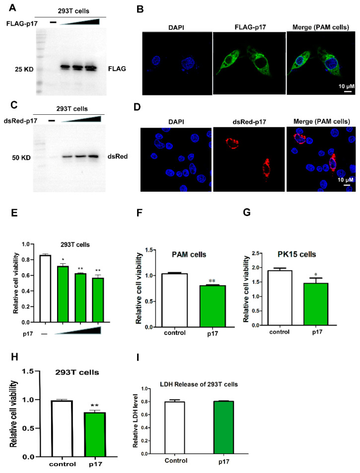
Figure 1.
Detecting the expression of ASFV p17 protein in 293T and PAM cells, and effects of p17 on cell proliferation and LDH release. (A) Expression of FLAG-p17 protein in 293T cells was detected by Western Blot using anti-FLAG after transfection with FLAG-p17 plasmids for 24 h. The FLAG-p17 plasmids for transfection were 0.25, 0.5, and 1 μg/mL, respectively. (B) Expression of FLAG-p17 protein in PAM cells by IF. (C) Expression of dsRed-p17 protein in 293T cells was detected by western blot using anti-dsRed after transfection with dsRed-p17 plasmids for 24 h. The dsRed-p17 plasmids for transfection were 0.25, 0.5, and 1 μg/mL, respectively. (D) Expression of dsRed-p17 protein in PAM cells was detected by IF. (E) Effects of p17 on cell proliferation in 293T cells were analyzed with a CCK-8 kit after transfection with FLAG-p17 plasmids for 24 h. The FLAG-p17 plasmids for transfection were 0.25, 0.5, and 1 μg/mL, respectively. (F,G) Effects of p17 on cell proliferation in PAM and PK15 cells were analyzed with a CCK-8 kit. The p17 groups were transfected with FLAG-p17 (1 μg/mL) for 24 h. (H) Effect of p17 on cell proliferation in 293T was detected with an MTT assay. The FLAG-p17 plasmid for transfection was 1 μg/mL. (I) Effect of p17 on LDH release in 293T cells. The p17 groups wasere transfected with FLAG-p17 (1 μg/mL) for 24 h. Control groups were transfected with empty plasmids. Values represent the mean ± S.D; * p < 0.05, ** p < 0.01 versus the control group.

