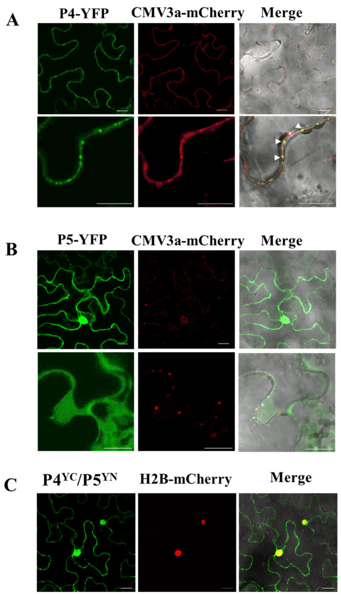
Figure 5.
Subcellular localization (A,B) and bimolecular fluorescence (BiFC) (C) assays of P4 and P5 of jujube yellow mottle-associated virus (JYMaV) in epidermal cells of wild-type Nicotiana benthamiana leaves. The fusion proteins CMV3a-mCherry and H2B-mCherry were used as plasmodesmata and nucleus markers, respectively. Colocalization dots of JYMaV-P4 with CMV3a-mCherry at PD were indicated by arrow heads. Images were acquired 3 days after agroinfiltration under a confocal microscope at 63x/1.20 WATER objective. Scale bar = 20 μm.

