Abstract
The aim of the study was to verify the effects of creatine (Cr) supplementation on functional capacity (walking capacity; primary outcome) and calf muscle oxygen saturation (StO2) (secondary outcome) in symptomatic peripheral arterial disease (PAD) patients. Twenty-nine patients, of both sexes, were randomized (1:1) in a double-blind manner for administration of placebo (PLA, n = 15) or creatine monohydrate (Cr, n = 14). The supplementation protocol consisted of 20 g/day for 1 week divided into four equal doses (loading phase), followed by single daily doses of 5 g in the subsequent 7 weeks (maintenance phase). Functional capacity (total walking distance) was assessed by the 6 min walk test, and calf muscle StO2 was assessed through near infrared spectroscopy. The measurements were collected before and after loading and after the maintenance phase. The level of significance was p < 0.05. No significant differences were found for function capacity (total walking distance (PLA: pre 389 ± 123 m vs. post loading 413 ± 131 m vs. post maintenance 382 ± 99 m; Cr: pre 373 ± 149 m vs. post loading 390 ± 115 m vs. post maintenance 369 ± 115 m, p = 0.170) and the calf muscle StO2 parameters (p > 0.05). Short- and long-term Cr supplementation does not influence functional capacity and calf muscle StO2 parameters in patients with symptomatic PAD.
Keywords: intermittent claudication, mobility limitation, dietary supplements
1. Introduction
Walking impairment is the main clinical concern in patients with symptomatic peripheral artery disease (PAD). In these patients, walking impairment has been associated with increased barriers to physical activity practice [1], reduced physical activity levels [2], impaired cardiovascular profile [3,4], increased risk of limb loss, and a higher risk of cardiovascular disease and mortality [5,6]. Creatine (Cr) is a natural bioenergetic compound, which is converted to creatine phosphate or phosphocreatine and stored in muscle, where it is used for energy production [7]. Previous studies showed that a short period of oral Cr supplementation increases the amount of Cr available in the muscle by as much as 20% [8] and improves muscle performance in athletes [9] and healthy individuals [10]. In addition, Cr supplementation promotes improvements in handgrip strength and reduces lower-limb muscle fatigue [11,12]. In addition, long-term Cr supplementation increased muscle glycogen content [13,14], leading to an improvement in walking ability in clinical populations [15]. Therefore, as Cr has the ability to increase the muscle content of both phosphocreatine and glycogen, Cr supplementation could improve walking tolerance in patients with PAD, especially after the onset of claudication pain when anaerobic metabolism becomes predominant. Furthermore, a recent review [16] demonstrated the potential effects of Cr supplementation on vascular function and the lack of randomized controlled trials on this topic. Thus, the aim of this study was to analyze the effects of oral Cr supplementation on functional capacity and calf muscle oxygen saturation (StO2) in patients with symptomatic PAD.
2. Materials and Methods
2.1. Experimental Design
This was a randomized control pilot trial with a pre-test and post-test design. A 7 day, double-blind, placebo-controlled study was conducted from December 2016 to October 2017 in São Paulo, Brazil (registered at clinicaltrials.gov as NCT02993874). This manuscript is reported according to the CONSORT guidelines [17].
Patients were randomly assigned to the experiment in a 1:1 ratio, to blocks of 4–6, considering sex and total walking distance, to receive either placebo or creatine monohydrate supplementation according to a computer-generated treatment sequence in a double-blind design. The randomization process was carried out by a researcher who was not involved in the project. The primary outcome was total walking distance, as measured by the 6 min walk test (6MWT). Secondary outcomes were upper-limb strength (handgrip strength test), lower-limb strength (sit-to-stand test), and calf muscle StO2 assessed by near-infrared spectroscopy (NIRS). Participants were assessed at baseline (pre intervention), after 1 week of supplementation (loading), and after 8 weeks (maintenance). Throughout the protocol, both groups received clinical recommendations for patients with PAD [18].
2.2. Participant Recruitment and Screening
Patients were recruited from a tertiary vascular center in Sao Paulo, Brazil. The sample consisted of 29 patients of both sexes with PAD and symptoms of intermittent claudication. The inclusion criteria were the presence of intermittent claudication symptoms during the 6MWT, ankle brachial index <0.90 in one or both lower limbs, and the absence of chronic renal insufficiency (creatinine clearance <30 mL/min). Patients were excluded if they presented any side effects caused by Cr supplementation (i.e., gastric discomfort or diarrhea) or did not comply with the supplementation procedures. This study was approved by the Ethics Committee of the Institutions (process 62601416.7.0000.0071). All patients gave informed consent prior to participation.
2.3. Clinical Characteristics
Demographic information, height, weight, smoking history, and comorbid conditions (hypertension, dyslipidemia, diabetes, and coronary artery disease) were obtained through medical history and physical examination. Body mass (kg) and stature (m) were measured (Welmy, São Paulo, Brazil), and body mass index was calculated. The ankle brachial index was calculated as the ratio between ankle systolic and brachial systolic blood pressure as previously described [19]. A trained researcher performed all measurements.
2.4. Creatine Supplementation Protocol and Blinding Procedure
Patients received plain packages containing placebo (PLA) (dextrose) (Probiotica, Sao Paulo, Brazil) or creatine monohydrate (Cr) supplementation (Creapure, AlzChem Trostberg GmbH, Germany), 20 g/day for 1 week divided into four equal doses (loading phase), followed by single daily doses of 5 g for the next 7 weeks (maintenance phase). During the first 7 days (loading phase), supplements were presented in four packages and patients were instructed to ingest the packages at breakfast, lunch, dinner, and before bedtime. During the maintenance phase, patients consumed the supplement as a single dose with their lunch. The supplement packages were coded so that neither the investigators nor the participants were aware of the contents until completion of the analyses. Quality control and purity of the Cr were guaranteed by the manufacturer. The supplements were provided by a staff member of our research team who did not participate in acquisition, analyses, or interpretation of the data. Adherence to the supplementation was determined in a subsample of nine patients through plasma creatine levels, using high-performance liquid chromatography (FL SPD-20A Shimadzu®, Kyoto, Japan), as previously described [20].
Patients in both groups received the recommendation to increase their physical activity levels as recommended by vascular disease guidelines [18,19]. Physical activity was monitored using a previously validated pedometer (polar A300, Finland) [21], measured throughout the 8 weeks of supplementation. Patients were instructed to wear the monitor while awake and to remove it before bed.
2.5. Primary Outcome—6 Min Walk Test (6MWT)
Patients performed a 6MWT supervised by a trained kinesiologist. Two cones were placed 30 m apart in a marked corridor as previously described [22]. Patients were instructed to walk as many laps around the cones as possible and to report when the claudication symptom occurred. It is worth mentioning that none of the patients stopped during the test. The kinesiologist recorded the initial claudication distance (ICD) and total walking distance (TWD) to evaluate the walking capacity. In PAD patients, the 6MWT presents high reliability, with the intraclass correlation coefficient ranging between 0.94 and 1.00 and the coefficient of variation ranging between 0.4% and 1.6% [22].
2.6. Secondary Outcomes
Calf Muscle Oxygen Saturation (Calf Muscle StO2)
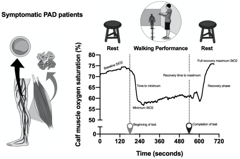
Calf muscle StO2 was assessed during the 6MWT using near-infrared spectroscopy (PortaMon, Artinis Medical Systems) through a sensor attached on the leg with the lowest ankle brachial index. The sensor was attached to the skin on the medial portion of the gastrocnemius muscle, and several calf muscle StO2 parameters were obtained before, during, and after the 6MWT (Figure 1). Before the test, a baseline measure of calf muscle StO2 was obtained at rest (sitting position) for 3 min to allow stabilization of the values (baseline StO2). During exercise, the minimum calf muscle StO2 value (minimum StO2) was obtained, as the time taken to reach the minimum value (time to minimum) and the absolute drop in calf muscle StO2 from rest (baseline StO2) to the minimum exercise value (minimum StO2). The end of the 6MWT was recorded (completion of test), as well as the maximal calf StO2 value during recovery (full recovery maximum StO2) in the sitting position. The recovery times for calf muscle StO2 to reach the full resting value (recovery phase time) and the maximum calf muscle StO2 value (recovery time to maximum) were calculated [23].
Figure 1.
Calf muscle oxygen saturation (StO2) parameters obtained before, during, and after the 6 min walk test (6MWT) test in symptomatic peripheral artery disease (PAD).
2.7. Handgrip Strength Test
Handgrip strength was measured using a digital hand dynamometer (Model HE101, WCT fitness). The measurement was performed with the participants in a sitting position, with the shoulder adducted in a neutral position and without rotation. The elbow joints were positioned at 90° flexion, with the forearm and wrist in a neutral position. Measurements were performed three times on both arms and the highest value was considered.
2.8. Sit-to-Stand Test
Participants were asked to start by sitting with their feet on the floor and their upper limbs bent over the chest, and then to stand up and sit down again without using their arms. Participants repeated the action five times as quickly as possible, and the time required to complete the five repetitions was recorded [24].
2.9. Statistical Analysis
The sample power was calculated using the G* Power software 3.19 statistics program. Thus, considering that the current sample included 29 patients (PLA, n = 15; Cr, n = 14) with an effect size of 1.91 and alpha error of 0.05, the sample power was 0.80. The normality of the data was verified using the Shapiro–Wilk test. The comparison between the general characteristics of the sample was performed using the t-test for independent samples. The categorical data were compared using the chi-square test. The baseline values were assessed using the Mann–Whitney U test. To verify the effect of supplementation, the generalized estimating equation model for repeated measures was performed. The level of significance was p < 0.05.
3. Results
The characteristics of the groups are presented in Table 1. Groups were similar at baseline in all clinical characteristics (p > 0.05).
Table 1.
Characteristics of participants at baseline (n = 29).
| PLA (n = 15) |
Cr (n = 14) |
p-Value | |
|---|---|---|---|
| Women (%) a | 54 | 46 | 0.56 |
| Age (years) a | 64 ± 8 | 64 ± 10 | 0.54 |
| Weight (kg) a | 77 ± 10 | 68 ± 17 | 0.18 |
| Height (m) a | 1.64 ± 0.09 | 1.60 ± 0.06 | 0.21 |
| Body mass index (kg/m2) a | 28.7 ± 3.1 | 26.7 ± 6.5 | 0.43 |
| Ankle-brachial index (mmHg) a | 0.50 ± 0.13 | 0.51 ± 0.16 | 1.00 |
| Initial claudication distance (m) a | 143 ± 84 | 143 ± 65 | 0.88 |
| Total walking distance (m) a | 371 ± 81 | 344 ± 82 | 0.65 |
| Comorbidities (%) | |||
| Hypertension b | 86.7 | 78.6 | 0.67 |
| Diabetes b | 60.0 | 50.0 | 0.43 |
| Dyslipidemia b | 6.7 | 7.1 | 0.74 |
| Current smoking b | 78.6 | 78.6 | 0.68 |
| Coronary artery disease b | 46.7 | 28.6 | 0.26 |
Data are presented as the mean and standard deviation for numerical variables and frequency for categorical variables. a t-Test for independent samples. b Chi-square test. PLA—placebo group; Cr—creatine group.
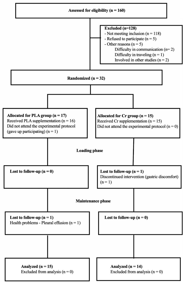
Initially, 160 patients were interviewed for eligibility; of these, 118 did not meet the inclusion criteria, five refused to participate, and five were not included for other reasons such as not answering phone calls (two subjects), difficulty in traveling because they lived far away (one patient), and being involved in another study (two patients). Thus, 32 patients started the study, with 17 allocated to the placebo group and 15 to the Cr group. Before the beginning of the protocol, one patient was excluded for not attending the evaluation (placebo; n = 1). During the loading period, one patient receiving Cr was excluded due to gastric discomfort (n = 1). During the maintenance supplementation period, one patient was excluded for presenting pleural perfusion (placebo group). Twenty-nine patients completed the study (PLA, n = 15; Cr, n = 14) (Figure 2).
Figure 2.
Study flow.
Patients taking Cr presented higher plasma creatine levels than the placebo group (PLA: pre 21.5 ± 38.9 μmol/L vs. post 30.7 ± 39.8 μmol/L; Cr: pre 32.1 ± 61.4 μmol/L vs. post 163.2 ± 42.65 μmol/L; time × intervention effect, p = 0.042).
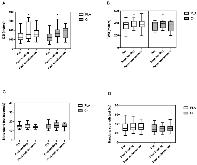
No significant interactions were found for ICD (Figure 3A) (PLA: pre 124 ± 72 m vs. post loading 150 ± 199 m vs. post maintenance 145 ± 90 m; Cr: pre 124 ± 73 m vs. post loading 168 ± 90 m vs. post maintenance 193 ± 110 m; time × intervention effect, p = 0.532) and TWD (Figure 3B) (PLA: pre 389 ± 123 m vs. post loading 413 ± 131 m vs. post maintenance 382 ± 99 m; Cr: pre 373 ± 149 m vs. post loading 390 ± 115 m vs. post maintenance 369 ± 115 m; time × intervention effect, p = 0.170). However, we observed a significant increase in ICD from pre to post loading in both groups (time effect, p = 0.009).
Figure 3.
Functional capacity parameters obtained during the 6MWT test, handgrip strength test, and sit-to-stand test before (pre) and after the loading and maintenance supplementation period in symptomatic peripheral arterial disease (n = 29; PLA = 15 and Cr = 14). Data are expressed as the median and interquartile range. * Significant difference pre vs. post loading supplementation period (p < 0.05). (A) ICD = initial claudication distance; (B) TWD = total walking distance; (C) sit-to-stand test; (D) handgrip strength test. PLA—placebo group; Cr—creatine group.
For physical activity levels (number of steps), no differences were found at pre vs. post supplementation (PLA: pre 457 ± 652 steps/day vs. post maintenance 465 ± 602 steps/day; Cr: pre 584 ± 360 steps/day vs. post maintenance 599 ± 314 steps/day; time effect = 0.258; time × intervention effect, p = 0.357).
There were no changes in the sit-to-stand test (Figure 3C) (PLA: pre 14.8 ± 2.9 s vs. post loading 15.1 ± 3.2 s vs. post maintenance 13.6 ± 1.5 s; Cr: pre 14.1 ± 5.1 s vs. post loading 15.6 ± 5.9 s vs. post maintenance 16.1 ± 2.2 s; time × intervention effect, p = 0.400) or handgrip strength test (Figure 3D) (PLA: 32.5 ± 16.0 kgf vs. post loading 34.3 ± 15.4 kgf vs. post maintenance 32.4 ± 13.7 kgf; Cr: pre 28.3 ± 15.2 kgf vs. post loading 29.2 ± 11.7 kgf vs. post maintenance 29.3 ± 10.2 kgf; time × intervention effect, p = 0.251).
The effects of supplementation on calf muscle StO2 parameters are presented in Table 2. No significant time × intervention interaction was found for any parameter (p > 0.05).
Table 2.
Calf muscle StO2 data before (pre) and after the short period (post loading) and long period (post maintenance) of supplementation (n = 29).
| PLA (n = 15) |
Cr (n = 14) |
p-Value Interaction | |
|---|---|---|---|
| Baseline StO2 (%) | |||
| Pre | 68.8 ± 3.7 | 70.0 ± 4.6 | 0.082 |
| Post loading | 68.2 ± 2.8 | 70.5 ± 6.1 | |
| Post maintenance | 71.3 ± 5.7 | 71.1 ± 4.8 | |
| Minimum StO2 (%) | |||
| Pre | 54.2 ± 10.0 | 61.6 ± 13.0 | 0.191 |
| Post loading | 52.9 ± 9.3 | 61.7 ± 14.8 | |
| Post maintenance | 54.2 ± 7.7 | 62.8 ± 12.3 | |
| Time to minimum StO2 (s) | |||
| Pre | 52.5 ± 34.3 | 56.5 ± 46.1 | 0.833 |
| Post loading | 46.8 ± 58.6 | 44.4 ± 26.2 | |
| Post maintenance | 50.1 ± 88.3 | 58.4 ± 43.5 | |
| Completion of test (%) | |||
| Pre | 12.2 ± 8.5 | 7.2 ± 9.7 | 0.691 |
| Post loading | 12.1 ± 8.2 | 9.5 ± 7.1 | |
| Post maintenance | 9.2 ± 10.5 | 7.6 ± 10.4 | |
| Recovery time to maximum StO2 (s) | |||
| Pre | 158.9 ± 43.1 | 160.1 ± 175.1 | 0.723 |
| Post loading | 146.4 ± 75.1 | 177.1 ± 202.9 | |
| Post maintenance | 160.9 ± 119.1 | 181.9 ±125.8 | |
| Recovery phase time StO2 (s) | |||
| Pre | 126.7 ± 112.4 | 132.9 ± 194.2 | 0.827 |
| Post loading | 93.0 ± 77.7 | 121.1 ± 248.3 | |
| Post maintenance | 87.1 ± 137.5 | 130.3 ± 103.7 | |
| Full recovery maximum StO2 (%) | |||
| Pre | 69.5 ± 15.8 | 72.6 ± 12.4 | 0.312 |
| Post loading | 68.6 ± 9.7 | 72.3 ± 14.5 | |
| Post maintenance | 74.5 ± 15.8 | 73.1 ± 15.8 | |
Data are expressed as the median and interquartile range. PLA—placebo group; Cr—creatine group.
4. Discussion
The main findings of this pilot study were that short- and long-term Cr supplementation did not improve functional capacity and calf muscle StO2 in symptomatic PAD patients.
Previous studies demonstrated that short- and long-term Cr supplementation increases physical performance in healthy subjects [10,25,26] and in patients with chronic diseases [15]. In contrast, our findings demonstrate no effects of Cr supplementation on 6MWT in symptomatic PAD patients. A potential explanation is that Cr rapidly increases the energy stores in type II fibers, and pain during walking in symptomatic PAD occurs mainly in calf muscles, predominantly composed of type I fibers [27]. Another factor may be related to the combination of Cr supplementation with other nutritional supplements. Benedetto et al. [15] analyzed the effects of 8 weeks of Cr supplementation combined with Coenzyme Q10 on walking capacity (6MWT) in patients with chronic obstructive pulmonary disease, and they demonstrated a significant increase of 51 m in the Cr supplementation group compared to the placebo group. In fact, Coenzyme Q10 is a compound that acts on the electron transport chain participating in cell respiration, helping to generate energy in the form of ATP [28]. As we only used Cr supplementation without the combination of other nutritional supplements, it is possible that the effects of Cr in the muscle were attenuated. Thus, future studies are necessary to investigate whether a combination of different nutritional supplements can improve physical performance in symptomatic PAD patients.
We also analyzed the microcirculation during the 6MWT using the calf muscle StO2 technique. Cr acts on microcirculation due to the increased activity of sensory nerves and epoxygenase metabolites, particularly epoxyethane diacetic acid, related to the endothelium-derived hyperpolarization factor [29]. Previous studies showed that Cr supplementation improved calf muscle StO2 in healthy young people [30] and vegan individuals [31]. However, in our study, Cr supplementation did not change any calf muscle StO2 parameters, which agrees with the results of the 6MWT. Thus, these results support that only Cr supplementation would not be enough to promote changes in microcirculation and physical performance in symptomatic PAD patients.
In the present study, Cr supplementation did not change other functional capacity tests, such as the sit-to-stand test and handgrip strength. The positive effects of Cr supplementation in tests involving muscle strength, fatigue, and endurance are controversial. For example, while Rawson et al. [11] demonstrated that 4 weeks of Cr supplementation reduced muscle fatigue assessed with isokinetic dynamometry in healthy older people, Lobo et al. [32] did not find any adjuvant effect after 1 year of Cr supplementation on lower-limb strength (sit-and-stand test) in postmenopausal older women. Taken together, these results suggest that the effects of Cr supplementation on muscle function are influenced by sample characteristics and methods to assess muscle function.
Our study presents some limitations. Although we analyzed the amount of creatine in the blood [33], we did not measure the content of creatine in the muscle and, thus, the impact of supplementation on the calf muscle was not determined. Our findings are restricted to the time of supplementation and the dosage of creatine used in this study. Furthermore, a limitation is the small sample size, and more studies are necessary with larger sample. Other tests to assess muscle function could be used, such as isokinetic dynamometry and a treadmill. However, we included tests to assess the strength in upper and lower limbs and the 6MWT, which represent greater clinical relevance in these patients [34,35,36]. Lastly, we only analyzed the effects of Cr supplementation for 8 weeks, and more studies with longer periods of supplementation are necessary.
5. Conclusions
Our results demonstrated that a short period (loading for 1 week) and a long period (maintenance for 8 weeks) of Cr supplementation do not increase functional capacity (walking distance, and upper- and lower-limb muscle strength) and calf muscle StO2 in patients with symptomatic PAD. This pilot study is applicable to the development of new Cr supplementation strategies to improve the performance and health outcomes in PAD population.
Acknowledgments
The authors thank CREAPURE for the supply of creatine.
Author Contributions
Conceptualization, W.J.R.D., R.M.R.-D., N.W., P.P.-L., P.M.N., and A.A.; methodology, W.J.R.D., R.M.R.-D., N.W., P.P.-L., P.M.N., and A.A.; formal analysis, R.M.R.-D. and D.B.C.; resources, W.J.R.D. and A.A.; data curation, R.M.R.-D., G.G.C., and D.B.C.; writing—original draft preparation, W.J.R.D., R.M.R.-D., A.E.Z., D.B.C., and A.A.; writing—review and editing, N.W., A.E.Z., P.P.-L., D.B.C., P.M.N., and A.A.M.; visualization, W.J.R.D. and R.M.R.-D.; supervision, N.W. and A.A.; project administration, W.J.D.R., R.M.R.-D., and A.A.; funding acquisition, W.J.D.R. and A.A. All authors have read and agreed to the published version of the manuscript.
Funding
This work was supported by the Foundation for Research Support of the State of Amazonas (FAPEAM) (grant number #062.03159.2014), the Araucaria Foundation (grant number #002/2017), and Coordination for the Improvement of Higher Education Personnel (CAPES) (grant number #001).
Conflicts of Interest
The authors declare no conflict of interest.
Footnotes
Publisher’s Note: MDPI stays neutral with regard to jurisdictional claims in published maps and institutional affiliations.
References
- 1.Barbosa J., Farah B., Chehuen M., Cucato G., Farias Júnior J., Wolosker N., Forjaz C., Gardner A., Ritti-Dias R. Barriers to Physical Activity in Patients with Intermittent Claudication. Int. J. Behav. Med. 2015;22:70–76. doi: 10.1007/s12529-014-9408-4. [DOI] [PubMed] [Google Scholar]
- 2.Gerage A.M., de Correia M.A., de Oliveira P.M.L., Palmeira A.C., Domingues W.J.R., Zeratti A.E., Puech-Leão P., Wolosker N., Ritti-Dias R.M., Cucato G.G. Physical Activity Levels in Peripheral Artery Disease Patients. Arq. Bras. Cardiol. 2019;113:410–416. doi: 10.5935/abc.20190142. [DOI] [PMC free article] [PubMed] [Google Scholar]
- 3.Germano-Soares A.H., Farah B.Q., Andrade-Lima A., Domingues W.R., Cavalcante B.R., de Almeida Correia M., Wolosker N., Cucato G.G., Ritti-Dias R.M. Factors Associated to Arterial Stiffness in Patients With Symptomatic Peripheral Artery Disease. Ann. Vasc. Surg. 2019;61:78–82. doi: 10.1016/j.avsg.2019.04.036. [DOI] [PubMed] [Google Scholar]
- 4.Germano-Soares A.H., Cucato G.G., Leicht A.S., Andrade-Lima A., Peçanha T., de Almeida Correia M., Zerati A.E., Wolosker N., Ritti-Dias R.M. Cardiac Autonomic Modulation Is Associated with Arterial Stiffness in Patients with Symptomatic Peripheral Artery Disease. Ann. Vasc. Surg. 2019;61:72–77. doi: 10.1016/j.avsg.2019.04.021. [DOI] [PubMed] [Google Scholar]
- 5.Cea Soriano L., Fowkes F.G.R., Johansson S., Allum A.M., García Rodriguez L.A. Cardiovascular outcomes for patients with symptomatic peripheral artery disease: A cohort study in The Health Improvement Network (THIN) in the UK. Eur. J. Prev. Cardiol. 2017;24:1927–1937. doi: 10.1177/2047487317736824. [DOI] [PubMed] [Google Scholar]
- 6.Morris D.R., Rodriguez A.J., Moxon J.V., Cunningham M.A., McDermott M.M., Myers J., Leeper N.J., Jones R.E., Golledge J. Association of lower extremity performance with cardiovascular and all-cause mortality in patients with peripheral artery disease: A systematic review and meta-analysis. J. Am. Heart Assoc. 2014;3 doi: 10.1161/JAHA.114.001105. [DOI] [PMC free article] [PubMed] [Google Scholar]
- 7.De Araujo Bonetti De Poli R., Roncada L.H., De Souza Malta E., Artioli G.G., Bertuzzi R., Zagatto A.M. Creatine supplementation improves phosphagen energy pathway during supramaximal effort, but does not improve anaerobic capacity or performance. Front. Physiol. 2019;10:352. doi: 10.3389/fphys.2019.00352. [DOI] [PMC free article] [PubMed] [Google Scholar]
- 8.Harris R.C., Söderlund K., Hultman E. Elevation of creatine in resting and exercised muscle of normal subjects by creatine supplementation. Clin. Sci. 1992;83:367–374. doi: 10.1042/cs0830367. [DOI] [PubMed] [Google Scholar]
- 9.Mielgo-Ayuso J., Calleja-Gonzalez J., Marqués-Jiménez D., Caballero-García A., Córdova A., Fernández-Lázaro D. Effects of creatine supplementation on athletic performance in soccer players: A systematic review and meta-analysis. Nutrients. 2019;11:757. doi: 10.3390/nu11040757. [DOI] [PMC free article] [PubMed] [Google Scholar]
- 10.Sculthorpe N., Grace F., Jones P., Fletcher I. The effect of short-term creatine loading on active range of movement. Appl. Physiol. Nutr. Metab. 2010;35:507–511. doi: 10.1139/H10-036. [DOI] [PubMed] [Google Scholar]
- 11.Rawson E.S., Wehnert M.L., Clarkson P.M. Effects of 30 days of creatine ingestion in older men. Eur. J. Appl. Physiol. Occup. Physiol. 1999;80:139–144. doi: 10.1007/s004210050570. [DOI] [PubMed] [Google Scholar]
- 12.Cañete S., San Juan A.F., Pérez M., Gómez-Gallego F., López-Mojares L.M., Earnest C.P., Fleck S.J., Lucia A. Does creatine supplementation improve functional capacity in elderly women? J. Strength Cond. Res. 2006;20:22–28. doi: 10.1519/R-17044.1. [DOI] [PubMed] [Google Scholar]
- 13.Pinto C.L., Botelho P.B., Pimentel G.D., Campos-Ferraz P.L., Mota J.F. Creatine supplementation and glycemic control: A systematic review. Amino Acids. 2016;48:2103–2129. doi: 10.1007/s00726-016-2277-1. [DOI] [PubMed] [Google Scholar]
- 14.Eijnde B.O., Richter E.A., Henquin J.C., Kiens B., Hespel P. Effect of creatine supplementation on creatine and glycogen content in rat skeletal muscle. Acta Physiol. Scand. 2001;171:169–176. doi: 10.1046/j.1365-201x.2001.00786.x. [DOI] [PubMed] [Google Scholar]
- 15.De Benedetto F., Pastorelli R., Ferrario M., de Blasio F., Marinari S., Brunelli L., Wouters E.F.M., Polverino F., Celli B.R. Supplementation with Qter® and Creatine improves functional performance in COPD patients on long term oxygen therapy. Respir. Med. 2018;142:86–93. doi: 10.1016/j.rmed.2018.08.002. [DOI] [PubMed] [Google Scholar]
- 16.Clarke H., Kim D.H., Meza C.A., Ormsbee M.J., Hickner R.C. The evolving applications of creatine supplementation: Could creatine improve vascular health? Nutrients. 2020;12:2834. doi: 10.3390/nu12092834. [DOI] [PMC free article] [PubMed] [Google Scholar]
- 17.Moher D., Hopewell S., Schulz K.F., Montori V., Gøtzsche P.C., Devereaux P.J., Elbourne D., Egger M., Altman D.G. CONSORT 2010 explanation and elaboration: Updated guidelines for reporting parallel group randomised trials. Int. J. Surg. 2012;10:28–55. doi: 10.1016/j.ijsu.2011.10.001. [DOI] [PubMed] [Google Scholar]
- 18.Gerhard-Herman M.D., Gornik H.L., Barrett C., Barshes N.R., Corriere M.A., Drachman D.E., Fleisher L.A., Fowkes F.G.R., Hamburg N.M., Lookstein R., et al. 2016 AHA/ACC Guideline on the Management of Patients with Lower Extremity Peripheral Artery Disease. Circulation. 2017;21:726–779. [Google Scholar]
- 19.Hirsch A.T., Haskal Z.J., Hertzer N.R., Bakal C.W., Creager M.A., Halperin J.L., Hiratzka L.F., Murphy W.R.C., Olin J.W., Puschett J.B., et al. ACC/AHA 2005 Practice Guidelines for the Management of Patients with Peripheral Arterial Disease (Lower Extremity, Renal, Mesenteric, and Abdominal Aortic) Circulation. 2006;113:e463–e654. doi: 10.1161/CIRCULATIONAHA.106.174526. [DOI] [PubMed] [Google Scholar]
- 20.Buchberger W., Ferdig M. Improved high-performance liquid chromatographic determination of guanidino compounds by pre-column dervatization with ninhydrin and fluorescence detection. J. Sep. Sci. 2004;27:1309–1312. doi: 10.1002/jssc.200401866. [DOI] [PubMed] [Google Scholar]
- 21.Boeselt T., Spielmanns M., Nell C., Storre J.H., Windisch W., Magerhans L., Beutel B., Kenn K., Greulich T., Alter P., et al. Validity and usability of physical activity monitoring in patients with Chronic Obstructive Pulmonary Disease (COPD) PLoS ONE. 2016;11:e0157229. doi: 10.1371/journal.pone.0157229. [DOI] [PMC free article] [PubMed] [Google Scholar]
- 22.Montgomery P.S., Gardner A.W. The clinical utility of a six-minute walk test in peripheral arterial occlusive disease patients. J. Am. Geriatr. Soc. 1998;46:706–711. doi: 10.1111/j.1532-5415.1998.tb03804.x. [DOI] [PubMed] [Google Scholar]
- 23.Andrade-Lima A., Cucato G.G., Domingues W.J.R., Germano-Soares A.H., Cavalcante B.R., Correia M.A., Saes G.F., Wolosker N., Gardner A.W., Zerati A.E., et al. Calf Muscle Oxygen Saturation during 6-min Walk Test and Its Relationship with Walking Impairment in Symptomatic Peripheral Artery Disease. Ann. Vasc. Surg. 2018;52:147–152. doi: 10.1016/j.avsg.2018.03.038. [DOI] [PubMed] [Google Scholar]
- 24.Jones S.E., Kon S.S.C., Canavan J.L., Patel M.S., Clark A.L., Nolan C.M., Polkey M.I., Man W.D.C. The five-repetition sit-to-stand test as a functional outcome measure in COPD. Thorax. 2013;68:1015–1020. doi: 10.1136/thoraxjnl-2013-203576. [DOI] [PubMed] [Google Scholar]
- 25.De Andrade Nemezio K.M., Bertuzzi R., Correia-Oliveira C.R., Gualano B., Bishop D.J., Lima-Silva A.E. Effect of Creatine Loading on Oxygen Uptake during a 1-km Cycling Time Trial. Med. Sci. Sports Exerc. 2015;47:2660–2668. doi: 10.1249/MSS.0000000000000718. [DOI] [PubMed] [Google Scholar]
- 26.Van Loon L.J.C., Oosterlaar A.M., Hartgens F., Hesselink M.K.C., Snow R.J., Wagenmakers A.J.M. Effects of creatine loading and prolonged creatine supplementation on body composition, fuel selection, sprint and endurance performance in humans. Clin. Sci. 2003;104:153–162. doi: 10.1042/CS20020159. [DOI] [PubMed] [Google Scholar]
- 27.Howden D., Boekenoogen C., Wagoner J., Miller S., Mendez-Guajardo B. The Effects of Creatine Supplementation on Muscular Strength and Endurance in Mice. 2015 NCUR. 2015 [Google Scholar]
- 28.Acosta M.J., Vazquez Fonseca L., Desbats M.A., Cerqua C., Zordan R., Trevisson E., Salviati L. Coenzyme Q biosynthesis in health and disease. Biochim. Biophys. Acta Bioenerg. 2016;1857:1079–1085. doi: 10.1016/j.bbabio.2016.03.036. [DOI] [PubMed] [Google Scholar]
- 29.Cracowski J.-L., Gaillard-Bigot F., Cracowski C., Sors C., Roustit M., Millet C. Involvement of cytochrome epoxygenase metabolites in cutaneous postocclusive hyperemia in humans. J. Appl. Physiol. 2013;114:245–251. doi: 10.1152/japplphysiol.01085.2012. [DOI] [PubMed] [Google Scholar]
- 30.De Moraes R., Van Bavel D., De Moraes B.S., Tibiriçá E. Effects of dietary creatine supplementation on systemic microvascular density and reactivity in healthy young adults. Nutr. J. 2014;13:115. doi: 10.1186/1475-2891-13-115. [DOI] [PMC free article] [PubMed] [Google Scholar]
- 31.Van Bavel D., de Moraes R., Tibirica E. Effects of dietary supplementation with creatine on homocysteinemia and systemic microvascular endothelial function in individuals adhering to vegan diets. Fundam. Clin. Pharmacol. 2018;33:428–440. doi: 10.1111/fcp.12442. [DOI] [PubMed] [Google Scholar]
- 32.Lobo D.M., Tritto A.C., da Silva L.R., de Oliveira P.B., Benatti F.B., Roschel H., Nie B., Gualano B., Pereira R.M.R. Effects of long-term low-dose dietary creatine supplementation in older women. Exp. Gerontol. 2015;70:97–104. doi: 10.1016/j.exger.2015.07.012. [DOI] [PubMed] [Google Scholar]
- 33.Domingues W.J.R., Ritti-Dias R.M., Cucato G.G., Wolosker N., Zerati A.E., Puech-Leão P., Nunhes P.M., Moliterno A.A., Avelar A. Does Creatine Supplementation Affect Renal Function in Patients with Peripheral Artery Disease? A Randomized, Double Blind, Placebo-controlled, Clinical Trial. Ann. Vasc. Surg. 2020;63:45–52. doi: 10.1016/j.avsg.2019.07.008. [DOI] [PubMed] [Google Scholar]
- 34.Van Lummel R.C., Walgaard S., Maier A.B., Ainsworth E., Beek P.J., Van Dieën J.H. The instrumented Sit-To-Stand test (iSTS) has greater clinical relevance than the manually recorded sit-to-stand test in older adults. PLoS ONE. 2016;11:e0157968. doi: 10.1371/journal.pone.0157968. [DOI] [PMC free article] [PubMed] [Google Scholar]
- 35.McDermott M.M., Guralnik J.M., Criqui M.H., Liu K., Kibbe M.R., Ferrucci L. Six-minute walk is a better outcome measure than treadmill walking tests in therapeutic trials of patients with peripheral artery disease. Circulation. 2014;130:61–68. doi: 10.1161/CIRCULATIONAHA.114.007002. [DOI] [PMC free article] [PubMed] [Google Scholar]
- 36.Reeve T.E., Ur R., Craven T.E., Kaan J.H., Goldman M.P., Edwards M.S., Hurie J.B., Velazquez-Ramirez G., Corriere M.A. Grip strength measurement for frailty assessment in patients with vascular disease and associations with comorbidity, cardiac risk, and sarcopenia. J. Vasc. Surg. 2018;67:1512–1520. doi: 10.1016/j.jvs.2017.08.078. [DOI] [PubMed] [Google Scholar]


