Abstract
Terfezia species are obligate symbiotic partners of several xerophytic host plants, mainly belonging to the Cistaceae. Yet, their mycorrhizal associations with members of the genus Cistus remain poorly characterized and their potential application in desert truffle cultivation remains unexplored. This work provides the first anatomic descriptions of the mycorrhizae formed in vitro by four Terfezia species (i.e., T. arenaria; T. extremadurensis; T. fanfani, T. pini) with C. ladanifer and C. salviifolius, two of the most widespread and common Cistus species in acidic soils. All the tested associations resulted in the formation of ectomycorrhizae with well-developed Hartig net, but with a varying degree of mantle development. Our results also demonstrate that all the experimented Terfezia-Cistus combinations expressed high mycorrhization rates. Moreover, the present work shows that C. salviifolius and C. ladanifer are suitable plant hosts for Terfezia species, including some that are, to date, known to be only associated with annual herbs or tree species. This new evidence might aid in broadening the number of situations whereby Terfezia spp. can be cultivated in acid soils.
Keywords: desert truffles, Terfezia cultivation, Cistus, putative plant hosts, mycorrhizae characte-rization, acid soils
1. Introduction
The term “desert truffle” commonly designates the fruitbodies produced by edible hypogeous Ascomycota (Pezizaceae), which includes several species of the genera Terfezia, Tirmania and Picoa, found in arid and semiarid areas throughout the world [1,2,3]. These fungi establish key mutualistic associations in arid and semiarid ecosystems with the roots of several xerophytic host plants [4], mainly belonging to the Cistaceae (e.g., Helianthemum spp. and Cistus spp.) but also to the Fagaceae and Pinaceae (i.e., oaks and pines) [2,5,6,7,8].
Many Terfezia species are among the most prized desert truffle species. Thus, in the last few decades, numerous research efforts have been made in order to enable their large-scale cultivation [2]. Still, desert truffle cultivation is only now leaving its infancy, and our knowledge on the ecology, physiology, and biochemistry of many Terfezia mycorrhizal associations still remains fragmentary [8]. One key issue that remains neglected is the choice of suitable putative hosts. In fact, so far, the only host plants that have been tested in experimental desert truffle cultivation are perennial and annual species of Helianthemum “sensu lato” and little or no attention has been given to the assessment of new potential hosts for desert truffle cultivation [9]. Within Cistaceae, several Cistus perennial species are considered putative hosts for Terfezia spp. [6,10,11,12] and therefore could be excellent candidates for desert truffle cultivation.
In fact, the genus Cistus L. (Cistaceae) includes about 20 perennial shrub species, distributed throughout the Mediterranean region and Canary Islands. In the Iberian Peninsula, the genus is represented by 13 species, all belonging to primary successional stages of many forest stands, growing readily in degraded areas or after disturbances such as fire [13,14], rendering their ectomycorrhizal ecology particularly interesting in the context of global warming and increasing desertification, in arid and semiarid areas worldwide.
As a whole, Terfezia mycorrhizae are known to display great structural versatility depending on certain factors (i.e., host species, concentration of auxins secreted by the fungi, root sensitivity to those auxins, phosphate concentrations, and drought conditions, [15,16]). Research on the association of Terfezia and various Cistaceae (mostly perennial and annual species of Helianthemum) has demonstrated the remarkable adaptability of these associations, which can result in the formation of (a) endomycorrhizae, characterized by undifferentiated coil-shaped or globular intracellular hyphae penetrating the plant cells [17,18,19,20]; (b) ectomycorrhizae, characterized by a Hartig net, but without a true sheath [18,21,22]; (c) ectendomycorrhizae, characterized by the presence of both intercellular Hartig net and intracellular hyphae penetrating the cortex cells [23,24]. Lately, it has been observed that in some instances, more than one of the above mycorrhizal types may be observed along the same root system of a single Helianthemum plant, a phenomenon that has been named “ectendomycorrhiza continuum” [23]. On the other hand, Terfezia-Cistus mycorrhizae seem to be consistently morphologically characterized as ectomycorrhizae with a well-developed Hartig net, however, the presence or absence of a true sheath is still subject of debate [5,16,25,26,27]. While most in vitro and ex vitro synthesis resulted in ectomycorrhizae with a well-developed Hartig net but without a true mantle [5], a more recent work provided evidence on the formation of ectomycorrhizae with a thin less-developed sheath using C. salvifolius, C. albidus, C. incanus, and three different Terfezia species [16]. In view of this new evidence, the main goal of the present work was to provide new insights on the association between Terfezia species and Cistus spp. by reporting on the mycorrhizae formed by four Terfezia species—namely T. arenaria; T. extremadurensis; T. fanfani, T. pini—along with C. ladanifer and C. salviifolius, two of the most widespread and common Cistus species in acidic soils. Furthermore, we aimed to assess which of the above Terfezia-Cistus combinations are the most compatible and open the possibility of mass production of Terfezia mycorrhized seedlings towards desert truffle cultivation in acid soils.
2. Materials and Methods
2.1. Fungal Material
Mature Terfezia ascocarps were harvested from different locations in the Alentejo (Southern Portugal), between February 2017 and April 2019. Ascocarps were identified to the species level (compared with the descriptions present in [28,29]) and fragments were isolated on LS medium [30] (in mg·L−1 475 KNO3, 110 CaCl2·2H2O, 92.5 MgSO4·7H2O, 42.5 KH2PO4, 412 NH4NO3, 9.31 Na2EDTA, 6.96 FeSO4·7H2O, 4.22 MnSO4·4H2O, 2.15 ZnSO4·7H2O, 0.006 CuSO4·5H2O, 0.006 CoCl2·6H2O, 0.06 Na2MoO4·2H2O, 1.55 H3BO3, 0.21 KI; 0.025 thiamine hydrochloride; 0.125 nicotinic acid; 0.125 pyridoxine hydrochloride; 25 myo-inositol, 0.5 glycine, 0.5 6-benzylaminopurine (BAP), 0.5 indole-3-acetic acid (IAA)) with sucrose 10 g·L−1 and solidified with 10 g·L−1 of agar and pH 5.5. All isolates were incubated in the dark at 25 ± 2 °C for 90 days. Four isolates, with better growth rate, were selected for mycorrhization trials and molecular characterization: T. arenaria (Moris) Trappe strain 220, T. extremadurensis Muñoz-Mohedano, Ant. Rodr. & Bordallo strain 271, T. fanfani Matt. strain 235 and T. pini Bordallo, Ant. Rodr. and Muñoz-Mohedano strain 278.
Molecular characterization was carried out by sequencing fragments of the nuclear ribosomal DNA region of selected Terfezia mycelial cultures. DNA extractions were performed by a modified CTAB method [31]. The internal transcribed spacer (ITS) region of the rDNA, including the 5.8 S ribosomal gene, was amplified using the ITS5 and ITS4 primers [32]. Amplifications of ITS rDNA sequences were performed using a Mastercycler Gradient thermocycler (Eppendorf, Hamburg, Germany) with the following cycling parameters: an initial denaturalization step for 3 min at 95 °C, followed by 35 cycles consis-ting of: 30 s at 95 °C, 30 s at 95 °C (annealing temp.), 1 min at 72 °C, and a final extension at 72 °C for 10 min. All reagents were acquired from NZYTech, Lda, sequencing was done commercially (STAB VIDA, Lda.) and all sequence alignments were performed with online MAFFT version 7, using the E-INS-i strategy [33]. Molecular identification was carried out by comparing our sequences with the existing ones in the GenBank database.
2.2. Plant Material
Seeds from C. salviifolius L. and C. ladanifer L. plants growing in Herdade da Mitra, near Évora (Alentejo, Portugal) (38°32′ N; 8°01′ W; 220 m a.s.l.), were collected on November 2013 in a Montado area with natural shrub undercover dominated by Cistus spp. The area belongs to the Mediterranean pluviseasonal-oceanic bioclimate and is located in the low mesomediterranean bioclimatic belt. It has a dry to subhumid ombrotype of climate with a mean annual temperature ranging from 9.2 °C to 21.5 °C and a mean annual rainfall of 664.6 mm [34,35]. The collected Cistus seeds were dried at 23 °C in a Memmert forced ventilation oven (Model 600) and kept at room temperature in the dark until use. The seeds were surface sterilized by immersion in 70% ethanol for 2 min, followed by another immersion in a 50% bleach solution for 10–15 min. Afterward, seeds were washed three times in sterilized tap water. To break seed dormancy, seeds were heated at 150 °C in a bi-distilled water bath for 5 min and left to cool down until room temperature was reached. Seeds were then placed in Petri dishes, on top of the filter paper moistened with bi-distilled water, and kept in a growth chamber in the dark with 24 °C/21 °C (±1 °C) day/night temperature. After germination, C. salviifolius and C. ladanifer seedlings were routinely micropropagated using the Cistus rapid multiplication protocol described in [36].
2.3. Mycorrhizal Synthesis
Mycorrhizal synthesis was performed in polypropylene transparent microboxes (90 mm Ø and 120 mm in height) with filtered polypropylene covers. Each box containing 200 mL of dried vermiculite and 100 mL of LS liquid medium was autoclaved at 121 °C and 18 psi for 20 min. Mycelium pure cultures of T. arenaria (strain 220, Genbank MW356871), T. extremadurensis (strain 271, Genbank MW356873), T. fanfani (strain 235, Genbank MW356872), and T. pini (strain 278, Genbank MW356874) were used in the experiment.
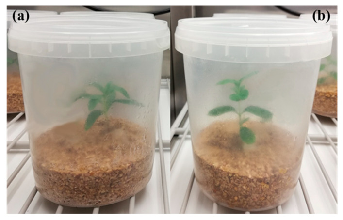
After a week (to check for possible contaminants), sixteen boxes were inoculated with two plugs dissected from one of the selected Terfezia strains and incubated in the dark (25 ± 2 °C). The process was repeated for the four Terfezia species, totaling sixty-four boxes. Two weeks later, one rooted Cistus micropropagated plantlet (C. salviifolius or C. ladanifer) was introduced in each box, near the active growing mycelia, totaling eight replicates for each Terfezia-Cistus pair (Figure 1).
Figure 1.
In vitro mycorrhizal system. (a) Detail of micropropagated C. ladanifer inoculated with T. arenaria mycelium; (b) detail of micropropagated C. salviifolius inoculated with T. arenaria mycelium.
The boxes were then placed in the grow chamber at 24 °C/21 °C (± 1 °C) day/night temperature and 15 h light period, under cool white fluorescent light (36 µmol·m−2·s−1). After two months, the Cistus plantlets were carefully retrieved from the growing medium and their roots gently washed to free them from adhering particles. The whole root system of each Cistus plantlet was separated from the aerial part, kept in 50 mL centrifuge tubes filled with a glutaraldehyde solution (4%), and stored at 5 °C until further examination.
2.4. Mycorrhizal Morphotyping and Colonization Assessment
Each Cistus plantlet root system was washed over a 2 mm sieve and cut into segments of approximately 1 cm in length. Afterwards, the root segments were spread in two Petri dishes containing bi-distilled water and all root tips were observed under a stereo microscope (WILD M3) to determine the existent morphological types. Characterization of the mycorrhizal root tips followed [37,38]. Prior to the microscopic observation, all root fragments were cleared with a 10% KOH solution and stained with 0.1% trypan blue in lactophenol following the method developed by [39]. Microscopic examination of the root fragments and characterization of the mycorrhizal system under the light microscope was done, using a Leica DM750 microscope equipped with a digital camera (Leica ICC50W), according to the methodology described in [40]. The percentage of fungal root colonization was estimated based on the frequency of infection expressed by: FI (%) = 100 (N−N0)/N, where N is the total number of observed root fragments and N0 is the number of root fragments uninfected [41].
2.5. Statistical Analysis
Data normality was assessed using Kolmogorov–Smirnov tests. Levene’s tests were employed to assess the variance homocedasticity assumption. Arcsine data transformation was necessary to perform the two-way ANOVA. Mean differences in frequency of infection between plant hosts and different Terfezia isolates were tested through a two-way ANOVA followed by Tukey post hoc tests. All calculations were performed with IBM SPSS Statistics V 24 [42].
3. Results
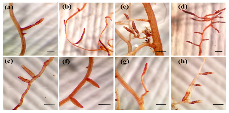
All Cistus plantlets, irrespective of the host plant–fungal species combination, formed mycorrhizal associations with all the Terfezia isolates after two months using the mycorrhizal system described in material and methods section. Concerning the macroscopic morphological characterization of the mycorrhizal root tips, a single morphotype was produced by every host plant–fungal species combination (Figure 2). Under the stereomicroscope, mycorrhizae were unbranched, unramified ends were straight to bent, more or less inflated, sometimes with a more enlarged apex (club shaped). Surface of unramified ends was smooth, color varied from brownish-yellow to rusty-brown ochre, with slightly darker tones on aged mycorrhizae. Emanating hyphae were infrequent, white, and shiny. No rhizomorphs were observed.
Figure 2.
External characteristics of Terfezia-Cistus mycorrhizae, each scale bar measure 0.5 mm: (a) T. arenaria × C. salviifolius mycorrhizae, (b) T. extremadurensis × C. salviifolius mycorrhizae, (c) T. fanfani × C. salviifolius mycorrhizae, (d) T. pini × C. salviifolius mycorrhizae, (e) T. arenaria × C. ladanifer mycorrhizae, (f) T. extremadurensis × C. ladanifer mycorrhizae, (g) T. fanfani × C. ladanifer mycorrhizae, (h) T. pini × C. ladanifer mycorrhizae.
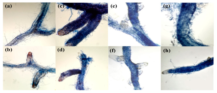
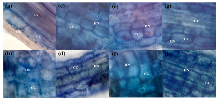
Additional microscopic examination of the root fragments revealed that all four Terfezia species (i.e., T. arenaria, T. fanfani, T. extremadurensis, and T. pini) formed ectomycorrhizae with a well-developed Hartig net but with varying degrees of mantle development (Figure 3, Figure 4 and Figure 5), both with C. salviifolius and C. ladanifer.
Figure 3.
Light microphotographs of Terfezia-Cistus mycorrhizal roots, (400×): (a) T. arenaria × C. salviifolius root showing rudimentary sheath, (b) T. arenaria × C. ladanifer root showing rudimentary sheath, (c) T. extremadurensis × C. salviifolius root surrounded by a well-developed sheath, (d) T. extremadurensis × C. ladanifer root showing well-developed sheath, (e) T. fanfani × C. salviifolius root showing a less-developed sheath, (f) T. fanfani × C. ladanifer root showing a less-developed sheath, (g) T. pini × C. salviifolius root surrounded by a diffuse sheath, (h) T. pini × C. ladanifer root showing a well-developed sheath.
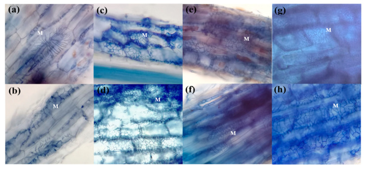
Figure 4.
Light microphotographs of Terfezia-Cistus mycorrhizal roots, (400×): (a) detail of T. arenaria × C. salviifolius ectomycorrhizae mantle structure (M), (b) detail of T. arenaria × C. ladanifer ectomycorrhizae mantle structure (M), (c) detail of T. extremadurensis × C. salviifolius ectomycorrhizae mantle structure (M), (d) detail of T. extremadurensis × C. ladanifer ectomycorrhizae mantle structure (M), (e) detail of T. fanfani × C. salviifolius ectomycorrhizae mantle structure (M), (f) detail of T. fanfani × C. ladanifer ectomycorrhizae mantle structure (M), (g) detail of T. pini × C. salviifolius ectomycorrhizae mantle structure (M), (h) detail of T. pini × C. ladanifer ectomycorrhizae mantle structure (M).
Figure 5.
Light microphotographs of Terfezia-Cistus mycorrhizal roots, (400×): (a) T. arenaria x C. salviifolius ectomycorrhizae showing Hartig net (HN) restricted to cortical cells (CC), (CY) central cylinder; (b) T. arenaria x C. ladanifer ectomycorrhizae showing well-developed Hartig net (HN) surrounding cortical cells (CC); (c) T. extremadurensis x C. salviifolius ectomycorrhizae showing intercellular hyphae pearl structure (Hartig net (HN)) between cortical cells (CC); (d) T. extremadurensis x C. ladanifer ectomycorrhizae showing well-developed Hartig net (HN) between cortical cells (CC); (e) T. fanfani x C. salviifolius ectomycorrhizae showing the characteristic pearl structure of the Hartig net (HN) surrounding root cortical cells (CC); (f) T. fanfani x C. ladanifer ectomycorrhizae with well-developed Hartig net (HN) between cortical cells (CC); (g) T. pini x C. salviifolius ectomycorrhizae showing well-developed Hartig net (HN) surrounding cortical cells (CC), (CY) central cylinder; (h) T. pini x C. ladanifer ectomycorrhizae showing a widespread Hartig net (HN) surrounding cortical cells (CC).
Overall, the mycorrhizae formed between the different Terfezia-Cistus associations displayed similar microscopic characteristics irrespective of host plant–fungal species association. A general description of the anatomic features of these associations is provided below.
The outer mantle structure with a densely plectenchymatous to nearly pseudoparenchymatous structure was composed of colorless angular cells, which were more marked in T. extremadurensis and T. pini, but also noticeable in T. fanfani. The inner mantle plectenchymatous was characterized by colorless hyphae forming a coarse net of irregularly shaped hyphae, tightly glued together, which sometimes begin as small star-like arrangements, as in the case of T. arenaria (Figure 4a,b).
The Hartig net is usually composed of a single row of hyphae that protrudes deeply towards the endodermis, enveloping completely one to three rows of cortical cells, but never touching the endodermis or the central cylinder. Hyphal segments around cortical cells are initially of constant width but later forming a beaded or pearl-like structure (Figure 5c,e).
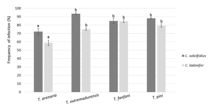
The two-way ANOVA showed that mean frequencies of infection were significantly influenced by the Terfezia species, the Cistus species, and their interaction (Figure 6). As such, regarding the mycosimbiont, T. arenaria showed significantly lower mean frequencies of infection then the other three Terfezia species. Concerning plant host, C. salviifolius revealed higher frequencies of infection with T. arenaria, T. extremadurensis, and T. pini, compared to C. ladanifer. However, no clear differences in the mean frequencies of infection were found between plant hosts for T. fanfani.
Figure 6.
Frequency of infection by the four Terfezia isolates (means ± SE, n = 8) on root tips of C. salviifolius and C. ladanifer. Different letters over the bars represent significant differences between the means among different Terfezia-Cistus combinations using two-way ANOVA followed by Tukey test (p < 0.05). Two-way ANOVA results for Terfezia (T) frequency of infection in Cistus (C), T: F = 27.42 ***, C: F = 41.59 ***, T X C: F = 6.36 ***. Significance level *** p < 0.001.
4. Discussion
The present work has brought forward compelling evidence on the ability of T. arenaria, T. extremadurensis, T. fanfani, and T. pini to engage in mycorrhizal association under in vitro culture conditions, with two of the most widespread Cistus species (i.e., C. salviifolius and C. ladanifer). Furthermore, it provides for the first time a comprehensive macro and microscopic descriptions of the mycorrhizae formed between T. arenaria, T. extremadurensis, and T. pini on the above mentioned Cistus species.
One interesting question that needed answering was the presence or absence of a true sheath in these mycorrhizal associations. We can now ascertain that all four Terfezia species analyzed (i.e., T. arenaria, T. fanfani, T. extremadurensis, and T. pini) do form ectomycorrhizae with a true sheath, and therefore agree with the work of [16]. However, differences in mantle development were observed between the mycorrhizae formed by the four mycosymbionts, with T. arenaria colonized roots showing only a sparsely rudimentary sheath, whereas on the other end, T. extremadurensis colonized roots where surrounded by a profuse well-developed sheath, under the same experimental conditions. Although these differences might represent true differences on the morphological characters of those particular associations, another possible explanation is that the observed differences might just be a reflection of the flexibility of each Terfezia species to colonize different plant hosts. In other words, although capable of entering into the mycorrhizal association, the time or the conditions required to form fully developed mycorrhizae may differ between Terfezia species. For instance, this might indicate that T. arenaria have a narrower putative host range than the remaining Terfezia in the analysis.
In summary, given that modern truffle cultivation is largely based on mass production of adequately colonized plants raised under controlled conditions [43], our results are encouraging since all eight Terfezia-Cistus combinations expressed high rates of mycorrhization (comparable to those obtained in previous works) [16,27,44]. Nevertheless, our experimental data seem to indicate that C. salviifolius is a better option than C. ladanifer as potential host for the production of T. arenaria inoculated plants, and its subsequent application on desert truffle cultivation. These results are encouraging since T. arenaria sporocarps are one of the most prized and traded desert truffle worldwide.
Yet, the choice of the fungal partner in truffle cultivation depends on various factors (e.g., sporocarp size, edibility, plantation purpose, interactions with other organisms, etc.). Regarding the tested mycosymbionts, T. fanfani seems to be the best option towards desert truffle cultivation in acid soils, mostly due to its ascocarp size, gastronomic value, and its capacity to infect both hosts.
Nevertheless, the introduction of T. extremadurensis and T. pini inoculated Cistus seedlings may also be interesting alternatives for reforestation programs and/or to prevent soil erosion after intense disturbances.
Though these results are promising for trufficulture, further studies are still needed to ascertain if the considered Terfezia-Cistus combinations will result in the formation of sporocarps under field conditions. In that regard, we already obtained some preliminary results in an experimental plot where we confirmed the persistence of Cistus inoculated mycorrhizae and sporocarp production [45] two years after plant installation on the plot.
Acknowledgments
We thank Tânia Nobre for her critical comments and help in improving the manuscript.
Author Contributions
Conceptualization, C.S.-S. and R.L.; methodology (field and laboratory work), R.L. and B.N.; formal analysis, C.S.-S. and R.L.; data curation, C.S.-S. and R.L.; writing—original draft preparation, R.L. and B.N.; writing—review and editing, C.S.-S. and R.L.; supervision, C.S.-S.; project administration, C.S.-S.; funding acquisition, C.S.-S. All authors have read and agreed to the published version of the manuscript.
Funding
This research was funded by Alentejo 2020 (Project ALT20-03-0145-FEDER-000006) and National Funds through FCT - Foundation for Science and Technology under the Project UIDB/05183/2020.
Institutional Review Board Statement
Not applicable.
Informed Consent Statement
Not applicable.
Data Availability Statement
Not applicable.
Conflicts of Interest
The authors declare no conflict of interest. The funders had no role in the design of the study; in the collection, analyses, or interpretation of data; in the writing of the manuscript; or in the decision to publish the results.
Footnotes
Publisher’s Note: MDPI stays neutral with regard to jurisdictional claims in published maps and institutional affiliations.
References
- 1.Moreno G., Alvarado P., Manjón J.P. Hypogeous desert fungi. In: Kagan-Zur V., Roth-Bejerano N., Sitrit Y., Morte A., editors. Desert Truffles, Phylogeny, Physiology, Distribution and Domestication. Springer; Berlin/Heidelberg, Germany: 2014. [Google Scholar]
- 2.Morte A., Honrubia M., Gutiérrez A. Biotechnology and cultivation of desert truffles. In: Varma A., editor. Mycorrhiza: State of the Art Genetics and Molecular Biology, Eco-Function, Biotechnology, Eco-Physiology, Structure and Systematics. Springer; Berlin/Heidelberg, Germany: 2008. [Google Scholar]
- 3.Navarro-Ródenas A., Lozano-Carrillo M.C., Pérez-Gilabert M., Morte A. Effect of water stress on in vitro mycelium cultures of two mycorrhizal desert truffles. Mycorrhiza. 2011;21:247–253. doi: 10.1007/s00572-010-0329-z. [DOI] [PubMed] [Google Scholar]
- 4.Pérez-Gilabert M., García-Carmona F., Morte A. Enzymes in Terfezia claveryi ascocarps. In: Kagan-Zur V., Roth-Bejerano N., Sitrit Y., Morte A., editors. Desert Truffles, Phylogeny, Physiology, Distribution and Domestication. Springer; Berlin/Heidelberg, Germany: 2014. [Google Scholar]
- 5.Alsheikh A.M. Ph.D. Thesis. Oregon State University; Corvallis, OR, USA: 1994. Taxonomy and Mycorrhizal Ecology of the Desert Truffles in the Genus. [Google Scholar]
- 6.Diez J., Manjon J.L., Martin F. Molecular phylogeny of the mycorrhizal desert truffles (Terfezia and Tirmania), host specificity and edaphic tolerance. Mycologia. 2002;94:247–259. doi: 10.1080/15572536.2003.11833230. [DOI] [PubMed] [Google Scholar]
- 7.Fortas Z., Chevalier G. Effet des conditions de culture sur la mycorhization de l’Helianthemum guttatum par trois espèces de terfez des genres Terfezia et Tirmania d’Algerie. Can. J. Bot. 1992;70:2453–2460. doi: 10.1139/b92-303. [DOI] [Google Scholar]
- 8.Kagan-Zur V., Roth-Bejerano N. Desert Truffles. Fungi. 2008;1:32–37. [Google Scholar]
- 9.Morte A., Andrino A. Domestication: Preparation of mycorrhizal seedlings. In: Kagan-Zur V., Roth-Bejerano N., Sitrit Y., Morte A., editors. Desert Truffles, Phylogeny, Physiology, Distribution and Domestication. Springer; Berlin/Heidelberg, Germany: 2014. [Google Scholar]
- 10.Bordallo J.J., Rodríguez A., Kaounas V., Camello F., Honrubia M., Morte A. Two new Terfezia species from southern Europe. Phytotaxa. 2015;230:239–249. doi: 10.11646/phytotaxa.230.3.2. [DOI] [Google Scholar]
- 11.Crous P.W., Wingfield M.J., Lombard L., Roets F., Swart W.J., Alvarado P., Carnegie A.J., Moreno G., Luangsaard J., Thangavel R., et al. Fungal Planet Description Sheets: 951–1041. Persoonia. 2019;43:223–425. doi: 10.3767/persoonia.2019.43.06. [DOI] [PMC free article] [PubMed] [Google Scholar]
- 12.Kovacs G.M., Balazs T.K., Calonge F.D., Martín M.P. The diversity of Terfezia desert truffles: New species and a highly variable species complex with intra-sporocarpic nrDNA ITS heterogeneity. Mycologia. 2011;103:841–853. doi: 10.3852/10-312. [DOI] [PubMed] [Google Scholar]
- 13.Águeda B., Parladé J., de Miguel A.M., Martínez-Peña F. Characterization and identification of field ectomycorrhizae of Boletus edulis and Cistus ladanifer. Mycologia. 2006;98:23–30. doi: 10.1080/15572536.2006.11832709. [DOI] [PubMed] [Google Scholar]
- 14.Nuytinck J., Verbeken A., Leonardi M., Pacioni G., Rinaldi A.C., Comandini O. Characterization of Lactarius tesquorum ectomycorrhizae on Cistus sp., and molecular phylogeny of related European Lactarius taxa. Mycologia. 2004;96:272–282. doi: 10.1080/15572536.2005.11832977. [DOI] [PubMed] [Google Scholar]
- 15.Roth-Bejerano N., Navarro-Ródenas A., Gutiérrez A. Types of mycorrhizal association. In: Kagan-Zur V., Roth-Bejerano N., Sitrit Y., Morte A., editors. Desert Truffles, Phylogeny, Physiology, Distribution and Domestication. Springer; Berlin/Heidelberg, Germany: 2014. [Google Scholar]
- 16.Zitouni-Haouar F.H., Fortas Z., Chevalier G. Morphological characterization of mycorrhizae formed between three Terfezia species (desert truffles) and several Cistaceae and Aleppo pine. Mycorrhiza. 2014;24:397–403. doi: 10.1007/s00572-013-0550-7. [DOI] [PubMed] [Google Scholar]
- 17.Awameh M.S. The response of Helianthemum salicifolium and H. ledifolium to infection by the desert truffle Terfezia boudieri. Mushroom. Sci. 1981;11:843–853. [Google Scholar]
- 18.Gutiérrez A., Morte A., Honrubia M. Morphological characterization of the mycorrhiza formed by Helianthemum almeriense Pau with Terfezia claveryi Chatin and Picoa lefebvrei (Pat.) Maire. Mycorrhiza. 2003;13:299–307. doi: 10.1007/s00572-003-0236-7. [DOI] [PubMed] [Google Scholar]
- 19.Kagan-Zur V., Kuang J., Tabak S., Taylor F.W., Roth-Bejerano N. Potential verification of a host plant for the desert truffle Terfezia pfeilii by molecular methods. Mycol. Res. 1999;103:1270–1274. doi: 10.1017/S0953756299008448. [DOI] [Google Scholar]
- 20.Slama A., Fortas Z., Boudabous A., Neffati M. Cultivation of an edible desert truffle (Terfezia boudieri Chatin) Afr. J. Microbio. Res. 2010;4:2350–2356. [Google Scholar]
- 21.Dexheimer J., Gerard J., Leduc J.P., Chevalier G. Etude ultrastructurale comparee des associations symbiotiques mycorrhiziennes Helianthemum salicifolium-Terfezia claveryi et Helianthemum salicifolium-Terfezia leptoderma. Can. J. Bot. 1985;63:582–591. doi: 10.1139/b85-073. [DOI] [Google Scholar]
- 22.Roth-Bejerano N., Livne D., Kagan-Zur V. Helianthemum-Terfezia relations in different growth media. New Phytol. 1990;114:235–238. doi: 10.1111/j.1469-8137.1990.tb00395.x. [DOI] [Google Scholar]
- 23.Navarro-Ródenas A., Pérez-Gilabert M., Torrente P., Morte A. The role of phosphorus in the ectendomycorrhiza continuum of desert truffle mycorrhizal plants. Mycorrhiza. 2012;22:565–575. doi: 10.1007/s00572-012-0434-2. [DOI] [PubMed] [Google Scholar]
- 24.Navarro-Ródenas A., Bárzana G., Nicolás E., Carra A., Schubert A., Morte A. Expression analysis of aquaporins from desert truffle mycorrhizal symbiosis reveals a fine-tuned regulation under drought. Mol. Plant Microbe Interact. 2013;26:1068–1078. doi: 10.1094/MPMI-07-12-0178-R. [DOI] [PubMed] [Google Scholar]
- 25.Chevalier G., Riousset L., Dexheimer J., Dupre C. Synthese mycorhizienne entre Terfezia leptoderma Tul. et diverses cistacés. Agronomie. 1984;4:210–211. [Google Scholar]
- 26.Leduc F.P., Dexheimer J., Chevalier G. Mycorrhizae: Physiology and Genetics. Institut National de la Recherche Agronomique; Paris, France: 1986. Etude ultrastructurale comparee des association de Terfezia leptoderma avec Helianthemum salicifolium, Cistus albidus, et C. salviifolius. [Google Scholar]
- 27.Zaretsky M., Kagan-Zur V., Mills D., Roth-Bejerano N. Analysis of mycorrhizal associations formed by Cistus incanus transformed root clones with Terfezia boudieri isolates. Plant Cell Rep. 2005;25:62–70. doi: 10.1007/s00299-005-0035-z. [DOI] [PubMed] [Google Scholar]
- 28.Mattirolo O. Gli ipogei di Sardegna e di Sicilia. Malpighia. 1900;14:39–110. [Google Scholar]
- 29.Bordallo J.J., Rodriguez A., Muñoz-Mohedano J.M., Suz L.M., Honrubia M., Morte A. Morphological and molecular characterization of five new Terfezia species from the Iberian Peninsula. Mycotaxon. 2013;124:189–208. doi: 10.5248/124.189. [DOI] [Google Scholar]
- 30.Louro R., Nobre T., Santos-Silva C. Terfezia solaris-libera sp. nov., A New Mycorrhizal Species within the Spiny-Spored Lineages. J. Mycol. Mycol. Sci. 2020;3:1–13. doi: 10.23880/OAJMMS-16000121. [DOI] [Google Scholar]
- 31.Nobre T., Gomes L., Rei F. Uncovered variability in olive moth (Prays oleae) questions species monophyly. PLoS ONE. 2018;13 doi: 10.1371/journal.pone.0207716. [DOI] [PMC free article] [PubMed] [Google Scholar]
- 32.White T.J., Bruns T., Lee S., Taylor J. Amplification and direct sequencing of fungal ribosomal RNA genes for phylogenetics. In: Innis M.A., Gelfand D.H., Sninsky J.J., White T.J., editors. PCR Protocols: A Guide to Methods and Applications. Academic Press; San Diego, CA, USA: 1990. [Google Scholar]
- 33.Katoh K., Rozewicki J., Yamada K.D. MAFFT online service: Multiple sequence alignment, interactive sequence choice and visualization. Brief. Bioinf. 2017;bbx118:1e7. doi: 10.1093/bib/bbx108. [DOI] [PMC free article] [PubMed] [Google Scholar]
- 34.INMG . The Climate of Portugal: Normais Climatológicas of the Region of Alentejo and Algarve, Corresponding to 1951–1980. INMG; Lisboa, Portugal: 1991. [Google Scholar]
- 35.Rivas-Martínez S. Advances in Geobotany-Opening Speech of the Academic Year of the Real National Academy of Pharmacy of the Year 2005. Royal National Academy of Pharmacy—Institute of Spain; Madrid, Spain: 2005. [Google Scholar]
- 36.Louro R., Peixe A., Santos-Silva C. New insights on Cistus salviifolius L. Micropropagation. J. Bot. Sci. 2017;6:10–14. [Google Scholar]
- 37.Agerer R. Colour Atlas of Ectomycorrhizae. Einhorn Verlag; Schwabisch Gmünd, Germany: 1987–2002. [Google Scholar]
- 38.Agerer R. Characterization of ectomycorrhiza. In: Norris J.R., Read D.J., Varma A.K., editors. Techniques for the Study of Mycorrhiza. Academic Press; London, UK: 1991. [Google Scholar]
- 39.Phillips J.M., Hayman D.S. Improved procedures for clearing roots and staining parasitic and vesicular-arbuscular mycorrhizal fungi for rapid assessment of infection. Trans. Br. Mycol. Soc. 1970;55:158–161. doi: 10.1016/S0007-1536(70)80110-3. [DOI] [Google Scholar]
- 40.Brundrett M., Bougher N., Dell B., Grove T., Malajczuk N. Working with Mycorrhizas in Forestry and Agriculture. Australian Centre for International Agricultural Research; Canberra, Australia: 1996. [Google Scholar]
- 41.Trouvelot A., Kough J.L., Gianinazzi-Pearson V. Mesure du taux de mycorhization VA d’un système radiculaire. Recherche de méthodes d’estimation ayant une signification fonctionnelle. In: Gianinazzi-Pearson V., Gianinazzi S., editors. Physiological and Genetical Aspects of Mycorrhizae. Institut National de la Recherche Agronomique; Paris, France: 1986. [Google Scholar]
- 42.IBM Corp . IBM SPSS Statistics for Windows. IBM Corp.; Armonk, NY, USA: 2016. Version 24.0. [Google Scholar]
- 43.Zambonelli A., Iotti M., Hall I.R. Current status of truffle cultivation: Recent results and future perspectives. Ital. J. Mycol. 2015;44:31–40. [Google Scholar]
- 44.Morte A., Andrino A., Honrubia M., Navarro-Ródenas A. Terfezia cultivation in arid and semiarid soils. In: Zambonelli A., Bonito G.M., editors. Edible Ectomycorrhizal Mushrooms. Springer; Berlin/Heidelberg, Germany: 2012. [Google Scholar]
- 45.Santos-Silva C., Louro R. Desert truffle cultivation in acid soils – Terfezia arenaria mass production. New Phytol. unpublished, manuscript in preparation. [Google Scholar]
Associated Data
This section collects any data citations, data availability statements, or supplementary materials included in this article.
Data Availability Statement
Not applicable.


