Abstract
A new lupane caffeoyl ester, lup-20(29)-ene 3β-caffeate-30-al (7), and a new oleanane-type triterpene, 3β-hydroxyolean-13(18)-en-12-one (17), were isolated from the aerial parts of Dobera glabra (Forssk), along with ten known triterpenes, including seven lupane-type lupeol (1), 30-nor-lup-3β-ol-20-one (2), ∆1-lupenone (3), lup-20(29)-en-3β,30-diol (4), lupeol caffeate (5), 30-hydroxy lup-20(29)-ene 3β-caffeate (6), and betunaldehyde (8); three oleanane-type compounds were also identified, comprising δ-amyrone (15), δ-amyrin (16), and 11-oxo-β-amyrin (18); together with six sterols, comprising β-sitosterol (9), stigmasterol (10), 7α-hydroxy-β-sitosterol (11), 7α-hydroxy-stigmasterol (12), 7-keto-β-sitosterol (13), and 7-keto-stigmasterol (14). Their structures were elucidated using a variety of spectroscopic techniques, including 1D (1H, 13C, and DEPT-135 13C) and 2D (1H–1H COSY, 1H–13C HSQC, and 1H–13C HMBC) nuclear magnetic resonance (NMR) and accurate mass spectroscopy. Subsequently, the different plant extracts and some of the isolated compounds (1–9, 11 and 13) were investigated for their possible cytotoxic activity in comparison to cisplatin against a wide array of aggressive cancer cell lines, such as colorectal cancer (HCT-116), hepatocellular carcinoma (HepG-2), and prostate cancer (PC-3) cell lines. Compound 11 displayed broad cytotoxicity against all of the tested cell lines (IC50 ≅ 8 µg/mL in all cases), and a high safety margin against normal Vero cells (IC50 = 70 µg/mL), suggesting that 11 may be a highly selective and effective anticancer agent candidate. Notably, the evidence indicated that the mode of action of compound 11 could possibly consist of the inhibition of phosphodiesterase I (80.2% enzyme inhibition observed at 2 µM compound concentration).
Keywords: Dobera glabra, salvadoraceae, triterpenes, steroids, cytotoxic activity, phosphodiesterase inhibition
1. Introduction
The Salvadoraceae plant family comprises three genera—Azima, Dobera, and Salvadora—with around twelve species, distributed in the hot and dry areas of mostly mainland Africa, Madagascar, Southeast Asia, the Indonesian island of Java, and Malaysia [1,2]. The Salvadoraceae family is represented in Saudi Arabia by two genera: Dobera and Salvadora; both are dominant near the foothills, where Wadi Jizan originate.
Dobera is a small genus comprising only two species, Dobera loranthifolia and Dobera glabra, which are endemic to East Africa and North West India. D. glabra (Forssk) is native to many African countries, such as Djibouti, Ethiopia, Somalia, and Sudan, but it is also found in India and Saudi Arabia. It is a highly valued plant, and it is the only Dobera species to be found in Saudi Arabia, usually in alluvial areas, on slopes, or in Wadis like Wadi Tashar and Wadi Kawbah, in the regions near the border with Yemen [3]. This plant species might be endangered, as the local people declared that there is no new generation of the trees, and only the old trees are sparingly distributed; in a comparatively recent study, the survival rate of seedlings and samplings was observed to be greatly decreasing, and the rate of fruit production was observed to be minimal [4]. D. glabra, known as Dobar in Arabic, is a fair-sized evergreen shrub or tree with thick, leathery, opposite leaves, sweet-scented white flowers, and purple ovate fruits with a layer of jelly-like edible fluid around a single flat seed. The fruits are characterized by a bad smell, and they are considered, together with the seeds, typical famine food consumed during times of drought in Ethiopia and many other African countries [5,6]. However, excessive ingestion causes stomach aches and intestinal problems [7]. Notably, the tree is highly valued in folkloric medicine, since its latex is applied to both eyes once daily for three days for the treatment of ophthalmic problems [8], whereas the plant’s flowers provide an essential oil used as perfume [9]. In Somalia, it is mainly used as shade for the farmer and his livestock. Its bush fallow is often used to maintain soil fertility [10], and in Kenya, it is used as a diet and fodder tree [11].
Despite the importance of D. glabra as an edible plant for humans and animals, our literature search revealed only one phytochemical study conducted on this plant; it reported the isolation of seven flavonoids from the leaves of D. glabra, which displayed antioxidant activity and genotoxic protection against CCl4-induced liver damage in male rats [12].
Cancer is a major public health problem worldwide and is the second leading cause of death in the United States. As part of our intensive search for new bioactive compounds in Saudi plants with a potential cytotoxic activity, in the current study we thoroughly investigated D. glabra grown in the wild in Saudi Arabia in order to identify its active constituents and assess their cytotoxic activity. All of the obtained fractions, as well as some of the isolated compounds chosen based on their availability and the reported activity of structurally-related steroids and triterpenes, were screened for cytotoxic activity [13,14]. The biologically-guided fractionations of the different active fractions were subjected to further chromatographic isolation and separation.
2. Results and Discussion
2.1. Structure Elucidation of the New Triterpenoids
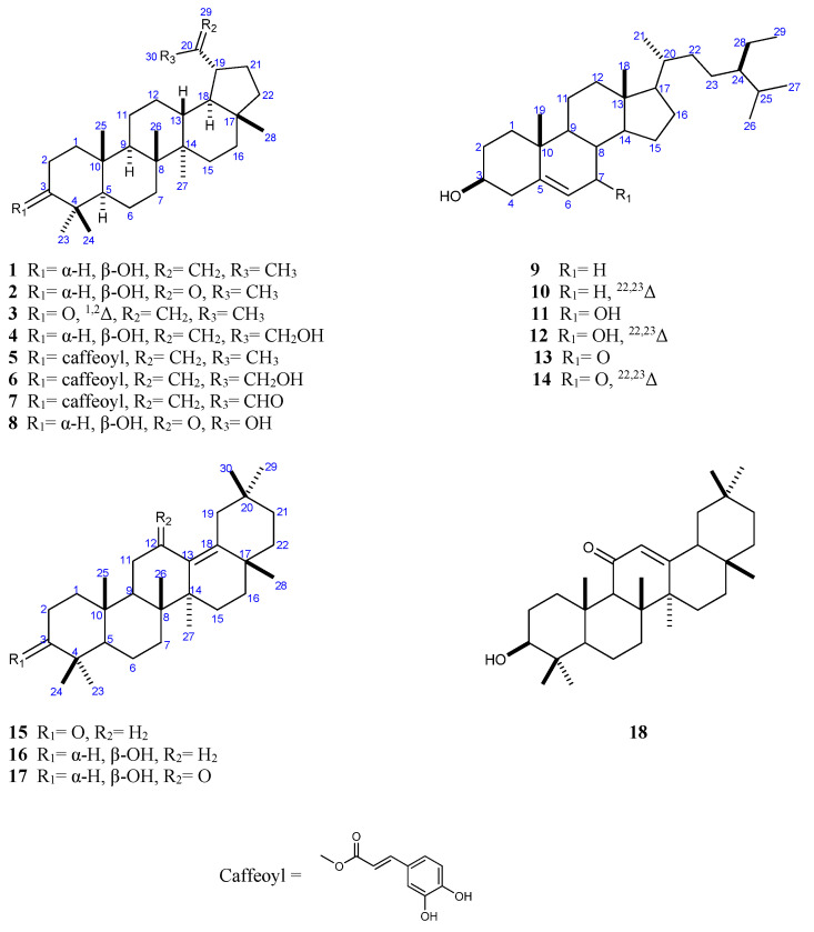
The investigation of the phytochemicals isolated from the aerial parts of D. glabra via a combination of different chromatographic methods led to the isolation of 18 triterpene and steroid compounds (1–18) (Figure 1). Two of the isolated compounds (7 and 17) are hereby reported for the first time as being isolated from a natural source, whereas the remaining compounds are hereby reported for the first time as being isolated from D. glabra [12]. The structures of the isolated compounds were elucidated via the extensive use of various spectroscopic techniques, including 1D (1H, 13C, and DEPT-135 13C) and 2D (1H–1H COSY, 1H–13C HSQC, and 1H–13C HMBC) nuclear magnetic resonance (NMR) (Figures S1–S46) and accurate mass measurements, as well as by comparing the compounds’ physical and spectral characteristics with those reported in the literature for previously-isolated compounds. The known compounds were identified as lupeol (1) [15,16,17], 30-norlup-3β-ol-20-one (2) [18], ∆1-lupenone (glochidone) (3) [19], lup-20(29)-ene-3β,30-diol (29-hydroxylupeol) (4) [20], lupeol caffeate (5) [17,21], 30-hydroxy lup-20(29)-ene 3β-caffeate (6) [22] and betunaldehyde (8) [23]; three oleanane-type compounds were also identified: δ-amyrone (15) [24], δ-amyrin (16) [24], and 11-oxo-β-amyrin (18) [25]; in addition, six sterols were identified: β-sitosterol (9) [26], stigmasterol (10) [27], 7α-hydroxy-β-sitosterol (11) [28], 7α-hydroxy-stigmasterol (12) [29], 7-keto-β-sitosterol (13) [30,31], and 7-keto-stigmasterol (14) [32]
Figure 1.
Structures of compounds (1–18) isolated from the aerial parts of Dobera glabra.
Compound 7 was obtained as a pale yellow amorphous solid. Based on the high-resolution electron ionization mass spectrometry (HREIMS) evidence, the molecular formula of this compound was determined to be C39H54O5, derived from the quasi-molecular ion peak (m/z 603.4048 [M + H]+), implying thirteen degrees of unsaturation. The infra-red (IR) spectrum of 7 included absorption bands at 3481, 1705, and 1688 cm−1, which were assigned to the OH, conjugated CO ester, and aldehydic C = O groups, respectively; the ultraviolet (UV) spectrum of 7 included absorption bands at λmax = 224, 246, 298, and 330 nm.
The 13C NMR spectrum of compound 7 (Table 1), with the aid of the Distortionless Enhancement of Polarization Transfer using a 135 degree decoupler pulse (DEPT-135) and 1H–13C heteronuclear single quantum coherence (HSQC) experiments, was comprised of the resonance signals of 39 carbons, which were identified as six methyls, 11 methylenes (ten aliphatic and one vinylic), 12 methines (five aliphatic, one O-bearing at δC 81.5, two olefinic at δC 115.8 and 145.2, three aromatic at δC 114.4, 115.5 and 122.4, and one aldehydic at δC 196.0), and 10 quaternary carbon atoms (five aliphatic carbons, one vinylic carbon at δC 157.5, three aromatic carbons at δC 127.3, 144.4, and 146.9, and one carbonyl carbon at δC 168.3). Moreover, the 1H NMR spectrum (Table 1) of 7 comprised one characteristic aldehyde proton signal at δH 9.65 (s), two deshielded exocyclic methylene protons at δH 6.48 and 6.11 (1H each, brs), one O-bearing methine proton at δH 4.72 (t, J = 8.1), and six tertiary methyl group protons at δH 0.96 (s), 0.99 (s), 1.01 (s), 1.04 (s), 1.06 (s), and 1.14 (s). In addition, two H-atom signals associated to a trans olefinic bond were observed at δH 6.39 (d, J = 15.5) and 7.70 (d, J = 15.5); these signals were accompanied by three aromatic signals assignable to a 1,3,4-trisubstitued phenyl ring at δH 7.02 (d, J = 7.5), 7.11 (d, J = 7.5), and 7.26 (brs), indicating the presence of a caffeoyl moiety [17]. The aforementioned data suggest that 7 is a pentacyclic triterpenoid caffeate.
Table 1.
1H (500 MHz) and 13C NMR (125 MHz) spectroscopic data of compounds 7 and 17 in CDCl3.
| No. | 7 | 17 | ||
|---|---|---|---|---|
| δ C | δ H | δ C | δ H | |
| 1 | 38.4 | 1.78, m | 39.5 | 1.62, m 1.01, m |
| 2 | 23.9 | 1.80, m | 27.8 | 1.61, m |
| 3 | 81.5 | 4.72, t (8.1) | 79.4 | 3.16, dd (11.0, 4.8) |
| 4 | 38.1 | - | 39.9 | - |
| 5 | 55.4 | 0.92, m | 56.7 | 0.82, m |
| 6 | 18.3 | 1.65, m 1.53, m |
19.4 | 1.67, m 1.52, m |
| 7 | 34.3 | 1.52, m | 35.0 | 1.52, m |
| 8 | 40.9 | - | 42.0 | - |
| 9 | 50.2 | 1.36, m | 51.6 | 1.74, m |
| 10 | 37.1 | - | 38.4 | - |
| 11 | 21.0 | 1.47, m 1.30, m |
41.5 | 2.30, m |
| 12 | 27.7 | 1.14, m | 209.9 | - |
| 13 | 37.8 | 1.07 | 140.6 | - |
| 14 | 42.8 | - | 46.6 | - |
| 15 | 27.4 | 1.82, m | 26.2 | 1.89, m 1.12, m |
| 16 | 35.4 | 1.65, m 1.57, m |
36.9 | 1.40, m |
| 17 | 43.4 | - | 36.2 | - |
| 18 | 50.2 | 1.36, m | 149.6 | - |
| 19 | 37.8 | 1.77, m | 40.5 | 1.67, d (12.7) |
| 20 | 157.5 | - | 34.7 | - |
| 21 | 29.8 | 1.40, m | 36.4 | 1.60, m 1.21, m |
| 22 | 40.0 | 1.58, m 1.52, m |
40.1 | 1.46, m |
| 23 | 28.1 | 1.01, s | 28.6 | 0.99, s |
| 24 | 16.8 | 1.04, s | 16.2 | 0.79, s |
| 25 | 16.2 | 0.99, s | 16.4 | 0.95, s |
| 26 | 16.0 | 1.14, s | 17.5 | 1.06, s |
| 27 | 14.5 | 1.06, s | 22.1 | 0.97, s |
| 28 | 17.9 | 0.96, s | 23.7 | 1.11, s |
| 29 | 134.2 | 6.48, brs 6.11, brs |
24.9 | 0.87, brs |
| 30 | 196.0 | 9.65, s | 32.7 | 0.93, s |
| 1’ | 168.3 | - | ||
| 2’ | 115.8 | 6.39, d (15.5) | ||
| 3’ | 145.2 | 7.70, d (15.5) | ||
| 4’ | 127.3 | - | ||
| 5’ | 114.4 | 7.26, brs | ||
| 6’ | 144.4 | - | ||
| 7’ | 146.9 | - | ||
| 8’ | 115.5 | 7.02, d (7.5) | ||
| 9’ | 122.4 | 7.11, d (7.5) | ||
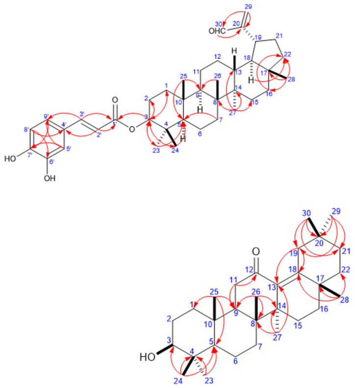
The assignments of the signals due to the methyl groups and the remaining proton and carbon signals were performed through 1H–1H COSY and 1H–13C HMBC experiments. Ultimately, these data enabled us to conclude the gross structure of 7 to be that of a lupane-type triterpenoid caffeate (Figure 2) [17,21,22]. Key 1H–13C HMBC cross-peaks were observed between H3-23, H3-24 (δH 1.01, 1.04, respectively) and C-3, C-5; H3-25 (δH 0.99) and C-5, C-9; H3-26 (δH 1.14) and C-9; H3-27 (δH 1.06) and C-8, C-13, C-14, and C-15; H3-28 (δH 0.96) and C-16, C-17, and C-22; and H2-29/H-30 and C-20. Based on the above-detailed observations, the planar structure of the triterpenoid moiety of 7 was identified to be 3β-hydroxylup-20(29)-en-30-al. The caffeoyl unit is proposed to be linked to the C-3 of the triterpenoid moiety, given that the NMR resonance of H-3 was downfield-shifted to δH 4.72 (t, J = 8.1) in 7 with respect to compound 1 (δH 3.16 (dd, J = 8.1)), and the resonance of C-3 was downfield-shifted to δC 81.5 in 7 compared to δC 79.04 in 1. This conclusion was ultimately confirmed by the observed HMBC correlation between H-3 and C-1’ (δC 168.3). Furthermore, the aldehyde group proton (δH 9.65 (s)) was assigned at C-30, as inferred via the 2JCH HMBC correlation between H-30 and C-20 (δC 157.5), as well as the 3JCH HMBC correlation between vinylic protons H2-29 and C-30. Accordingly, 7 was elucidated to be lup-20(29)-en-30-al 3β-caffeate.
Figure 2.
Key Heteronuclear multiple-bond correlation spectroscopy correlations (H → C) in compounds 7 and 11.
Compound 17 was obtained as a white amorphous solid. Its molecular formula was determined to be C30H48O2, based on the HR-ESI-MS data (m/z 441.3733 ([M + H]+; calc. 441.3733), implying seven degrees of unsaturation. The IR spectrum of 17 included absorption bands at 3436 and 1663 cm−1, which were assigned to a hydroxyl group and a conjugated ketone, respectively.
The 1H NMR spectrum of 17 included resonance signals due to one oxygenated methine at δH 3.16 (1H, dd, J = 11.0, 4.8 Hz, H-3), and eight methyl protons at δH 0.79 (3H, s, H-24), 0.87 (3H, s, H-29), 0.93 (3H, s, H-30), 0.95 (3H, s, H-25), 0.97 (3H, s, H-27), 0.99 (3H, s, H-23), 1.06 (3H, s, H-26), and 1.11 (3H, s, H-28). The 13C NMR spectrum of 17 was comprised of resonance signals due to 30 carbons, including a conjugated ketone carbonyl group at δC 209.9 (C-12), two olefinic carbons at δC 140.6 and 149.6 of four substituted double bonds, eight methyls at δC 32.7 (C-30), 28.6 (C-23), 24.9 (C-29), 23.7 (C-28), 22.1 (C-27), 17.5 (C-26), 16.4 (C-25), and 16.2 (C-24), three methines (two aliphatic and one O-bearing at δC 79.4), ten methylenes, and six aliphatic quaternary carbons (Table 1). The above-mentioned spectroscopic data indicate that compound 17 is a pentacyclic triterpene.
The assignment of the signals of the methyl groups and the remaining of proton and carbon signals was performed through 1H–1H COSY and 1H–13C HMBC experiments; the data collected in this way indicated the gross structure of 17 to be that of an oleane-type triterpenoid [16] (Figure 2). Key HMBC correlations were observed between H3-23, H3-24 (δH 0.99, 0.79, respectively) and C-3 (δC 79.4), C-5 (δC 56.7); H3-25 (δH 0.95) and C-1 (δC 39.5), C-5 (δC 56.7), and C-9 (δC 51.6); H3-26 (δH 1.06) and C-8 (δC 42.0), C-9 (δC 51.6), and C-14 (δC 46.6); H3-27 (δH 0.97) and C-8 (δC 42.0), C-13 (δC 140.6), C-14 (δC 46.6), and C-15 (δC 26.2); H3-28 (δH 1.11) and C-17 (δC 36.2), C-18 (δC 149.6), and C-22 (δC 40.1); H3-29 (δH 0.87)/H3-30 (δH 0.93) and C-19 (δC 40.5), C-20 (δC 34.7), and C-21 (δC 36.4). Furthermore, a key 2JCH HMBC correlation between H2-11 (δH 2.30) and C-9/C-12 (δC 209.9), accompanied by a 2JCH HMBC correlation from H2-19 (δH 1.67) to C-18, and a 3JCH HMBC correlation from H2-19/H3-27 and C-13, and from H3-28 to C-18 indicated the presence of a 12-oxo-olean 13(18)-ene system [24,33]. Based on the above observations, the structure of 17 was deduced to be that of 12-oxo-olean-13(18)-en-3β-ol (12-oxo-δ-amyrin).
2.2. Cytotoxic Activity
The antitumor activities of the different fractions and isolated compounds were tested against different cancer cell lines (HCT-116, PC-3, and HepG-2). The data indicated that compound 11 showed broad-spectrum activity on three different cell lines. Interestingly, all of the extracts exhibited good (CHCl3 extract) to moderate (n-hexane, butanol, and ethanol extracts) activity. On the other hand, most of the pure compounds (1–4, 8, and 9) displayed no activity, whereas compounds 5–7 and 13 displayed moderate activity (Table 2). Perhaps the higher activity of all of the extracts against the pure compounds was due to synergistic effect of all of the secondary metabolites in the whole extracts, rather than one compound. According to the obtained results (Table 2), the sterol nucleus, in general, exhibited stronger activity than that of lupane triterpene. For the sterol, the maximum activity was observed with the 7α-enol system. The oxidation of the 7α-hydroxy group to form the 7-enone system reduced the activity, while the activity of the lupane nucleus is improved by acylation with caffeic acid at OH-3 [13,14].
Table 2.
Cytotoxic activities of the different plant extract fractions and isolated compounds.
| Fractions/Compounds | HCT-116 IC50 (µg/mL) |
PC-3 IC50 (µg/mL) |
HepG-2 IC50 (µg/mL) |
VERO-B IC50 (µg/mL) |
|---|---|---|---|---|
| Hexane fraction | 24 ± 0.34 | 24 ± 0.34 | 24 ± 0.34 | 82 ± 1.4 |
| CHCl3 fraction | 7.1 ± 0.23 | 7.1 ± 0.23 | 7.1 ± 0.23 | 80 ± 0.2 |
| n-Butanol fraction | 16 ± 1.31 | 16 ± 1.31 | 16 ± 1.31 | 78 ± 1.4 |
| Crude ethanolic extract of stems | 18 ± 0.02 | 18 ± 0.02 | 18 ± 0.02 | 86 ± 0.5 |
| Crude ethanolic extract of leaves | 19 ± 1.46 | 19 ± 1.46 | 19 ± 1.46 | 80 ± 1.4 |
| 1 | >100 | >100 | >100 | >100 |
| 2 | >100 | >100 | >100 | >100 |
| 3 | >100 | >100 | >100 | >100 |
| 4 | >100 | >100 | >100 | >100 |
| 5 | 59 ± 0.45 | 59 ± 0.45 | 59 ± 0.45 | 81 ± 1.1 |
| 6 | 68 ± 1.72 | 68 ± 1.72 | 68 ± 1.72 | 68 ± 0.3 |
| 7 | 62 ± 1.42 | 62 ± 1.42 | 62 ± 1.42 | >100 |
| 8 | >100 | >100 | >100 | >100 |
| 9 | >100 | >100 | >100 | >100 |
| 11 | 8 ± 1.02 | 8 ± 1.02 | 8 ± 1.02 | 70 ± 0.5 |
| 13 | 22.3 ± 0.24 | 22.3 ± 0.24 | 22.3 ± 0.24 | 64 ± 1.1 |
| Cisplatin | 12.6 ± 2 | 5 ± 0.45 | 5 ± 1.5 | 11 ± 1.3 |
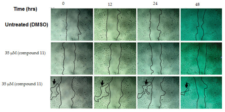
In order to investigate the selectivity of the most active compounds for cancer cell lines, and to demonstrate that they had no cytotoxic effects on normal (non-cancerous) cells, viability and wound-healing assays were performed. Over 95% cell viability (in Vero cells) was obtained at a 70 µM concentration of the most active compounds, so the biocompatibility was confirmed by conducting the wound-healing assay at either 35 or 70 µM concentrations of each compound. The cells treated with compound 11 were able to heal the wound at a rate closer to that observed for untreated cells (Figure 3). These data indicated that 11, the most active compound, is not cytotoxic to normal cells, and that it is worth investigating further for use as a safe anticancer agent.
Figure 3.
Effect of compound 11 on the wound healing of normal cells (Vero cell line). The cells were able to migrate in order to decrease the space between each other (black arrow), so that the wound size decreased after 48 h of incubation.
2.3. Possible Cytotoxicity Mechanism of Compound 11
The bioactivity of compound 11 was further investigated using phosphodiesterase, and this compound displayed an 80.2% inhibitory activity toward the mentioned enzyme in the used condition. Although high intracellular levels of cAMP have been reported to effectively inhibit the proliferation of cancer cells, compounds that cause cAMP levels to be elevated are not recommended as anticancer drugs, due to their high cytotoxicity [34,35,36].
All of the known phosphodiesterase inhibitors operate via three main types of interactions: interactions with metal ions mediated through water, H-bond interactions with protein residues involved in nucleotide recognition, and, most importantly, interactions with the enzyme’s hydrophobic residues; therefore, these interactions should guide the design of new classes of inhibitors [37].
Our research team, which consists of a multidisciplinary collection of international scientists, is developing more selective and effective anticancer agents than those that are available today, spurred on by the increasing need for safe and efficacious agents for cancer therapy. In this context, compound 11 proved to be an efficient and selective agent that is worth considering for further in vivo studies as a cancer treatment.
Phosphodiestrase Inhibition Investigation
Compound 11, which proved to be the most active and selective anticancer agent among all of the compounds tested in the present study, showed a remarkable inhibitory activity against phosphodiesterase I (PDE1), with an 80.2% inhibition of this enzyme at a 2 µM concentration of the compound.
The in vitro assay revealed 11 to be a highly selective anticancer agent, with a noticeable cytotoxic activity against colorectal cancer (HCT-116), hepatocellular carcinoma (HepG-2), and prostate cancer (PC-3) cells compared to the commonly-used chemotherapeutic cisplatin drug.
Additionally, in the PDE1 inhibition tests, compound 11 exhibited a high cytotoxic activity against colorectal, prostate, liver cancers, with high selectivity. This high selectivity and promising activity against three aggressive cancer cell lines renders compound 11 a promising anticancer agent candidate. Therefore, the use of this compound in combination with other chemotherapeutic drugs should be carefully investigated as a way to explore the possibility of developing chemotherapeutic cancer treatments with increased efficacy and reduced undesired side-effects.
3. Materials and Methods
3.1. Instrumentation and Chemicals
The Fourier-transform infrared (FT-IR) spectra were recorded on a Nicolet 5700 FT-IR Microscope spectrometer (FT-IR Microscope Transmission, company, Waltham, MA, USA). The optical rotations were measured using a Perkin-Elmer Model 341 LC polarimeter (PerkinElmer, MA, USA). The accurate mass determination was achieved with a JEOL JMS-700 High-Resolution Mass Spectrophotometer (JEOL USA Inc., Peabody, MA, USA) with a positive and negative mode. The NMR spectroscopy experiments were carried out using deuterated chloroform and an UltraShield Plus 500 (Bruker) spectrometer operating at 500 MHz for 1H and 125 MHz for 13C at the College of Pharmacy, Sattam Bin Abdulaziz University. The microplate reader used was a Coming Inc., NY, USA, ELISA BioTek L × 800 microplate. The thin layer chromatography (TLC) was performed on normal and reversed phase silica gel (Merck, Darmstadt, Germany) with a layer thickness of 250 μm and a mean particle size of 10–12 μm, with different dimentions. n-hexane:EtOAc, CHCl3:EtOAc and CHCl3:MeOH were used for the normal phase TLC, while H2O: MeOH (10:90) was used for reversed phase RP-18 TLC. Additionally, the compounds were visualized by spraying the TLC plates with 15% H2SO4/ethanol, or with anisaldehyde-sulfuric acid, followed by heating. The column chromatography was carried out on silica gel (Merck 60 A, 230–400 mesh ASTM, Darmstadt, Germany), while LiChrorep RP-18 (25–40 µm) was used for the reversed phase column chromatography.
The centrifugal preparative TLC (CPTLC) was performed on a chromatotron instrument (Harrison Research, Palo Alto, California, CA, USA). Plates coated with 1 and 2 mm of silica gel 60, 0.04–0.06 mm were used. The reagents, chemicals, and solvents were of analytical grade, and they were purchased from Sigma-Aldrich, Loba Chemie Pvt. Ltd., and SD Fine Chem. Ltd. The water was doubly distilled before use [38,39].
3.2. Plant Material
The aerial parts of D. glabra were collected from the Shoqaiq in February 2013. The specimen of the plant was identified by Mohamed Yousef, Professor of Taxonomy at the Department of Pharmacognosy, College of Pharmacy, King Saud University. A voucher specimen No. 16,036 of D. glabra was deposited at the herbarium of the Pharmacognosy Department, College of Pharmacy, King Saud University, Kingdom of Saudi Arabia. The undesirable parts of plant material were removed. The aerial parts were dried in air-shade until they reached a constant weight, after which they were ground using a toothed mill, followed by sifting using suitable mesh in order to give a homogenous particle size powder for the subsequent efficient extraction.
3.3. Extraction and Isolation
The air-dried and powdered aerial parts of D. glabra (1200 g) were extracted by maceration with 96% ethanol. The extract thus obtained was evaporated in vacuo to yield a brownish residue (28 g), which was suspended in water and subsequently partitioned with n-hexane (3 g), chloroform (4.5 g), and n-butanol (0.5 g), in succession.
The n-hexane fraction was purified by chromatography over a silica gel column and eluted with CHCl3:EtOAc mixtures of increasing polarity. On the basis of the TLC behavior, the appropriate fractions were combined in order to give seven main fractions (H1–H7). The columns were monitored by examination under a UV lamp 254/366 nm, followed by spraying with 15% H2SO4 in ethanol, followed by heating at 120 °C. Fractions H1–H3 were eluted with CHCl3:EtOAc (98:2–95:5), and yielded compounds 1 (50.0 mg), 9 (54.0 mg), and 10 (40.0 mg), after solvent treatment. Fraction H4 was purified by chromatography over an RP-18 column using CH3CN only as the eluent to afford compounds 2 (11.5 mg) and 3 (9.5 mg). Fraction H5 was eluted with CHCl3:EtOAc (98:2), and was further purified using a silica gel RP-18 column using methanol (MeOH) as the eluent to afford compounds 15 (33.6 mg) and 16 (18.7 mg). Fraction H6 was subjected to a chromatotron (CPTL, silica gel 60 GF254, 1mm, solvent: CHCl3:EtOAc (97:3)); this procedure yielded 31.0 mg of compound 13 and 12.7 mg of compound 14. Fraction H7 was eluted with CHCl3:EtOAc (90:10), and was subjected to chromatography over a silica gel RP-18 column using MeOH only as the eluent, to yield compounds 11 (3.0 mg) and 12 (1.6 mg).
The CHCl3 fraction was purified by chromatography over a silica gel column, and was eluted with CHCl3: MeOH mixtures of increasing polarity in order to yield five main fractions (C1–C5). The crystallization of C1 in CHCl3:MeOH yielded compound 4 (13.0 mg); on the other hand, the filtrate of this fraction was subjected to CPTLC (silica gel 60 GF254, 1 mm, solvent: hexane:EtOAc (80:10)) to yield 5 mg of compound 18. Fraction C2 was purified by CPTLC (silica gel 60 GF254, 1 mm, solvent: hexane:EtOAc (70:30)) to give compound 8 (8.7 mg).
Fractions C3 and C4 were re-chromatographed separately over a silica gel RP-18 column, using MeOH only as the eluent, to yield compounds 5 (14.0 mg), 6 (10.1 mg), and 7 (10.5 mg). The elution of the silica gel RP-18 column of fraction C5 with H2O: MeOH (10:90) afforded compound 17 (5.7 mg).
3.4. Cytotoxic Activity
3.4.1. Cell Lines and Tested Compounds
The cytotoxic activity of the different fractions and the isolated compounds was tested against different human cancer cells—that is, prostate carcinoma cells (PC-3), hepatocellular carcinoma cells (HepG-2), and colorectal cancer cell (HCT-116)—as well as against the African green monkey kidney cell line (Vero-B). The cell lines were obtained from the American Type Culture Collection. The cells were cultivated at 37 °C and 10% CO2 in Dulbecco’s Modified Eagle Medium (Lonza, 12-604F) supplemented with 10% fetal bovine serum (Lonza, Cat. No.14-801E), 100 IU/mL pencillin and 100 µg/mL streptomycin (Lonza, 17-602E). Cisplatin (cis-diamineplatinum (II) dichloride), obtained from sigma, was dissolved in 0.9% saline, then stored as an 8 mM stock solution at −20 °C and used as the positive control.
The tested compounds were solubilized in dimethyl sulfoxide (DMSO) and stored at −20 °C. A 0.5% solution of crystal violet was prepared in MeOH and used to stain the viable cells [40,41,42,43]. Notably, crystal violet binds to proteins and DNA in adherent and viable cells, so this staining is indicative of the viability of the treated cells. The viability of the cells was quantified using the MTT reagent, which contains 3-(4,5-dimethylthiazol-2-yl)-2,5-diphenyl tetrazolium bromide, and measures the activity of mitochondrial dehydrogenase in viable cells.
3.4.2. Cell Cultures
The cells were seeded in a 96-well plate as 5 × 10⁴ cells/mL (100 µL/well). In total, 100 µL/well of from the serial dilutions of the tested compounds and cisplatin (100, 30, 10, 3.3, 1.1, or 0.37 µM) were added to the plate after the overnight incubation of the cells at 37 °C and 5% CO2. DMSO was used as a control (0.1%). The cells were incubated for 48 h. Subsequently, 15 µl MTT (5 mg/mL PBS, phosphate buffered saline) was added to each well, and the plate was incubated for another 4 h. The formazan crystals were solubilized in 100 µL acidified sodium dodecyl sulfate (SDS) solution (10% SDS/0.01 M HCl). After 14 h of incubation at 37 °C and 5% CO2, the absorbance of the wells was measured at 570 nm using a Biotech plate reader. Each experiment was repeated three times, and the standard deviation was calculated (±). The concentration that caused a 50% inhibition of the cell growth (IC50) was calculated for each compound or fraction. The growth of the cells was monitored and the images were acquired using Gx microscopes (GXMGXD202 Inverted Microscope) (10x Eyepiece) after staining with crystal violet [44].
3.5. Phosphodiestrase Inhibition Investigation
The phosphodiesterase I inhibition assay was performed using snake venom according to a previously-reported method, with minute variations. Briefly, Tris–HC1 buffer 33 mM at pH 8.8 (97 μL), 30 mM Mg acetate with an enzyme concentration of 0.742 µU well-1, and 0.33 mM bis-(p-nitrophenyl) phosphate (Sigma N-3002, 60 μL) as the substrate were taken. An EDTA solution characterized by an IC50 ± SD value of 274 ± 0.007 µM was used as the positive control. After a pre-incubation period of 30 min, the enzyme with the test samples was observed spectrophotometrically in order to detect its enzymatic activity on a microtitre plate reader at 37 °C. In particular, the rate at which the optical density of the sample changed (in min−1) was followed at 410 nm, which is a wavelength absorbed by the p-nitrophenol released from p-nitrophenyl phosphate, a reaction known to be catalyzed by phosphodiesterase I. All of the assays were processed in triplicate [34,35,36,37].
3.6. Wound-Healing Assay
WI-38 cells were seeded in a 6-well plate at 20 × 104 cells per ml (2 mL in each well), which was incubated overnight at 37 °C and 5% CO2. During the second day, a scratch was created in each well with a p200 tip; the medium was then replaced with fresh medium containing either DMSO or different concentrations of the most active compounds. Images were recorded at different time points (0, 4, 24, and 48 h) in order to monitor the wound closure. Subsequently, the cells were washed twice with ice-cold 1X PBS and fixed with ice-cold MeOH for 20 min at 4 °C. The fixed cells were washed twice with 1X PBS and stained with 0.5% crystal violet for 30 min. Any unreacted crystal violet was washed off with distilled H2O until no color was observed in the washing. The size of the wound was measured using Image J1.47 software [45].
3.7. Analytical Data for Compound 7
Compoud 7 is a pale yellow amorphous solid (10.5 mg); [α]23D +17.4° (c 0.5, MeOH); UV λmax MeOH nm (log ε): 224 (4.13), 246 (3.76), 298 (3.55), and 330 (3.82). IR (KBr) vmax 3481, 2938, 1705, 1688, 1617, 1522, 1447, 1385, 1267, 1183, 871 cm−1; 1H and 13C NMR (see Table 1 and Table 2); HR-ESI-MS [M + H]+ m/z 603.4048 (calculated for C39H54O5, 603.4049).
3.8. Analytical Data for Compound 17
Compound 17 is a white amorphous solid (5.7 mg); [α]23D +31° (c 0.8, CHCl3); UV λmax MeOH nm (log ε): 253 (3.84). IR (KBr) vmax 3436, 2941, 2865, 1663, 1610, 1460, 1385, 1253, 1177 cm−1; 1H and 13C NMR (see Table 1 and Table 2); HR-ESI-MS [M + H]+ m/z 4413.3733 (calculated for C30H48O2, 441.3733).
4. Conclusions
Two compounds (7 and 17) were isolated for the first time from the aerial parts of D. glabra, and from a natural source. Additionally, a series of cytotoxic triterpenes and sterols, which were not previously known to be found in D. glabra, were also isolated from the mentioned plant parts. The structures of the new triterpenes were determined using a range of spectroscopic techniques, including high-resolution mass spectrometry. The different plant extracts and some of the isolated compounds were tested for their cytotoxic activity against colorectal cancer (HCT-116), hepatocellular carcinoma (HepG-2), and prostate cancer (PC-3) cell lines. The cytotoxic potency and selectivity of the bioactive compounds were investigated by the phosphodiestrase enzyme inhibition method. Compound 11 displayed a remarkable inhibitory activity against PDE1. The use of this compound in combination with other chemotherapeutic drugs should thus be investigated, in order to explore the possibility of producing chemotherapeutic cancer treatments characterized by increased efficacy and reduced undesired side effects.
Currently, a more rigorous in vivo study is underway, which is directed at obtaining more preclinical information, such as oral stability, bioavailability, and pharmacokinetic data, with the anticipation of better activity and a high safety margin.
Acknowledgments
The authors would like to thank the Deanship of Scientific Research at King Saud University for funding the work through the Research Group Project No. RG 1437-021.
Supplementary Materials
The following are available online at https://www.mdpi.com/2223-7747/10/1/119/s1, Figure S1: Structures of compounds (1–18) isolated from D. glabra.; Figures S2–S46 NMR spectral data (1D and 2D for compounds 1–8 and 13–18.
Author Contributions
W.M.A.-M. isolated the compounds, interpreted the NMR data, revised the manuscript, and prepared the supplementary material. A.A.E.-G. and S.M.A.-M. isolated the compounds, wrote the manuscript, performed the investigation, assigned the spectral data, and designed the practical section. O.A.B. and M.S.A.-K. measured, interpreted, and assigned the NMR data, and helped in preparing/revising the manuscript. F.A.B. measured and discussed the biological activity section and revised the manuscript. A.J.A.-R. collected the plant, and wrote and revised the manuscript. H.Y.A. prepared and revised the manuscript. All authors have read and agreed to the published version of the manuscript.
Funding
This work was funded by the Deanship of Scientific Research at King Saud University through the Research Group Project No. RG 1437-021.
Conflicts of Interest
The authors declare no conflict of interest.
Footnotes
Publisher’s Note: MDPI stays neutral with regard to jurisdictional claims in published maps and institutional affiliations.
References
- 1.Vogt K. A Field Worker’s Guide to the Identification, Propagation and Uses of Common Trees and Shrubs of Dryland Sudan. SOS Sahel International; London, UK: 1996. p. 167. [Google Scholar]
- 2.Group A.P. An update of the Angiosperm Phylogeny Group classification for the orders and families of flowering plants: APG III. Bot. J. Linn. Soc. 2009;161:105–121. [Google Scholar]
- 3.Al-Aklabi A., Al-Khulaidi A.W., Hussain A., Al-Sagheer N. Main vegetation types and plant species diversity along an altitudinal gradient of Al-Baha region, Saudi Arabia. Saudi J. Biol. Sci. 2016;23:687–697. doi: 10.1016/j.sjbs.2016.02.007. [DOI] [PMC free article] [PubMed] [Google Scholar]
- 4.Aref I., El Atta H., Al Ghtani A. Ecological study on Dobera glabra Forssk. At Jazan region in Saudi Arabia. J. Hortic. For. 2009;1:198–204. [Google Scholar]
- 5.Alfarhan A., Al-Turki T., Basahy A. Flora of Jizan Region. Volume 1. King Abdulaziz City for Science and Technology (KACST); Riyadh, Saudi Arabia: 2005. p. 237. [Google Scholar]
- 6.Tsegaye D., Balehgn M., Gebrehiwot K., Haile M., Gebresamuel G., Aynekulu E. The Role of ‘Garsa’ (Dobera glabra) for Household Food Security at Times of Food Shortage in Aba’ala Wereda, North Afar: Ecological Adaptation and Socio-Economic Value: A Study from Ethiopia. DCG; New York City, NY, USA: 2007. [Google Scholar]
- 7.Guinand Y., Lemessa D. Wild Food Plants in Southern Ethiopia: Reflections on the Role of “Famine Foods” at a Time of Drought. UNDP-EUE Field Mission Report; Addis Ababa, Ethiopia: 2000. [Google Scholar]
- 8.Swaleh A. Ethnoveterinary Medicine, in Ormaland-Kenya. Dissertation Submitted in Partial Fulfillment of the MSc. Centre for Tropical Veterinary Medicine (CTVM), University of Edinburgh; Edinburgh, UK: 1999. [Google Scholar]
- 9.Heywood V.H., Moore D.M., Richardson I.B.K., Sterrn W.T. Salvadoraceae. Flowering Plants of the World, Croom Helm London & Sydney. Kew Science; Wakehust, UK: 2001. [Google Scholar]
- 10.Leslie A.A., Jarvis P.G. Agroforesty practices in Somalia. For. Ecol. Manag. 1991;45:298–308. doi: 10.1016/0378-1127(91)90224-J. [DOI] [Google Scholar]
- 11.Barrow E.G.C. Evaluating the effectiveness of participatory agroforestry extension programmes in a pastoral system, based on existing traditional values. A case study of the Turkana in Kenya. Agroforesty Syst. 1991;14:1–21. doi: 10.1007/BF00141594. [DOI] [Google Scholar]
- 12.Elkhateeb A., Abdel Latif R.R., Marzouk M.M., Hussein S.R., Kassem M.E.S., Khalil W.K.B., El-Ansari M.A. Flavonoid constituents of Dobera glabra leaves: Amelioration impact against CCl4-induced changes in the genetic materials in male rats. Pharm. Biol. 2017;55:139–145. doi: 10.1080/13880209.2016.1230879. [DOI] [PMC free article] [PubMed] [Google Scholar]
- 13.Wang W.-S., Gao K., Wang C.M., Jia Z.-J. Cytotoxic triterpenes from Ligulariopsis shichuana. Pharmazie. 2003;58:148–150. doi: 10.1002/chin.200324151. [DOI] [PubMed] [Google Scholar]
- 14.Wu S.-B., Bao Q.-Y., Wang W.-X., Zhao Y., Xia G., Zhao Z., Zeng H., Hu J.-F. Cytotoxic Triterpenoids and Steroids from the Bark of Melia azedarach. Planta Med. 2011;77:922–928. doi: 10.1055/s-0030-1250673. [DOI] [PubMed] [Google Scholar]
- 15.Reynolds W.F., McLean S., Poplawski J., Enriquez R.G., Escobar L.I., Leon I. Total assignment of carbon-13 and proton spectra of three isomeric triterpenol derivatives by 2D NMR: An investigation of the potential utility of proton chemical shifts in structural investigations of complex natural products. Tetrahedron. 1986;42:3419–3428. doi: 10.1016/S0040-4020(01)87309-9. [DOI] [Google Scholar]
- 16.Mahato S.B., Kundu A.P. 13C NMR Spectra of pentacyclic triterpenoids–A compilation and some salient features. Phytochemistry. 1994;37:1517–1575. doi: 10.1016/S0031-9422(00)89569-2. [DOI] [Google Scholar]
- 17.Fuchino H., Satoh T., Tanaka N. Chemical evaluation of Betula species in Japan. I. Constituents of Betula ermanii. Chem. Pharm. Bull. 1995;43:1937–1942. doi: 10.1248/cpb.43.1937. [DOI] [Google Scholar]
- 18.Koul S., Razdan T.K., Andotra C.S., Kalla A.K., Koul S., Taneja S.C., Dhar K.L. Koelpinin-A, B and C– three triterpenoids from Koelpinia linearis. Phytochemistry. 2000;53:305–309. doi: 10.1016/S0031-9422(99)00490-2. [DOI] [PubMed] [Google Scholar]
- 19.Puapairoj P., Naengchomnong W., Kijjoa A., Pinto M.M., Pedro M., Nascimento M.S.J., Silva A.M.S., Herz W. Cytotoxic activity of lupane-type triterpenes from Glochidion sphaerogynum and Glochidion eriocarpum two of which induce apoptosis. Planta Med. 2005;71:208–213. doi: 10.1055/s-2005-837818. [DOI] [PubMed] [Google Scholar]
- 20.Betancor C., Freire R., Gonzalez A.G., Salazar J.A., Pascard C., Prange T. Three triterpenes and other terpenoids from Catha cassinoides. Phytochemistry. 1980;19:1989–1993. doi: 10.1016/0031-9422(80)83019-6. [DOI] [Google Scholar]
- 21.Alvarenga N., Ferro E.A. A new lupane caffeoyl ester from Hippocratea volubilis. Fitoterapia. 2000;71:719–721. doi: 10.1016/S0367-326X(00)00217-3. [DOI] [PubMed] [Google Scholar]
- 22.Ying Y.-M., Li C.-Y., Chen Y., Xiang J.-G., Fang L., Yao J.-B., Wang F.-S., Wang R.-W., Shan W.-G., Zhan Z.-J. Lupane- and Friedelane-type Triterpenoids from Celastrus stylosus. Chem. Biodivers. 2015;12:1222–1228. doi: 10.1002/cbdv.201400269. [DOI] [PubMed] [Google Scholar]
- 23.Kamble S.M., Patel H.M., Goyal S.N., Noolvi M.N., Umesh B., Mahajan U.B., Ojha S., Patil C.R. In-Silico Evidence for Binding of Pentacyclic Triterpenoids to Keap1-Nrf2 Protein-Protein Binding Site. Comb. Chem. High Throughput Screen. 2017;20:215–234. doi: 10.2174/1386207319666161214111822. [DOI] [PubMed] [Google Scholar]
- 24.He A., Wang M., Hao H., Zhang D., Lee K. Hepatoprotective triterpenes from Sedum sarmentosum. Phytochemistry. 1998;49:2607–2610. doi: 10.1016/S0031-9422(98)00434-8. [DOI] [Google Scholar]
- 25.Herath H.M.T.B., Athukoralage P.S. Oleanane Triterpenoids from Gordonia ceylanica. Nat. Prod. Sci. 1998;4:253–256. doi: 10.1080/10575630108041301. [DOI] [PubMed] [Google Scholar]
- 26.Nyemb J.N., Magnibou L.M., Talla E., Tchinda A.T., Tchuenguem R.T., Henoumont C., Laurent S., Mbafor J.T. Lipids constituents from Gardenia aqualla stapf & hutch. Open Chem. 2018;16:371–376. [Google Scholar]
- 27.Pierre L.L., Moses M.N. Isolation and Characterisation of Stigmasterol and β-Sitosterol from Odontonema strictum (Acanthaceae) J. Innov. Pharm. Biol. Sci. 2015;2:88–96. [Google Scholar]
- 28.Tasyriq M., Najmuldeen I.A., In L.L., Mohamad K., Awang K., Hasima N. 7α-Hydroxy-β-Sitosterol from Chisocheton tomentosus Induces apoptosis via dysregulation of cellular Bax/Bcl-2 ratio and cell cycle arrest by downregulating ERK1/2 activation. Evid. Based Complement. Alternat. Med. 2012;2012:765316. doi: 10.1155/2012/765316. [DOI] [PMC free article] [PubMed] [Google Scholar]
- 29.Achenbach H., Benirschke G. Joannesialactone and other compounds from Joannesia princeps. Phytochemistry. 1997;45:149–157. doi: 10.1016/S0031-9422(96)00777-7. [DOI] [Google Scholar]
- 30.Della Greca M., Monaco P., Previtera L. Studies on aquaticplants. Part XVI. Stigmasterols from Typha latifolia. J. Nat. Prod. 1990;53:1430–1435. doi: 10.1021/np50072a005. [DOI] [Google Scholar]
- 31.Pettit G.R., Numata A., Cragg G.M., Herald D.L., Takada T., Iwamoto C., Riesen R., Schmidt J.M., Doubek D.L., Goswami A. Isolation and structures of schleicherastatins 1–7 and schleicheols 1 and 2 from the teak forest medicinal tree Schleichera oleosa. J. Nat. Prod. 2000;63:72–78. doi: 10.1021/np990346r. [DOI] [PubMed] [Google Scholar]
- 32.Shu Y., Jones S.R., Kinney W.A., Selinsky B.S. The synthesis of spermine analogs of the shark aminosterol squalamine. Steroids. 2002;67:291–304. doi: 10.1016/S0039-128X(01)00161-1. [DOI] [PubMed] [Google Scholar]
- 33.Tanaka R., Matsunaga S. Triterpene constituents from Euphorbia Supina. Phytochemistry. 1988;27:3579–3584. doi: 10.1016/0031-9422(88)80772-6. [DOI] [Google Scholar]
- 34.Hirsh L., Dantes A., Suh B.S., Yoshida Y., Hosokawa K., Tajima K., Kotsuji F., Merimsky O., Amsterdam A. Phosphodiesterase inhibitors as anti-cancer drugs. Biochem. Pharmacol. 2004;68:981–988. doi: 10.1016/j.bcp.2004.05.026. [DOI] [PubMed] [Google Scholar]
- 35.Lugnier C. Cyclic nucleotide phosphodiesterase (PDE) superfamily: A new target for the development of specific therapeutic agents. Pharmacol. Ther. 2006;109:366–398. doi: 10.1016/j.pharmthera.2005.07.003. [DOI] [PubMed] [Google Scholar]
- 36.Bischoff E. Potency, selectivity, and consequences of non-selectivity of PDE inhibition. Int. J. Impot. Res. 2004;16(Suppl. S1):S11–S14. doi: 10.1038/sj.ijir.3901208. [DOI] [PubMed] [Google Scholar]
- 37.Card G.L., England B.P., Suzuki Y., Fong D., Powell B., Lee B., Luu C., Tabrizizad M., Gillette S., Ibrahim P.N., et al. Structural basis for the activity of drugs that inhibit phosphodiesterases. Structure. 2004;12:2233–2247. doi: 10.1016/j.str.2004.10.004. [DOI] [PubMed] [Google Scholar]
- 38.Muhammad I., Samoylenko V., Machumi F., Zakia M.A., Mohammed R., Hetta M.H., Gillum V. Preparation and application of reversed phase Chromatorotor for the isolation of natural products by centrifugal preparative chromatography. Nat. Prod. Commun. 2013;8:311–314. [PubMed] [Google Scholar]
- 39.Kulkarni N., Mandhanya M., Jain D.K. Centrifugal thin layer chromatography. Asian J. Pharm. Life Sci. 2011;1:294–300. [Google Scholar]
- 40.Mohamed N.H., Liu M., Abdel-Mageed W.M., Alwahibi L.H., Dai H., Ismail M.A., Badr G., Quinn R.J., Liu X., Zhang L., et al. Cytotoxic cardenolides from the latex of Calotropis procera. Bioorg. Med. Chem. Lett. 2015;25:4615–4620. doi: 10.1016/j.bmcl.2015.08.044. [DOI] [PubMed] [Google Scholar]
- 41.Feoktistova M., Geserick P., Leverkus M. Crystal violet assay for determining viability of cultured cells. Cold Spring Harb. Protoc. 2016;2016:pdb.prot087379. doi: 10.1101/pdb.prot087379. [DOI] [PubMed] [Google Scholar]
- 42.El-Gamal A.A., Al-Massarani S.M., Shaala L.A., Alahdald A.M., Al-Said M.S., Ashour A.E., Kumar A., Abdel-Kader M.S., Abdel-Mageed W.M., Youssef D.T. Cytotoxic compounds from the Saudi Red Sea Sponge Xestospongia testudinaria. Mar. Drugs. 2016;14:82. doi: 10.3390/md14050082. [DOI] [PMC free article] [PubMed] [Google Scholar]
- 43.El-Gamal A.A., Al-Massarani S.M., Abdel-Mageed W.M., El-Shaibany A., Al-Mahbashi H.M., Basudan O.A., Badria F.A., Al-Said M.S., Abdel-Kader M.S. Prenylated flavonoids from Commiphora opobalsamum stem bark. Phytochemistry. 2017;141:80–85. doi: 10.1016/j.phytochem.2017.05.014. [DOI] [PubMed] [Google Scholar]
- 44.El-Naggar M.H., Mira A., Bar F.M.A., Shimizu K., Amer M.M., Badria F.A. Synthesis, docking, cytotoxicity, and LTA 4 H inhibitory activity of new gingerol derivatives as potential colorectal cancer therapy. Bioorg. Med. Chem. 2017;25:1277–1285. doi: 10.1016/j.bmc.2016.12.048. [DOI] [PubMed] [Google Scholar]
- 45.Lipton A., Klinger I., Paul D., Holley R.W. Migration of mouse 3T3 fibroblasts in response to a serum factor. Proc. Natl. Acad. Sci. USA. 1971;68:2799–2801. doi: 10.1073/pnas.68.11.2799. [DOI] [PMC free article] [PubMed] [Google Scholar]
Associated Data
This section collects any data citations, data availability statements, or supplementary materials included in this article.


