Figure 1.
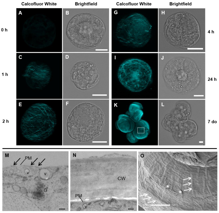
Images of the cell wall of Lolium suspension culture cells (SCC) during regeneration using the general cell wall stain Calcofluor White and laser scanning microscopy (A–L) and the cell wall detail using transmission (M,N) and scanning electron microscopy (O). The fluorescent images have had their brightness enhanced for easier visualization. Image K is a maximum projection of a z-stack of images and A, C, E, G, and I are single confocal sections of the cell wall. At the 0 h time point (A,B), minimal autofluorescence is observed. By 1 h (C,D), a number of fine filaments cover approximately half the protoplast. At 2 h (E,F), a loose fibrillar network encases the whole cell. At 4 h (G,H), the fibrillar network becomes more dense, which continues over the next 24 h (I, J). Actively growing Lolium SCC clump together in culture with a thick wall (K,L). Electron microscopy sections of the cell wall at 1 h after protoplasting (M) and in an unprotoplasted cell (N) show the difference in the cell wall structure. At 1 h, only nascent cell material is present (black arrows) at the plasma membrane (PM), while in unprotoplasted cells, the cell wall (cw) is approximately 1 μm thick. Vesicles (v) and Golgi (G) are observed near the plasma membrane (PM). Detailed structure of the wall surface (O) from a position equivalent to the box in K, showing a variety of ridges (white arrows) and mucilage (asterisks). Scale bars = 10 μm (A–L), 200 nm (M,N), and 5 μm (O).

