A 12-year-old boy presented to the oncology clinic with Ewing sarcoma of the ulna. The patient had a wide resection of the ulnar diaphysis, and we harvested his ipsilateral fibula as an autologous nonvascularized graft. During resection, we meticulously preserved the periosteal layer through a longitudinal incision with circumferential detachment from the bone, taking care to avoid interference to the local vascularization. We did not suture the periosteum after harvesting but approximated its flaps by accurate fascial closure. The fibular graft was 11 cm in length.
We were able to reconstruct the ulna in the same surgical session because contemporaneous histological examination of the residual medullary canal of the ulna showed no tumour.
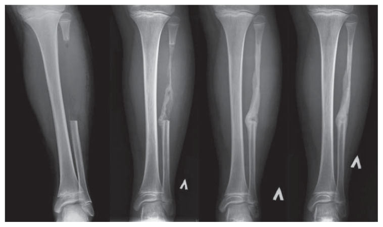
The patient was able to walk with 1 crutch and partially weight bear a few days after surgery. Full weight bearing was permitted after 3 weeks. Radiographs taken at 1, 2, 4 and 6 months after surgery showed rapid fibular bone regrowth with complete regeneration after 6 months (Figure 1).
Figure 1:
Conventional radiographs, taken at 1, 2, 4 and 6 months after surgery in a 12-year-old boy with Ewing sarcoma of the ulna, showing complete fibular regeneration after harvesting of an autologous bone graft.
Periosteum preservation is important in the bone-healing process. Agarwal and colleagues1 report similar findings, describing regeneration in 21 cases of harvested fibula in pediatric patients in whom the periosteum was preserved. Chemotherapy may harm bone healing,2 but in this case we show complete bone regeneration that occurred during postoperative chemotherapy and close to adolescence.
The periosteum is well recognized as a source of osteoprogenitor cells, and with careful preservation, large bone defects may be repaired, especially in children, in whom it is thicker and more functional than in adults.3
Clinical images are chosen because they are particularly intriguing, classic or dramatic. Submissions of clear, appropriately labelled high-resolution images must be accompanied by a figure caption. A brief explanation (300 words maximum) of the educational importance of the images with minimal references is required. The patient’s written consent for publication must be obtained before submission.
Footnotes
Competing interests: None declared.
This article has been peer reviewed.
The authors have obtained consent from the patient’s family.
References
- 1.Agarwal A, Kumar A. Fibula regeneration following non-vascularized graft harvest in children. Int Orthop 2016;40:2191–7. [DOI] [PubMed] [Google Scholar]
- 2.Virolainen P, Inoue N, Nagao M, et al. The effect of a doxorubicin, cisplatin and ifosfamide combination chemotherapy on bone turnover. Anticancer Res 2002;22:1971–5. [PubMed] [Google Scholar]
- 3.Steiger CN, Journeau P, Lascombes P. The role of the periosteal sleeve in the reconstruction of bone defects using a non-vascularized fibula graft in the pediatric population. Orthop Traumatol Surg Res 2017;103:1115–20. [DOI] [PubMed] [Google Scholar]


