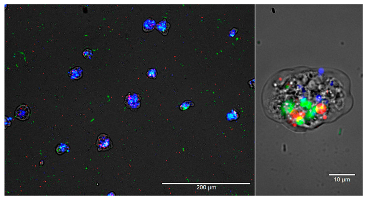
Figure 3.
Intracellular L. pneumophila interferes with W. magna’s digestion process. W. magna and L. pneumophila were co-cultured at RT, and after 24 h, E. coli TOP10 and E. coli MG1655 were added to the culture. The images represent overlaid images of the same field of view under four light channels, Monocolor transmission light channel, Green fluorescent channel, Texas-Red channel, and DAPI channel at 200× and 1000× magnification after 26 h of co-culture. L. pneumophila cells are green, E. coli MG1655 cells are red, and E. coli TOP10 cells are blue in color.

