Abstract
Introduction
Esthetic medicine is a buoyant field of medicine. As the number of performed procedures – mainly injections of botulin toxin and dermal fillers – is increasing, the number of complications is rising as well. The most popular dermal filler is hyaluronic acid. Injection of hyaluronic acid dermal fillers is considered a minimally invasive procedure, but complications in the form of skin nodules and lumps are being encountered more and frequently. Esthetic medicine does not currently offer its own diagnostic methods that would allow one to diagnose complications. In these circumstances, the implementation of objective diagnostic methods from other fields of medicine becomes significant. High-frequency ultrasound is one of such methods.
Aim of the study
The aim of this study was to implement high-frequency ultrasound for the diagnosis of palpable nodules after the administration of dermal fillers.
Material and method
The study group included 15 women who developed palpable nodules in the region of hyaluronic acid injection. The study includes both early and late complications. An EPIQ 5 (Philips, Bothell, USA) ultrasound machine and a L5–18 transducer were used to examine the nodules. Ultrasound images were evaluated qualitatively by 2 independent investigators.
Results
Ultrasound enabled the diagnosis of hyaluronic acid deposition in 9 women, granulomas in 3 women, fibrosis in 2 women and a deposition with inflammation in 1 case. Each of the diagnosed structures presented a typical ultrasound appearance.
Conclusions
High-frequency ultrasound is a useful diagnostic method that has a chance to become a widespread tool to diagnose and treat complications. Because of a low number of study reports in this area, continued research is warranted.
Keywords: high-frequency ultrasound, granulomas, deposition, hyaluronic acid, complications
Introduction
Esthetic medicine procedures are becoming more and more popular. The global market size was estimated at USD 52.5 billion in 2018, and an anticipated annual increase is 8.9%. The predicted 2026 market size is USD 103.4 billion. The causes of such dynamic development lie in the growing awareness regarding the value of an attractive physical appearance and in an increase in the population aged 25–65 years. The observed trend indicates that minimally invasive procedures are becoming more and more popular. This tendency has been confirmed in a German market report, which notes a marked increase in minimally invasive procedures compared with invasive ones. As expected, the most common minimally invasive procedures in 2015–2026 will be Botox injections followed by the use of soft tissue fillers, which unfortunately carry a risk of complications(1,2). With an increase in the popularity and number of procedures, the number of complications will increase as well, which prompts the search for the methods of their diagnosis and treatment.
Currently, the most common are biodegradable hyaluronic acid fillers. Complications that may develop after the administration of dermal fillers can be divided into early complications, i.e. occurring directly after the procedure or several days to several weeks after injection, and late complications, i.e. occurring several months and even years after injection(2,3). Complications usually develop in the region of the cheeks, lips, nasolabial folds, orbital rim, forehead, marionette lines and nose. The most common symptoms are in these cases edema, palpable nodules and pain(3,4). These symptoms are unfortunately non-specific for a given cause. Some complications, for example intra-arterial administration or vascular compression by filler deposition, skin necrosis, inflammation or blindness, are severe and may cause deformities and permanent esthetic defects. The available recommendations concerning the management of dermal filler-related complications lack guidelines on the diagnosis of the causes of these events. It is stated, however, that diagnosis and differential diagnosis of a complication can be highly challenging(3,5). This is probably associated with the fact that, apart from physical examination and medical history, no other commonly objective diagnostic methods to evaluate subcutaneous tissues are used in esthetic medicine. The dynamic technology development, including imaging techniques, that has occurred in the past years, has resulted in the emergence of diagnostic methods that enable one to assess complications. These techniques include magnetic resonance imaging (MRI) or ultrasound. However, taking into account the possibilities and limitations of the novel methods, it can be stated that only the latter has a chance to enter widespread use in the field of esthetic medicine.
The aim of this article is to present the authors’ experience in using high-frequency ultrasound in the diagnosis of palpable skin nodules after the administration of hyaluronic acid dermal fillers.
Material and method
The study group comprised 15 women aged between 30 and 70 (mean age: 43 years), who reported to a medical esthetician with symptoms following esthetic procedures involving administration of dermal fillers in the facial region. Most cases (11) were late complications that occurred several months or years after the procedure. Four cases were early complications that developed within several weeks after the procedure. In 13 cases, the clinical examination revealed the presence of multiple palpable nodules, lumps or single nodules. One woman presented with cheek overcorrection that developed immediately after the procedure and did not resolve for the next 3 years. One woman developed massive, hard thickening in the region of the lips, which occurred 10 years after the procedure. Detailed study group characteristics are presented in Tab. 1.
Tab. 1.
Study group characteristics
| No | Age (at administration) | Site of administration | Type of filler | Onset of complication | Signs and symptoms | Ultrasound diagnosis |
|---|---|---|---|---|---|---|
| 1 | 38 | lips | HA | 12 years | small, hard nodules | massive fibrosis |
| 2 | 39 | lips | HA | 3 months | hard nodules | depositions |
| 3 | 70 | cheeks | HA | 1 month | skin redness, pain and palpable nodules | depositions |
| 4 | 50 | glabella, nasolabial folds | Princess Filler | 6 months | multiple lumps | granulomas |
| 5 | 46 | lips, nasolabial folds | HA | 8 months | hard nodules, edema | granulomas |
| 6 | 30 | lips | Teosyal Kiss | 3 months | multiple, small, hard nodules | granulomas |
| 7 | 38 | cheeks | Teosyal Kiss | 7 years | hard nodules with varying size | depositions |
| 8 | 64 | lips | HA | 10 years | subtle paleness of the white roll of the lips, upper lip nodule | fibrosis in the upper lip and persistent depositions |
| 9 | 44 | chin | Juvederm Voluma | 1.5 years | chin nodule | depositions |
| 10 | 32 | cheeks, nasolabial folds | Neuvia | 2 years | multiple nodules palpable under the skin | depositions |
| 11 | 38 | lips | HA | 10 years | hard lip thickening | fibrosis |
| 12 | 34 | lips | HA | 3 weeks | dry lips and stinging sensation | inflammation, depositions |
| 13 | 47 | cheeks | HA | 3 years | overcorrection on the right side since administration | multiple depositions |
| 14 | 38 | cheeks, temporal region | HA | 1 week | Edema of the right cheek and temple | large deposition bulging the temporal muscle |
| 15 | 37 | chin | HA | 1 week | chin nodule | massive inflammatory reaction in the subcutaneous tissue |
All patients underwent skin ultrasound of the affected location. Because dermal fillers are administered at various depths, ranging from several millimeters to 1–3 cm, the examinations were performed with an EPIQ 5 (Philips, Bothell, USA) ultrasound machine with a broadband linear L18-5 transducer of variable frequency up to 18 MHz. The equipment was set to enable the highest possible image resolution. The images were evaluated in real time during the examinations and stored in the DICOM (Digital Imaging and Communications in Medicine) format in the US scanner system. In doubtful cases and for the purposes of this study, the images were re-evaluated on a working station equipped with a dedicated DICOM browser (QLAB). Because of group heterogeneity and the unavailability of quantitative parameters, the ultrasound images were evaluated qualitatively. This was done by 2 independent investigators with long experience in performing ultrasound examinations. It was aimed to search for areas with acute external borders that were well-delineated from adjacent tissues. These regions were considered depositions of hyaluronic acid (HA) dermal fillers. Moreover, it was attempted to differentiate HA deposition from granuloma based on an ultrasound presentation. Granulomas were defined as hypoechoic regions of uneven external borders or areas with a nonhomogeneous hypoechoic rim and anechoic center.
Results
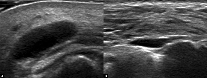
The qualitative analysis of the collected material showed that dermal filler deposition was present in 9 cases. Depositions were seen on ultrasound as anechoic, usually round regions with an even outline. When combining the medical history data with ultrasound images, the observed depositions could be divided into: persistent HA depositions that failed to absorb over time and depositions that developed shortly after the procedure and were visible and/or palpable as nodules and thus unaccepted by the patients. These depositions developed shortly after the procedure and were probably linked with improper HA administration. Unfortunately, ultrasound did not differentiate between persistent and early depositions (Fig. 1).
Fig. 1.
Ultrasound presentation of HA depositions. A.Persistent deposition within the cheek, 2 years after filling. B.Deposition that developed 1 month after filling; located within the cheek
In one case, ultrasound showed anechoic regions surrounded by hyperechoic fat; these were classified as dermal filler depositions with an inflammatory reaction.
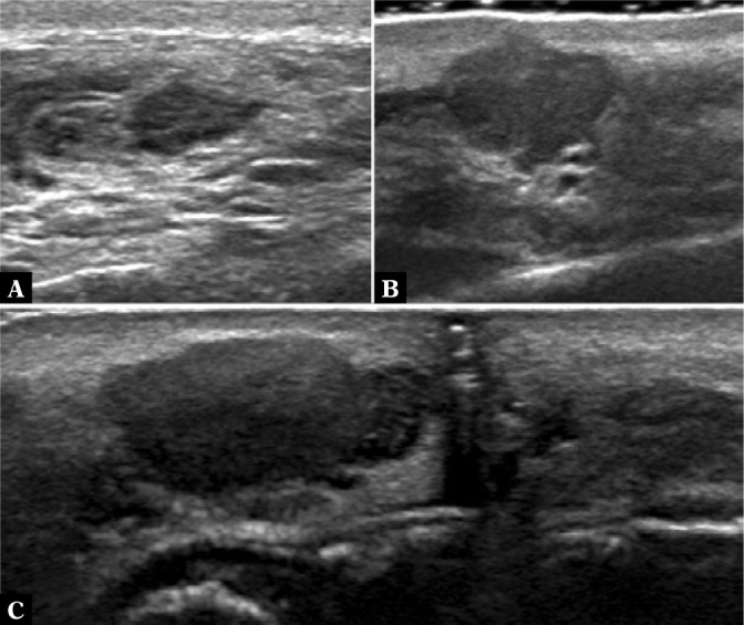
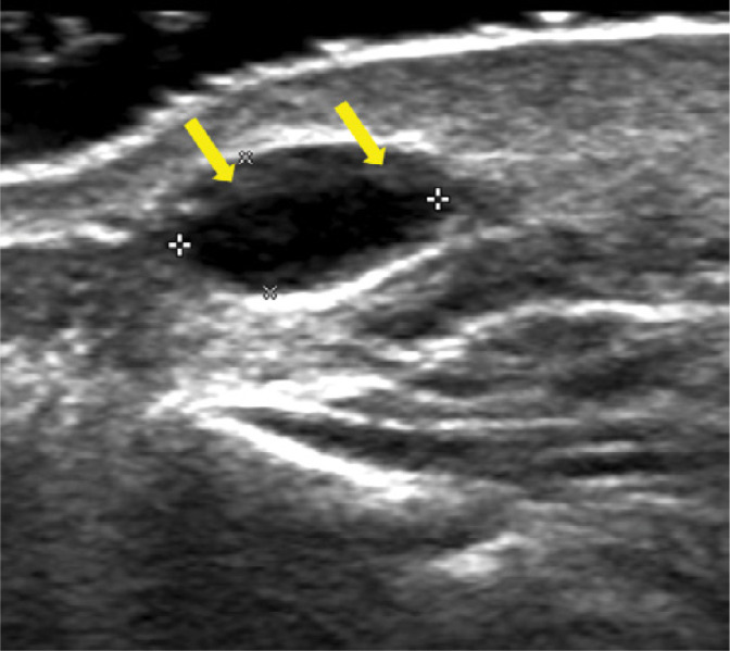
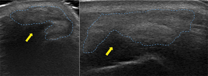
Heterogeneous hypoechoic regions of uneven external borders or areas with a nonhomogeneous hypoechoic rim and an anechoic center were classified as granulomas. These were found in 3 cases (Fig. 2). Sometimes, granulomas that arise in the inflammatory mechanism, may initially show a hypoechoic periphery and an anechoic center (Fig. 3). Two women with palpable nodules occurred to have massive fibrosis within the subcutaneous tissue. In one of these patients, only fibrosis was found on ultrasound, while the other presented with fibrosis and persistent deposition. These were visible as hyperechoic regions within the subcutaneous tissue but presented no echotexture typical of the subcutaneous tissue (Fig. 4).
Fig. 2.

Ultrasound presentation of granulomas after HA injection. A.Nasolabial folds 8 months after filling. B.Mouth corner region 6 months after filling. C.Lips 3 months after filling
Fig. 3.

A developing granuloma that arises in the mechanism of inflammation (yellow arrows)
Fig. 4.
Fibrosis (encircled with a dashed line) that developed after HA administration; acoustic shadow under the fibrotic area (yellow arrow)
Discussion
The aim of this study was to assess the performance of high-frequency ultrasound in the diagnosis of palpable nodules that developed after the administration of dermal fillers. A palpable nodule is often accompanied by other symptoms, such as skin redness, edema and pain. These are the symptoms that are troublesome to patients and prompt them to report to the physician. For further management, it is significant to determine the type of a palpable lesion.
Esthetic medicine has become a vast market and sees a continuous buoyant development. Even so, there are few reports on the diagnosis of complications in this field. The literature features few papers and recommendations with discussions devoted to the management of specific complications. However, the diagnostic issues are not included(2–5). In these circumstances, the search for and development of uniform algorithms for both diagnosis and treatment of complications after dermal filler administration remain an open issue.
To date, it has been attempted to visualize dermal fillers using MRI and ultrasound, including high-frequency ultrasound. The research results from MRI studies suggest the utility of this modality in the diagnosis of complications after dermal filler injection in the form of palpable nodules. Grippaudo et al.(6) showed that MRI is useful for assessing the filler location and for distinguishing between fillers, granulomas and fibrosis. It must be underlined that the authors also compared MR images with high-frequency ultrasound. This comparison demonstrated that MRI is not the only modality capable of illustrating complications, since ultrasound also led to the identification of filler deposits, granulomas and fibrosis. Di Girolamo et al.(7) demonstrated a statistically significant difference between granulomas and increased contrast accumulation in MRI. Recently, Niasme et al.(8) found that individual types of fillers differ in T2 relaxation, especially in MRI examinations performed with the use of 3.0T scanners, which enables their differentiation. These cited works unambiguously indicate that MRI is useful in the assessment of the skin and complications. This modality is characterized by high resolution, which helps obtain detailed images. It should be remembered, however, that MRI has a number of limitations, such as its high cost, scanning duration, artifacts or very limited access to MRI laboratories. This currently renders the usability of this method negligible in the field of esthetic medicine. There is no way to make MRI more widespread and enable broad and sometimes urgent access to this imaging examination in the case of early complications.
Considering these circumstances, the search for an alternative tool for diagnosing complications becomes justified. High-frequency ultrasound is the method that could fill the gap. This modality was selected for the present study. Ultrasound images obtained at the frequency of over 15 MHz may be useful for skin assessment due to their high resolution, which enables one to evaluate the epidermis, dermis or deeper tissue. This allows simultaneous diagnosis and implementation of immediate treatment, which is particularly significant in the case of early complications. Moreover, ultrasonography is non-invasive, safe for patients, inexpensive and broadly available. There are some studies in which ultrasound has been applied to evaluate dermal fillers or in which dermal fillers have been administered under ultrasound guidance. Some studies have also evaluated complications(9–13). It must be emphasized that all these studies draw attention to the usefulness of high-frequency ultrasound for the evaluation of dermal fillers.
Hyaluronic acid-based dermal fillers are capable of binding water, which makes them hydrophilic. Since water does not reflect ultrasounds, their image is typical and has been described by various authors. According to the proposed ultrasound nomenclature for dermal filler studies, HA depositions are visible on ultrasound images as well-delineated, round or oval, anechoic lesions without internal echoes(9–15). The results obtained by the authors of this study support the observations of other authors. The authors of this paper found it more difficult to evaluate granulomas and differentiate them from depositions based on ultrasound criteria. Unfortunately, this problem has been devoted little attention in research. The available reports describe granulomas generally as hypoechoic lesions of uneven, poorly visible borders(6,9,14). In this study, the authors distinguished granulomas from HA depositions only based on ultrasound features that had been previously reported in the literature.
The authors also diagnosed fibrosis in the examined patients. It presents on ultrasound as a large hyperechoic region that reflects ultrasound waves. Depending on the grade of fibrosis, one may observe hyperechoic regions of different sizes within normal tissues with posterior acoustic shadowing. It is also likely that ultrasound wave reflection by a massive fibrotic area is so large that the deeper structures remain concealed(6,12).
The study group also includes patients with inflammation that was visible on ultrasound as a diffuse area of increased echogenicity. Within these regions, one could also observe HA depositions. According to the recommendations found in the literature concerning the ultrasound imaging of inflammatory reactions, the color Doppler mode should be utilized(16,17). The authors did not use this mode of imaging on purpose and applied only grey-scale imaging. This form of examination was selected because esthetic medicine offices possess and use mainly high-frequency systems with single-crystal sector transducers with frequencies of 20 MHz and higher, and are not equipped with Doppler modes.
Conclusion
The studies showed that the authors managed to determine the causes of complications in the form of palpable nodules following dermal filler injection. The ultrasound images obtained in this study enabled the authors to detect all nodules that were HA depositions or granulomas, and were able to observe fibrosis and inflammation. To sum up, it can be stated that skin ultrasound is a useful method to evaluate the cause of a given complication and to institute appropriate therapeutic management. It is, however, necessary to continue research to specify standards for ultrasound and develop criteria for differential diagnosis in ultrasound.
Footnotes
Conflict of interest
Autorzy nie zgłaszają żadnych finansowych ani osobistych powiązań z innymi osobami lub organizacjami, które mogłyby negatywnie wpłynąć na treść niniejszej publikacji oraz rościć sobie do niej prawo.
References
- 1.Ugalmugle S, Swain R: Aesthetic Medicine Market Size, Share and Trends Analysis Report By Procedure Type (Invasive Procedures, Noninvasive Procedures), By Region (North America, Europe, APAC, MEA, LATAM), And Segment Forecasts, 2019–2026. Online: https://www. grandviewresearch.com/industry-analysis/medical-aesthetics-market [available: 21.10.2020].
- 2.Chiang YZ, Pierone G, Al-Niaimi F: Dermal fillers: pathophysiology, prevention and treatment of complications. J Eur Acad Dermatol Venereol 2017; 31: 405–413. [DOI] [PubMed] [Google Scholar]
- 3.Urdiales-Gálvez F, Delgado N, Figueiredo V, Lajo-Plaza J, Mira M, Moreno A. et al. : Treatment of soft tissue filler complications: expert consensus recommendations. Aesthetic Plast Surg 2018; 42: 498–510. [DOI] [PMC free article] [PubMed] [Google Scholar]
- 4.Beauvais D, Ferneini EM: Complications and litigation associated with injectable facial fillers: a cross-sectional study. J Oral Maxillofac Surg 2020; 78: 133–140. [DOI] [PubMed] [Google Scholar]
- 5.Signorini M, Liew S, Sundaram H, De Boulle K, Goodman G, Monheit G. et al. : Global aesthetics consensus: avoidance and management of complications from hyaluronic acid fillers-evidence- and opinion-based review and consensus recommendations. Plast Reconstr Surg 2016; 137: 961e–971e. [DOI] [PMC free article] [PubMed] [Google Scholar]
- 6.Grippaudo FR, Di Girolamo M, Mattei M, Pucci E, Grippaudo C: Diagnosis and management of dermal filler complications in the perioral region. J Cosmet Laser Ther 2014; 16: 246–252. [DOI] [PubMed] [Google Scholar]
- 7.Di Girolamo M, Mattei M, Signore A, Grippaudo FR: MRI in the evaluation of facial dermal fillers in normal and complicated cases. Eur Radiol 2015; 25: 1431–1442. [DOI] [PubMed] [Google Scholar]
- 8.Niasme E, Delattre BMA, Lenoir V, Modarressi A, Poletti PA, Becker M, Boudabbous S: Quantitative magnetic resonance imaging: differentiating soft tissue implants and fillers used in cosmetic and reconstructive surgery. Skeletal Radiol 2020. [DOI] [PubMed]
- 9.Schelke LW, Decates TS, Velthuis PJ: Ultrasound to improve the safety of hyaluronic acid filler treatments. J Cosmet Dermatol 2018; 17: 1019–1024. [DOI] [PubMed] [Google Scholar]
- 10.Wortsman X, Wortsman J, Orlandi C, Cardenas G, Sazunic I, Jemec GB: Ultrasound detection and identification of cosmetic fillers in the skin. J Eur Acad Dermatol Venereol 2012; 26: 292–301. [DOI] [PubMed] [Google Scholar]
- 11.Iwayama T, Hashikawa K, Osaki T, Yamashiro K, Horita N, Fukumoto T: Ultrasonography-guided cannula method for hyaluronic acid filler injection with evaluation using laser speckle flowgraphy. Plast Reconstr Surg Glob Open 2018; 6: e1776. [DOI] [PMC free article] [PubMed] [Google Scholar]
- 12.Schelke LW, Van Den Elzen HJ, Erkamp PP, Neumann HA: Use of ultrasound to provide overall information on facial fillers and surrounding tissue. Dermatol Surg 2010; 36 Suppl 3: 1843–1851. [DOI] [PubMed] [Google Scholar]
- 13.Rocha LPC, de Carvalho Rocha T, de Cássia Carvalho Rocha S, Henrique PV, Manzi FR, Silva MRMA: Ultrasonography for long-term evaluation of hyaluronic acid filler in the face: A technical report of 180 days of follow-up. Imaging Sci Dent 2020; 50: 175–180. [DOI] [PMC free article] [PubMed] [Google Scholar]
- 14.Scotto di Santolo M, Massimo C, Tortora G, Romeo V, Amitrano M, Brunetti A. et al. : Clinical value of high-resolution (5–17 MHz) echocolor Doppler (ECD) for identifying filling materials and assessment of damage or complications in aesthetic medicine/surgery. Radiol Med 2019; 124: 568–574. [DOI] [PubMed] [Google Scholar]
- 15.Schelke LW, Cassuto D, Velthuis P, Wortsman X: Nomenclature proposal for the sonographic description and reporting of soft tissue fillers. J Cosmet Dermatol 2020; 19: 282–288. [DOI] [PubMed] [Google Scholar]
- 16.Wortsman X: Ultrasound of the subcutaneous tissue. In: Humbert P, Fanian F, Maibach H, Agache A: Agache’s Measuring the Skin. Springer, 2017. [Google Scholar]
- 17.O’Rourke K, Kibbee N, Stubbs A: Ultrasound for the evaluation of skin and soft tissue infections. Mo Med 2015; 112: 202–205. [PMC free article] [PubMed] [Google Scholar]


