Abstract
Objective
Considering the high prevalence of leiomyoma and endometrial polyps, investigating the contributing factors and determining the pathophysiology of these lesions are essential. Target therapy is now an acceptable method for the treatment of some diseases. We aimed to determine the expression of transforming growth factor (TGF)-β1 in endometrial polyps and leiomyomas to discover a drug-based method to overcome surgical treatments.
Methods
In this cross-sectional study, 55 patients with leiomyoma and 55 patients with polyps were included. Prepared slides from leiomyoma and adjacent myometrium or polyp lesions and adjacent endometrium were obtained and investigated for TGF-β1. Then, data were collected and analyzed using SPSS version 22.
Results
The mean age of participants was 40.6±5.8 years. Based on their reports, 88.2% (n=97) of patients in the study population had abnormal uterine bleeding with similar distributions among both groups. In contrast, 63.5% of the leiomyoma group did not express TGF-β1. However, in normal myometrium, 23.6% had the highest degree of TGF-β1 expression. Polyp tissue did not show staining for TGF-β1 in any patients. Additionally, 89.1% of non-polypoid endometrium did not express TGF-β1. Normal tissue had a significantly greater amount of TGF-β1 compared to leiomyoma and endometrial polyps.
Conclusion
TGF-β1 is expressed more prominently in normal myometrium with mostly high-intensity features compared to leiomyoma. Additionally, polyps showed no staining for TGF-β1, while normal endometrium showed a low-density staining pattern.
Keywords: Cytokines, Polyps, Leiomyoma, Transforming growth factor beta, Uterus
Introduction
Uterine leiomyoma, known as fibroids, is experienced by many women after reaching the age of 50. It affects patients’ quality of life by causing abnormal uterine bleeding (AUB), pain, and infertility [1–3]. Uterine fibroids originate from mesenchymal cells that are affected by gonadal hormones, such as progesterone and 17 beta estradiol, in addition to cytokines including transforming growth factor (TGF)-β [1,4–8]. Uterine polyps are benign lesions of the uterus derived from overgrowth of the endometrial gland and stroma that can cause AUB [9]. Although its definite cause is unknown, some hormonal imbalance, immune mechanisms, and cytokines such as TGF-β (mainly TGF-β1) are attributed to polyp development [10–13].
TGF-β, which is released from cells to the extracellular matrix, has 3 isoforms: TGF-β1, TGF-β2, and TGF-β3. Binding of TGF-β to its receptor followed by activation of this cytokine by proteases leads to phosphorylation of proteins such as Smads, which are known as signal transducers involved in TGF-β receptor pathways. Consequently, the transmission of transcription messages to the cell nucleus takes place [7]. Moreover, TGF-β can play a key role in both SMAD-mediated and non-SMAD-mediated pathways [14]. TGF-β1 is recognized to play a role in fibrotic processes, proliferation progression, and alteration of the fibroblast phenotype, all of which affect both the myometrium and endometrium, leading to leiomyoma and polyp development, respectively [7,13,15,16]. The development of uterine leiomyoma is directly or secondarily affected by TGF-β via altering environmental estrogen interactions [17]. This cytokine has 3 isoforms with TGF-β3 as the major subtype expressed in fibroids [4]. Meanwhile, TGF-β1 is more highly expressed in endometrial polyps than in normal endometrium [13]. It is recognized that among patients with adenomyosis, TGF-β1, which is expressed in endometrial stromal cells, leads to increased collagen production by affecting endometrial fibroblasts [15].
To the best of our knowledge, there is limited data on the role of TGF-β1 in polyps [12,13,15]. In addition, there are some controversies regarding the levels of TGF-β1 and other subtypes of TGF-β in leiomyoma [4–6,18]. By considering the role of TGF-β as a target therapy agent in endometrial cancer [19] and recognizing the role of TGF-β in the development of leiomyoma and polyps, a door can be opened to non-surgical treatment of these diseases in order to overcome the financial, economic, social, and marital burden of hysterectomy as the distinct approach to manage leiomyoma [1] and hysteroscopy as the major method to treat polyps [11]. The aim of this study was to determine the association of TGF-β1 expression with endometrial polyps and leiomyoma to discover a drug-based method to overcome burdensome surgical treatments that cause concern for patients.
Materials and methods
1. Study design
In this cross-sectional study, 110 pathologically documented patients were studied (55 patients with leiomyoma and 55 patients with polyps). Based on Xuebing et al.’s study [20], considering a mean difference of 1.6, standard deviation (SD) of 2.8 in each group, and type 1 and type 2 errors of 0.05 and 0.20, respectively, the minimum required sample size was calculated as 50 in each group by applying the following formula:
where alpha and beta are type one and type 2 errors, S represents standard deviation, and μ denotes the mean of the 2 groups.
All patients were referred to the obstetric clinic of Hazrat Zeinab Hospital affiliated with Shiraz University of Medical Sciences from November 2016 to May 2017. After diagnosis of leiomyoma or polyps by ultrasonography, pathology sampling by surgery and immunohistochemical (IHC) staining for TGF-β were performed.
Patients with any type of malignancy, comorbidity, chronic liver or renal diseases, any organ failure, atypical change in pathological report of polyp or leiomyoma, leiomyoma and polyp coexistence, and previous administration of hormonal therapy or other treatments were excluded from this study.
Demographic data including age, history of AUB, parity, and body mass index were recorded in data collection forms. Then, prepared slides of leiomyoma and adjacent myometrium or polyp lesions and adjacent endometrium were obtained.
2. Immunohistochemical staining
Formalin-fixed, paraffin-embedded blocks were sectioned at 5 μm thickness. Then, IHC staining for TGF-β was performed using the avidin-biotin peroxidase complex method. The TGF-β1 primary antibody was a mouse monoclonal antibody (TGF β1 (3C11): SC-130348, dilution 1:200; Santa Cruz Biotechnology, Dallas, TX, USA). The peroxidase system was applied for secondary antibody using envision (biotinylated link anti-mouse and anti-rabbit) and diaminobenzidine (DAB 3000; DAKO, Glostrup Kommune, Denmark).
3. Immunohistochemical scoring
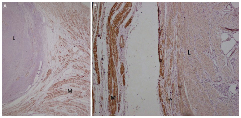
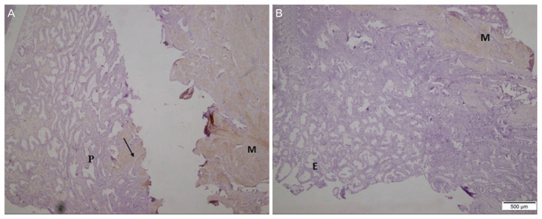
The sections stained by IHC were examined by 2 blinded pathologists. Cytoplasmic staining of TGF-β was described semi-quantitatively, where grade 0 showed less than 1% of cells as positive; grade 1, 1–10% of cells; grade 2, 11–25% of cells; grade 3, 26–50% of cells; grade 4, 51–75% of cells; and grade 5, more than 75% of cells were positive. The intensity of staining was categorized as strong (third degree), average (second degree), and poor (first degree). In this way, positive or negative results of TGF-β protein were compared between leiomyoma and normal myometrium or between polyp lesions and normal endometrium (Figs. 1 and 2).
Fig. 1.
Transforming growth factor (TGF)-β1 in myometrium and leiomyoma. (A) Expression of TGF-β1 in myometrial smooth muscle (M) and non-expression in leiomyoma cells (L). Hematoxylin and eosin (H&E), ×100. (B) Expression of TGF-β1 in myometrial smooth muscle with +3 staining intensity (M) and low staining intensity (+1) in leiomyoma (L). H&E, ×100.
Fig. 2.
Transforming growth factor (TGF)-β1 in the endometrium and polyps. (A) Expression of TGF-β1 in the smooth muscle of the arterial wall (arrow) and myometrium (M) and non-expression in the endometrial polyp (P). Hematoxylin and eosin, ×100. (B) Expression of TGF-β1 in the myometrium and non-expression in the endometrium.
4. Statistical analysis
Data were collected and analyzed using SPSS version 22 (IBM Corporation, Armonk, NY, USA). Mean, median, and SD data were analyzed descriptively. To compare groups and study the relationship between qualitative variables, χ2 and independent t-tests were used.
Results
Our study population consisted of 110 patients: 55 subjects with leiomyoma and 55 patients with polyps. The mean age of the participants was 41.709±4.562 in the leiomyoma group and 39.781±5.886 in the polyp lesion group. Based on patient reports, 88.2% (n=97) of patients in the study population had AUB. The distribution of AUB was similar in both groups in that 85.45% (n=47) of patients in the leiomyoma group and 90.9% (n=50) in the polyp group had AUB (P-value=0.55) (Table 1).
Table 1.
Age, abnormal uterine bleeding, body mass index and parity distribution among leiomyoma and polyp groups
| Characteristics | Leiomyoma | Polyp |
|---|---|---|
| Age | 43.1±4.7 | 38.1±5.8 |
| Abnormal uterine bleeding | 47 (85.45) | 50 (90.9) |
| Body mass index | 27.44±4.67 | 26.21±4.74 |
| Low weight: under18.5 | 0 (0) | 1 (1.8) |
| Normal weight: 18.5–24.9 | 15 (27.3) | 24 (43.63) |
| Over weight: 25–29.9 | 28 (50.9) | 18 (32.7) |
| Moderately obese: 30–34.9 | 7 (12.7) | 10 (18.2) |
| Severely obese: over 35 | 5 (9.1) | 2 (3.6) |
| No. of parity | ||
| 0 | 14 (25.5) | 8 (14.5) |
| 1 | 7 (12.7) | 10 (18.2) |
| 2 | 9 (16.4) | 16 (29.1) |
| 3 | 11 (20) | 14 (25.4) |
| Above 3 | 14 (25.4) | 7 (12.8) |
Values are presented as mean±standard deviation or number (%).
The degree of TGF-β1 staining in lesions of leiomyoma patients and in polyp lesions is shown in Table 2. As can be seen, 63.5% of leiomyoma lesions did not stain for TGF-β1, while all normal myometrium showed TGF-β1 staining. In addition, 23.6% of normal tissue samples demonstrated the highest degree of TGF-β1 expression with no reported cases among the leiomyoma group. Meanwhile, polyp tissue did not express TGF-β1 in any patients, while 10.9% of normal endometrium samples presented expression of TGF-β.
Table 2.
Degree of transforming growth factor-β1 staining in leiomyoma and polyp lesions compared to normal tissue in both groups
| Title | 0 degree | First degree | Second degree | Third degree | Fourth degree | Fifth degree | P-value |
|---|---|---|---|---|---|---|---|
| Leiomyoma lesion | 35 (63.5) | 4 (7.3) | 9 (16.4) | 5 (9.1) | 2 (3.6) | 0 | 0.001 |
| Normal myometrium | 0 | 0 | 5 (9.1) | 20 (36.4) | 17 (30.9) | 13 (23.6) | |
| Polyp lesion | 55 (100) | 0 | 0 | 0 | 0 | 0 | 0.012 |
| Normal endometrium | 49 (89.1) | 0 | 0 | 0 | 1 (1.8) | 5 (9.1) |
Values are presented as number (%).
The intensity of staining was also classified as described above. Most (65.5%) leiomyoma lesions presented with 0 degree of staining intensity, whereas normal myometrium had a higher prevalence of third-degree intensity (41.8%). The details are shown in Table 3.
Table 3.
Intensity of transforming growth factor-β staining in leiomyoma and polyp lesions compared to normal tissue in both groups
| Title | 0 degree | First degree (poor) | Second degree (average) | Third degree (strong) | P-value |
|---|---|---|---|---|---|
| Leiomyoma lesion | 36 (65.5) | 16 (29.1) | 2 (3.6) | 1 (1.8) | 0.001 |
| Normal myometrium | 1 (1.8) | 17 (30.9) | 14 (25.5) | 23 (41.8) | |
| Polyp lesion | 55 (100.0) | 0 | 0 | 0 | 0.014 |
| Normal endometrium | 49 (89.1) | 0 | 2 (3.6) | 4 (7.3) |
Values are presented as number (%).
Discussion
Uterine leiomyoma is an important subject due to the prevalence of the disease, financial and economic impacts on society, and problems caused for patients. Some medical treatments with the superior effectiveness of gonadotropin-releasing hormone analogs, such as nonsteroidal anti-inflammatory drugs, tranexamic acid, hormonal therapy, and ulipristal acetate, were introduced by Lewis et al. [1] as alternatives to surgery as the major treatment of the disease. The main purpose of some of these drugs is to counter the gonadal hormonal effects that produce and maintain leiomyoma by modifying the extracellular matrix. As a target therapy, anti-fibrotic agents affect not only the extracellular matrix but also the cytokines, hormones, and microRNA involved in the leiomyoma formation processes [21]. To alter the hormonal imbalance among patients with polyps, levonorgestrel-releasing intrauterine devices are presented as a non-surgical target therapy method [11].
To focus on TGF-β as the target therapy, Halder and his colleague [22] verified the major role of elevated levels of TGF-β3 isoform and TGF-β receptor 2 in leiomyoma tissues in comparison to normal myometrium. They presented the role of vitamin D therapy in interfering with TGF-β as a target therapy treatment option. Additionally, Liarozole is another agent that interferes with the TGF-β pathway in fibroids [23]. Shen et al. [16] examined uterine artery embolization as a successful alternative therapy to hysterectomy. They reported an improvement in the volume of the fibroma and relief of symptoms in addition to the decreased level of TGF-β as a serum marker that reflects leiomyoma growth after the procedure.
To determine the possible role of TGF-β, Chegini et al. [24] presented TGF-β receptors and Smad proteins as signal transducers of TGF-β in leiomyoma and normal adjacent myometrium, which are prominently expressed in leiomyoma. In our study, we checked the TGF-β1 level directly in both tissues that were prominent in the myometrium rather than in leiomyoma. In 2003, Arici and Sozen [6] showed a dual dose-dependent role of the existing concentration of TGF-β1 in promoting the expression of both fibroma and adjacent normal myometrium. They showed that the presence of TGF-β1 RNA was 1.2-fold higher in adjacent myometrium than in leiomyoma. We found the same result for TGF-β1 instead of TGF-β1 RNA. What is more, Lee and his colleague [5] presented that although TGF-β3 is increased in leiomyoma in comparison to normal myometrium, TGF-β1 has the same concentration in both tissues regardless of the menstrual cycle. In contrast to Lee and his colleague [5], in this survey, our analysis revealed that normal myometrium had a higher degree and intensity of TGF-β1 compared to leiomyoma. For further explanation, in another article in 2000, Arici and Sozen [4] showed a higher level of TGF-β3 in leiomyoma than in the myometrium depending on the menstrual cycle of the patient. They also emphasized that TGF-β3 has more dominant proliferative effects on the extracellular matrix compared to TGF-β1. This proliferative effect was correlated with a low concentration of TGF-β3 in both myometrial and leiomyoma tissues, while high doses of TGF-β3 did not show proliferative effects. However, analysis of fluid irrigation of the uterus in patients with leiomyoma showed high levels of TGF-β1 and metalloproteinase in the irrigation sample [18]. One of the limitations of their study is that the analysis of the irrigation fluid is doubtful because the unsecure origin of the cells makes it unclear whether it is a true indicator of leiomyoma, endometrium, or adjacent normal tissues.
Xuebing et al. [20] reported the overexpression of the TGF-β1 isoform in the glandular cells of the endometrium, especially in the secretory phase of menstruation in comparison to polyp lesions. They also demonstrated that stromal cells of the endometrium express this cytokine in a lower amount than glandular cells independent of menstrual cycles. Moreover, Zhu et al. [10] presented low concentrations of TGF-β among patients with polyps. Inagaki et al. [18] surveyed the level of cytokines in uterine cavity irrigation samples in patients with polyps. They reported a correlation between high levels of metalloproteinase and interleukin 1, but not TGF-β, in polyp irrigation samples. In line with Zhu et al. [10] and incongruent to Xuebing et al. [20], based on our study, polyp lesions showed no TGF-β1 presentation, while 10.9% of normal endometrium expressed this cytokine with no significant statistical difference.
Target therapy is crucial for leiomyoma because it has different bothersome presentations, leading to the acceptance of some major surgeries. Target therapy may not be applicable for polyps. One problem for target therapy of polyps is that they are mostly asymptomatic; thus, they are hormonally inactive, and hormonal therapy cannot be used to treat them. In addition, hysteroscopic resection of the lesion is a safe and easy method to overcome the disease [11,25,26].
Previous studies focusing on target therapy for leiomyoma were based on the level of TGF-β3 alteration. Furthermore, previous studies did not measure TGF-β1, except for Lee and Novak, and our result was incongruent with theirs [5]. This measurement is one of the strengths of our study. Measuring this cytokine in the polyps and endometrium is another positive point of our study. In addition, we performed this study to detect the level of this cytokine in the tissue itself, while some studies, such as Inagaki et al. [18], measured cytokines in irrigation samples rather than in tissue. In this study, we did not measure TGF-β3, which is considered a limitation of our study. Another limitation of this study is that the level of TGF-β1 was not checked among women who had neither leiomyoma nor polyps. As a control for this study, it is necessary to compare the expression of TGF-b1 in the normal myometrial and endometrial tissues of women without leiomyoma or endometrial polyps.
Matching the normal group is difficult because hysterectomy is a procedure performed in patients mostly due to adenomyosis, cancer, or other pathologic features. Young patients with polyps or myoma are treated by other fertility-preserving methods rather than hysterectomy used to treat these types of gynecologic problems that are usually found accidentally during hysterectomy as a result of other issues.
In conclusion, despite the controversies about the role of TGF-β1, our study revealed that TGF-β1 is expressed more prominently in normal myometrium with mostly high intensity features compared to leiomyoma. In addition, polyps showed no staining for TGF-β1, while normal endometrium showed a low-density staining pattern. Further studies are recommended to detect the exact role of TGF-β1 in both fibroids and polyps, leading to insights for target therapy for both diseases.
Acknowledgments
This article has been obtained from a thesis (registered No. 11797) submitted to the Shiraz University of Medical Sciences to obtain the degree of specialty in obstetrics and gynecology surgery. The project was sponsored by the Maternal-Fetal Medicine Research Center, Shiraz University of Medical Sciences.
Footnotes
Conflict of interest
No potential conflict of interest relevant to this article was reported.
Ethical approval
The study was approved by the Institutional Review Board of Shiraz University of Medical Sciences (IR.sums.med.rec. 1396. s32) and performed in accordance with the principles of the Declaration of Helsinki.
Patient consent
The patients provided written informed consent for publication and the use of their images. Moreover, the authors have fully anonymized them.
Funding information
It was sponsered by Shiraz University of Medical Sciences.
References
- 1.Lewis TD, Malik M, Britten J, San Pablo AM, Catherino WH. A comprehensive review of the pharmacologic management of uterine leiomyoma. BioMed Res Int. 2018;2018 doi: 10.1155/2018/2414609. 2414609. [DOI] [PMC free article] [PubMed] [Google Scholar]
- 2.Soliman AM, Margolis MK, Castelli-Haley J, Fuldeore MJ, Owens CD, Coyne KS. Impact of uterine fibroid symptoms on health-related quality of life of US women: evidence from a cross-sectional survey. Curr Med Res Opin. 2017;33:1971–8. doi: 10.1080/03007995.2017.1372107. [DOI] [PubMed] [Google Scholar]
- 3.Parker WH. Etiology, symptomatology, and diagnosis of uterine myomas. Fertil Steril. 2007;87:725–36. doi: 10.1016/j.fertnstert.2007.01.093. [DOI] [PubMed] [Google Scholar]
- 4.Arici A, Sozen I. Transforming growth factor-β3 is expressed at high levels in leiomyoma where it stimulates fibronectin expression and cell proliferation. Fertil Steril. 2000;73:1006–11. doi: 10.1016/s0015-0282(00)00418-0. [DOI] [PubMed] [Google Scholar]
- 5.Lee BS, Nowak RA. Human leiomyoma smooth muscle cells show increased expression of transforming growth factor-β 3 (TGF β 3) and altered responses to the anti-proliferative effects of TGF β. J Clin Endocrinol Metab. 2001;86:913–20. doi: 10.1210/jcem.86.2.7237. [DOI] [PubMed] [Google Scholar]
- 6.Arici A, Sozen I. Expression, menstrual cycle-dependent activation, and bimodal mitogenic effect of transforming growth factor-beta1 in human myometrium and leiomyoma. Am J Obstet Gynecol. 2003;188:76–83. doi: 10.1067/mob.2003.118. [DOI] [PubMed] [Google Scholar]
- 7.Borahay MA, Al-Hendy A, Kilic GS, Boehning D. Signaling pathways in leiomyoma: understanding pathobiology and implications for therapy. Mol Med. 2015;21:242–56. doi: 10.2119/molmed.2014.00053. [DOI] [PMC free article] [PubMed] [Google Scholar]
- 8.Ciarmela P, Islam MS, Reis FM, Gray PC, Bloise E, Petraglia F, et al. Growth factors and myometrium: biological effects in uterine fibroid and possible clinical implications. Hum Reprod Update. 2011;17:772–90. doi: 10.1093/humupd/dmr031. [DOI] [PMC free article] [PubMed] [Google Scholar]
- 9.Oguz S, Sargin A, Kelekci S, Aytan H, Tapisiz OL, Mollamahmutoglu L. The role of hormone replacement therapy in endometrial polyp formation. Maturitas. 2005;50:231–6. doi: 10.1016/j.maturitas.2004.06.002. [DOI] [PubMed] [Google Scholar]
- 10.Zhu Y, Du M, Yi L, Liu Z, Gong G, Tang X. CD4+ T cell imbalance is associated with recurrent endometrial polyps. Clin Exp Pharmacol Physiol. 2018;45:507–13. doi: 10.1111/1440-1681.12913. [DOI] [PubMed] [Google Scholar]
- 11.Kossaï M, Penault-Llorca F. Role of hormones in common benign uterine lesions: endometrial polyps, leiomyomas, and adenomyosis. Adv Exp Med Biol. 2020;1242:37–58. doi: 10.1007/978-3-030-38474-6_3. [DOI] [PubMed] [Google Scholar]
- 12.Mansour T, Chowdhury YS. Stat-Pearls. Treasure. Island (FL): StatPearls Publishing; 2020. Endometrial polyp. [PubMed] [Google Scholar]
- 13.Nijkang NP, Anderson L, Markham R, Manconi F. Endometrial polyps: pathogenesis, sequelae and treatment. SAGE Open Med. 2019;7:2050312119848247. doi: 10.1177/2050312119848247. [DOI] [PMC free article] [PubMed] [Google Scholar]
- 14.Salama SA, Diaz-Arrastia CR, Kilic GS, Kamel MW. 2-Methoxyestradiol causes functional repression of transforming growth factor β3 signaling by ameliorating Smad and non-Smad signaling pathways in immortalized uterine fibroid cells. Fertil Steril. 2012;98:178–184.e1. doi: 10.1016/j.fertnstert.2012.04.002. [DOI] [PubMed] [Google Scholar]
- 15.Cheong ML, Lai TH, Wu WB. Connective tissue growth factor mediates transforming growth factor β-induced collagen expression in human endometrial stromal cells. PLoS One. 2019;14:e0210765. doi: 10.1371/journal.pone.0210765. [DOI] [PMC free article] [PubMed] [Google Scholar]
- 16.Shen T, Shi H, Xu Q, Song Q, Xu Y, Huang Y. Effects of TGF-β on uterine fibroids of women of childbearing age and uterine artery embolization. Minim Invasive Ther Allied Technol. 2017;26:292–9. doi: 10.1080/13645706.2017.1300174. [DOI] [PubMed] [Google Scholar]
- 17.Shen Y, Lu Q, Zhang P, Wu Y, Ren M. The effect of TGF-β signaling on regulating proliferation of uterine leiomyoma cell via ERα signaling activated by bisphenol A, octylphenol and nonylphenol in vitro. J Cancer Res Ther. 2018;14:S276–81. doi: 10.4103/0973-1482.235342. [DOI] [PubMed] [Google Scholar]
- 18.Inagaki N, Ung L, Otani T, Wilkinson D, Lopata A. Uterine cavity matrix metalloproteinases and cytokines in patients with leiomyoma, adenomyosis or endometrial polyp. Eur J Obstet Gynecol Reprod Biol. 2003;111:197–203. doi: 10.1016/s0301-2115(03)00244-6. [DOI] [PubMed] [Google Scholar]
- 19.Mhawech-Fauceglia P, Kesterson J, Wang D, Akers S, DuPont NC, Clark K, et al. Expression and clinical significance of the transforming growth factor-β signalling pathway in endometrial cancer. Histopathology. 2011;59:63–72. doi: 10.1111/j.1365-2559.2011.03892.x. [DOI] [PMC free article] [PubMed] [Google Scholar]
- 20.Xuebing P, TinChiu L, Enlan X, Jing L, Xiaowu H. Is endometrial polyp formation associated with increased expression of vascular endothelial growth factor and transforming growth factor-beta1? Eur J Obstet Gynecol Reprod Biol. 2011;159:198–203. doi: 10.1016/j.ejogrb.2011.06.036. [DOI] [PubMed] [Google Scholar]
- 21.Islam MS, Ciavattini A, Petraglia F, Castellucci M, Ciarmela P. Extracellular matrix in uterine leiomyoma pathogenesis: a potential target for future therapeutics. Hum Reprod Update. 2018;24:59–85. doi: 10.1093/humupd/dmx032. [DOI] [PubMed] [Google Scholar]
- 22.Halder S, Al-Hendy A. Hypovitaminosis D and high serum transforming growth factor beta-3: important biomarkers for uterine fibroids risk. Fertil Steril. 2016;106:1648–9. doi: 10.1016/j.fertnstert.2016.09.048. [DOI] [PMC free article] [PubMed] [Google Scholar]
- 23.Levy G, Malik M, Britten J, Gilden M, Segars J, Catherino WH. Liarozole inhibits transforming growth factor-β3--mediated extracellular matrix formation in human three-dimensional leiomyoma cultures. Fertil Steril. 2014;102:272–281.e2. doi: 10.1016/j.fertnstert.2014.03.042. [DOI] [PMC free article] [PubMed] [Google Scholar]
- 24.Chegini N, Luo X, Ding L, Ripley D. The expression of Smads and transforming growth factor beta receptors in leiomyoma and myometrium and the effect of gonadotropin releasing hormone analogue therapy. Mol Cell Endocrinol. 2003;209:9–16. doi: 10.1016/j.mce.2003.08.007. [DOI] [PubMed] [Google Scholar]
- 25.Dibi RP, Zettler CG, Vanin CM, Ribeiro RV, Oliveira JM, Kremer TG, et al. Immunohistochemical assessment of symptomatic postmenopausal endometrial polyps in tamoxifen users and nonusers: a case control study. Sao Paulo Med J. 2020;138:64–8. doi: 10.1590/1516-3180.2018.0346.R4.19112019. [DOI] [PMC free article] [PubMed] [Google Scholar]
- 26.Stewart EA, Laughlin-Tommaso SK, Catherino WH, Lalitkumar S, Gupta D, Vollenhoven B. Uterine fibroids. Nat Rev Dis Primers. 2016;2:16043. doi: 10.1038/nrdp.2016.43. [DOI] [PubMed] [Google Scholar]


