

Abstract
The recent emergence of coronavirus disease-2019 (COVID-19) as a pandemic affecting millions of individuals has raised great concern throughout the world, and the severe acute respiratory syndrome coronavirus-2 (SARS-CoV-2) was identified as the causative agent for COVID-19. The multifunctional protein angiotensin converting enzyme 2 (ACE2) is accepted as its primary target for entry into host cells. In its enzymatic function, ACE2, like its homologue ACE, regulates the renin-angiotensin system (RAS) critical for cardiovascular and renal homeostasis in mammals. Unlike ACE, however, ACE2 drives an alternative RAS pathway by degrading Ang-II and thus operates to balance RAS homeostasis in the context of hypertension, heart failure, and cardiovascular as well as renal complications of diabetes. Outside the RAS, ACE2 hydrolyzes key peptides, such as amyloid-β, apelin, and [des-Arg9]-bradykinin. In addition to its enzymatic functions, ACE2 is found to regulate intestinal amino acid homeostasis and the gut microbiome. Although the non-enzymatic function of ACE2 as the entry receptor for SARS-CoV-2 has been well established, the contribution of enzymatic functions of ACE2 to the pathogenesis of COVID-19-related lung injury has been a matter of debate. A complete understanding of this central enzyme may begin to explain the various symptoms and pathologies seen in SARS-CoV-2 infected individuals, and may aid in the development of novel treatments for COVID-19.
Keywords: ACE2, Angiotensin II, ARDS, Coronavirus, COVID-19, SARS
1. Introduction
Two decades ago, two research groups, Donoghue et al. [1], attempting to discover new genes involved in heart failure, and Tipnis et al. [2], screening a human lymphoma cDNA library, independently discovered a new homologue of angiotensin converting enzyme (ACE), which they named ACE2. The human ACE2, like ACE, is a zinc metallopeptidase, which comprises 805 amino acids and is 42 % identical with ACE. Both types of ACE are type I integral membrane glycoproteins with an extracellular N-terminus and catalytic regions that metabolize circulating peptides [3]. ACE2, however, contains only one catalytic domain, while ACE contains two. A small C-terminal cytoplasmic domain, present in both ACE proteins, is the site of intracellular regulation and signaling, having a number of potential regulatory sites [3,4]. Thus, while ACE and ACE2 are related in name and overall topology, the juxtamembrane, transmembrane, and cytoplasmic C-terminal domains of ACE2 share a greater sequence identity (48 %) with the renal non-catalytic transmembrane protein collectrin [4,5].
ACE and ACE2 are also functionally different, and ACE2 activity is not affected by classical ACE inhibitors used in the treatment of cardiovascular diseases [1,2]. ACE2 acts exclusively as a carboxypeptidase, removing a single C-terminal amino acid from the octapeptide Angiotensin-II (Ang-II), thereby generating a heptapeptide Angiotensin-(1-7) [Ang-(1-7)] or, much less efficiently, from the decapeptide Angiotensin I (Ang-I), thereby forming a nonapeptide Angiotensin-(1-9) [Ang-(1-9)]. In contrast, ACE principally acts as a carboxydipeptidase (peptidyldipeptidase) removing the C-terminal dipeptide from Ang-I to form Ang-II. While ACE metabolizes bradykinin to [des-Arg9]-bradykinin, ACE2 degrades [des-Arg9]-bradykinin to biologically inactive breakdown products [1,2,6]. Other substrates for ACE2 include apelin-13/17, neurotensin (1-11), dynorphin A (1-13), amyloid-β peptides, β-casomorphin-(1-7), and ghrelin [3,6]. A comparison of the structure, pharmacology, and substrate specificities of ACE and ACE2 is provided in Table 1 .
Table 1.
Comparison of structural and functional characteristics of ACE and ACE2.
| ACE | ACE2 | References | |
|---|---|---|---|
| Structure | Zinc metallopeptidase with two catalytical sites; 1306 a.a.; glycosylated, | Zinc metallopeptidase with one catalytical site; 805 a,a.; glycosylated; 42 % homology to ACE, | Donoghue et al. 2000 [1] ; Tipnis et al. 2000 [2] |
| Function |
|
|
Kuba et al., 2010; 2013 [4,96] |
| Location | Ubiquitously distributed in the vasculature and mammalian tissues | Predominantly in the gastrointestinal tract, kidneys, heart, testes, and lungs | Hamming et al., 2004 [198] |
| Enzyme activity | Dipeptidyl carboxypeptidase Ang-I (1-10) to Ang-II (1-8) Ang-(1-7) to Ang-(1-5) Bradykinin to Des-Arg bradykinin Aβ-(1-42) → Aβ-(1-40) Aβ-(1-43) → Aβ-(1-41) | Monocarboxypeptidase Ang-I (1-10) to Ang-(1-9) Ang-II (1-8) to Ang-(1-7) Des-Arg bradykinin to inactive metabolites Aβ-(1-43) → Aβ-(1-42) | Turner, 2015 [3] |
| Ectodomain shedding | Alpha-Secretase | ADAM17 | Lambert et al., 2005 [11]; Iwata and Greenberg, 2011 [7] |
| Pharmacology | Inhibitors: Captopril, enalapril, lisinopril, moexipril, perindopril, quinapril, ramipril, | Inhibitors: DX600, MLN-4760, Insensitive to ACE inhibitors, Activators: Resorcinolnaphthalein, Xanthenone, Diminazine aceturate, | Tamargo and Tamargo, 2017; [317] Hernández Prada et al., 2008 [326] |
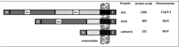 |
Singer and Camargo, 2011 [5] | ||
ACE, ACE2 and collectrin are type I integral proteins with a signal peptide in the N-terminal, transmembrane and short C-terminal domains. ACE2 and collectrin share identity in the C-terminus (white, shaded), whereas ACE and ACE2 are identical in the N-terminal extracellular domain (grey, vertical lines). ACE2 and ACE have one or two zinc-binding motif (HEMGH) (bm) in the extracellular domain.
2. Proteolytic cleavage of ACE2
ACE2 exists in a membrane-bound and an unbound soluble form, with the former constituting the majority of ACE2 in the body. Membrane-bound ACE2 contains a transmembrane domain that anchors a cleavable N-terminal ecto-domain, which retains its enzymatic activity in the secreted, soluble form. ACE2 is cleaved by a membrane-bound protease, also called secretase or sheddase, which is different from the secretase acting on ACE [7], since ACE2 lacks the ACE amino-acid sequence recognized by this enzyme [1,2]. Thus, in its soluble form, ACE2 is found in very low concentrations in the circulation [8,9]. In fact, while serum ACE levels were found to be 7 nM in over 500 subjects [9], the ACE2 content was 200-fold lower (33 pM). In recent years, it has become increasingly apparent that the proteolytic shedding of cell surface ACE2 is an important mechanism regulating its expression, functions, and soluble concentrations in biological fluids [10]. The major protease mediating proteolysis and ectodomain shedding of ACE2 is a type I transmembrane protein belonging to the adamalysin subfamily of zinc-dependent metalloproteases (A Disintegrin And Metalloprotease17; ADAM17). It is also known as “tumor necrosis factor-α (TNF-α) cleavage enzyme” (TACE) [11,12], since it mediates extracellular domain shedding and activation of TNF-α, a pro-inflammatory cytokine [13,14]. The activity of ADAM17 is regulated by phosphorylation through the formation of reactive oxygen species (ROS), which are induced by p38 mitogen-activated protein (MAP) kinase and NAPDH oxidase 2 [10,15,16].
Phosphorylation enhances the catalytic activity of ADAM17 and thus increases ACE2 shedding, resulting in the loss of ACE2 activity at the cell membrane. Phorbol esters, ionomycin, endotoxin and the proinflammatory cytokines interleukin-1β and TNF-α can also promote ectodomain release by ADAM17 [7,17]. In addition, calmodulin binding sites within the short cytoplasmic tail of ACE2 appear to regulate shedding. Studies of HEK-293 cells using calmodulin inhibitors indicate that calmodulin inhibits ACE2 shedding through proteases of the ADAM family [18]. Importantly, the activity of ADAM17 is regulated not only by endogenous peptides, such as Ang-II [12] and insulin [19], but also through pharmacological agents, e.g., rosiglitazone [20] and paricalcitol [21]. In addition, it is altered in diseases, such as hypertension and diabetes [19,20,22,23], suggesting that shedding is an important property of ACE2 function. Thus, ACE2 levels in circulation and biological fluids, such as CSF and urine, are dynamically determined by both the cell surface expression and shedding of ACE2. To that end, circulating ACE2 levels appear to serve as possible biomarkers in the progression of renal and cardiovascular diseases, such as hypertension, heart failure, and diabetes [24].
3. Enzymatic functions of ACE2 in plasma
3.1. Renin-angiotensin system
As mentioned earlier, ACE2 converts Ang-I into Ang-(1-9), and Ang-II into Ang-(1-7). The beneficial effects of ACE2 are attributed mainly to three factors: 1. degradation of Ang-II, thereby diminishing its detrimental effects, 2. degradation of Ang-I to Ang-(1-9), thereby limiting actions of ACE on its substrate, and 3. Formation of Ang-(1-7). These enzymatic functions of ACE2 are of particular significance in pathological conditions, where Ang-I and Ang-II overstimulate the RAS [[25], [26], [27]]. The cellular actions of Ang-(1-7) are mediated by Mas receptor (MasR) signaling, which appears to oppose the effects of Ang-II on the Ang-II type 1 receptor (AT1-R) [25,28]. Ang-(1-9), on the other hand, also promotes beneficial cardiovascular and renal effects through AT2-R activation. However, the expression of AT2-Rs is low in adults, but can be upregulated in diseases [25,27]. Thus, the ACE2/Ang-(1-7)/MasR axis has emerged as a physiological counter measure to the classical RAS pathway [25,28]. Conversely, diminished ACE2 activity results in activation of the Ang-II/AT-1-R axis, and is associated with increased progression of cardiovascular and renal pathologies [[25], [26], [27]]. Fig. 1 illustrates a summary of the classical and alternative RAS pathways.
Fig. 1.

The renin angiotensin system (RAS). The classical RAS consists of the breakdown of Angiotensin I (Ang-I) into Ang-II via ACE, which can bind either to the AT1 (angiotensin type 1) or the AT2 (angiotensin type 2) receptor. Ang-II has a higher affinity for the AT1 receptor. The non-classical RAS consists of conversion of Ang-I into Ang-(1-9) and Ang-II into Ang-(1-7) by ACE2. Ang-(1-7) stimulates the Mas receptor. Bradykinin and [des-Arg9]-bradykinin are degraded by ACE and ACE2, respectively, into pharmacologically inactive peptides.
Some of the endogenous peptides and proteins involved in the RAS also regulate cell surface expression of ACE2 by altering transcription of this protein, regulating the shedding process or acting on both mechanisms. Ang-II downregulates ACE2 expression by activating AT1-R-mediated upregulation of extracellular regulated (ERK)1/2 and p38 MAP kinases in human tubular kidney cells [29], rat aortic vascular smooth muscle cells [30], cardiomyocytes [31], and catecholaminergic neurons [32]. In Neuro-2A cells transfected with ACE2, AT1-R activation by Ang-II leads to internalization and subsequent destruction of ACE2 in lysosomes [33]. In addition, Ang-II activation of the AT1-R induces ADAM17-mediated proteolytic cleavage of ACE2 in cardiomyocytes [12] and hypothalamic neurons [10,34]. Furthermore, Ang-II stimulates phosphorylation of three mitogen-activated protein kinases (MAPK): p38, extracellular signal-regulated kinases (ERK) 1/2, and c-Jun N-terminal kinase (JNK), and increases the production of TGF-β1, which further suppresses ACE2 expression [[35], [36], [37]]. Thus, Ang-II-induced down-regulation of ACE2 expression eventually leads to impaired conversion of Ang-II into Ang-(1-7) and further accumulation of Ang-II and RAS-mediated detrimental effects in a probable positive feedback cycle. Similar to Ang-II, another vaso-constrictive peptide, Endothelin-1, also downregulates ACE2 transcription by activating p38 MAP kinase and ERK1/2 pathways in human bronchial epithelial cells [38] and rat cardiomyocytes [31].
As opposed to Ang-II, Ang-(1-7) and atrial natriuretic peptide (ANP) do not affect ACE2 expression [33]. However, both peptides counteract Ang-II-AT1-R-mediated downregulation of ACE2 by inhibiting MAP kinase in kidney tubular cells [39] and activating MAP kinase phosphatase in rat aortic vascular smooth muscle cells [30], astrocytes [40], cardiomyocytes [31,41], and in rat [42] but not human mesangial cells [43]. In addition, Ang-(1-7) and ANP inhibit ADAM17 activity [44,45].
3.2. Apelin
This endogenous peptide is synthesized as a precursor 77 amino acid pre-pro-peptide and is subsequently cleaved to produce three main apelin fragments: apelin-36, apelin-17, and apelin-13. The latter can undergo cyclization of the glutamine at its N terminus to produce pyroglutamated apelin-13, [Pyr1]apelin-13 [46]. Apelin peptides have been identified as substrates for ACE2, which cleaves some of these peptides with similar efficiency as its primary substrate Ang-II [6,47]. Apelin is an inotropic, vasodilator, and cardioprotective peptide that exhibits beneficial cardiovascular effects through activation of the G-protein coupled APJ receptor, which, in its transmembrane domains, shares 54 % sequence similarity with the AT1 receptor. However, it does not bind Ang-II; instead it negatively regulates Ang-II-mediated adverse cardiovascular actions [[46], [47], [48]]. Apelin upregulates ACE2 gene transcription in murine heart [49,50], cardiomyocytes and adipose tissue of diabetic rats [51]. It also promotes vascular repair following immune-mediated injury [52]. Accordingly, genetic deletion of apelin causes downregulation of cardiac ACE2 transcription and ACE2 protein levels, which can be reversed by infusion of apelin, indicating an important regulatory role of apelin in ACE2 gene expression [50,53]. Thus, while Ang-II, endothelin-1 and TGF-β1 downregulate ACE2 expression and promote Ang-II-related deleterious effects, apelin peptides, ANP, and Ang-(1-7) counteract these effects.
3.3. Kinin-kallikrein system
In addition to the RAS, ACE2 is involved in the regulation of the kinin-kallikrein system (KKS). The KKS constitutes the precursor kininogen, the proteolytic kallikrein enzymes, effector peptides bradykinin (BK) and its active metabolite [des-Arg9]-BK (DABK). These peptides recognize two pharmacologically distinct G protein-coupled receptors: the B1 receptor, whose main agonist is DABK, and the B2 receptor, whose ligand is BK [54]. The RAS enzymes ACE and ACE2 degrade BK and DABK, respectively [1,6]. In addition, Ang-(1-7) produced by ACE2 downregulates ACE activity [55,56] and potentiates BK-induced vasodilatations [57]. Of note, increased bradykinin levels are believed to be responsible for the dry cough which is a known side effect observed in some patients treated with ACE inhibitors. Thus, downregulation of ACE2 activity by disease conditions, such as lung injury and SARS-CoV infections, may increase DABK levels, and over-activation of B1 receptors can contribute to the pathogenesis of these diseases. In endotoxin-induced lung inflammation models, the loss of ACE2 function leads to accumulation of DABK, activation of B1 receptors, and the release of proinflammatory chemokines, such as C-X-C motif chemokine 1 and 5, macrophage inflammatory protein-2, and cytokines, such as TNF-α, from airway epithelia, increasing neutrophil infiltration and promoting lung inflammation and injury [58]. In addition, vasoconstrictions induced by DABK can contribute to endotoxin-related disease pathology [59]. Importantly, B1 receptor expression is upregulated in disease conditions, and its levels are low under normal circumstances, whereas B2 is the main constitutive receptor in homeostasis. Interestingly, DABK [60,61] and BK [62], acting through B1 and B2 receptors, respectively, have been shown to activate ADAM17, the major protease involved in ACE2 shedding.
4. Enzymatic functions of ACE2 in the central nervous system
4.1. Amyloid-β peptide and Alzheimer disease
In the central nervous system (CNS), both ACE and ACE2 have the ability to hydrolyze the amyloid-β (Aβ) peptide, which plays a key role in the pathogenesis of Alzheimer disease (AD), for review, see [63]. In the Japanese population, there is a significant association between the ACE genotype and AD [64]. This is explained by reduced plaque formation, the hallmark of AD, due to inhibition of Aβ aggregation by ACE [65]. ACE can cleave the amyloidogenic Aβ40, thereby reducing its aggregation [65], and it can also hydrolyze the more hydrophobic Aβ-(1-42) to the less neurotoxic Aβ40 form [66]. On the other hand, ACE2 can efficiently hydrolyze Aβ43 (the highly amyloidogenic form of Aβ that seeds plaque formation) to Aβ42, which is then further degraded by ACE to the less toxic Aβ40 [3,67]. ACE2 activity is significantly reduced in the frontal cortex and hypothalamus of OXYS AD rats and in the mid-frontal cortex of AD patients, which is associated with an elevated parenchymal Aβ and tau load [68,69]. In the cerebrospinal fluid of AD patients, the ACE2 activity is positively correlated with disease pathology [70]. In addition, ACE2 deficiency causes deterioration of cognitive functions in ACE2 knockout mice [71]. Conversely, activation of ACE2 by diminazene aceturate (DIZE) or overexpression of ACE2 reduces brain Aβ pathology, improves the cognitive performance [72,73] and ameliorates Aβ-induced inflammatory responses [74,75].
ACE2 is primarily responsible for converting Ang-II to Ang-(1-7). Ang-(1-7) has been shown to reduce AD-related disease pathologies in rat [76] and mouse [77] AD models. Decreased Ang-(1-7) levels in the plasma of AD patients [78] and brain tissue of AD model mice have been shown, accompanied by an inverse correlation between Ang-(1-7) and Tau hyper-phosphorylation [79] inducing neurofibrillary tangles, another histopathological hallmark of AD [63]. In addition, AVE0991, a nonpeptide analogue of Ang-(1-7), and glycosylated Ang-(1-7) peptide have been reported to attenuate aging-related neuroinflammation [80] and cognitive impairment in mouse models [81]. Interestingly, hypercholesterolemia, a known risk factor for AD, and increased oxidized oxysterols, a result of disturbed cholesterol metabolism in AD patients, have been shown to upregulate ACE2 expression in rat primary neurons [82]. Recent studies of the hippocampi of AD patients [83,84] or of AD mouse models [84] report increased ACE2 protein expression in association with oxidative stress. The role of ACE2 activity in the pathogenesis of AD has been reviewed recently [85,86].
4.2. Neurogenic hypertension
Hypertension and increased oxidative stress are known causative factors for neurodegenerative diseases. ACE2 counteracts over-activity of the RAS and oxidative stress [87] caused by increased Ang-II activity in the hypothalamus and brain stem and plays a regulatory role in neurogenic hypertension [23,88,89]. Activation of neuronal AT1-Rs promotes internalization and degradation of ACE2 and stimulates ADAM17-mediated ACE2 cleavage [10,33,90]. Indeed, a doubling of soluble ACE2 in the cerebrospinal fluid of hypertensive patients has been reported [10]. Overexpression of ACE2 in the brain attenuates neurogenic hypertension with enhanced brain RAS activity in animal models [89,91,92]. Similarly, in a number of hypertension models, reduction in Ang-II-mediated signaling or upregulation of the central ACE2/Ang-1-7 pathway have been shown to reduce hypertension through downregulation of sympathetic signaling and increased parasympathetic tone [88,89,93].
5. Non-enzymatic functions of ACE2
5.1. Interaction with neutral amino acid transporters
Structurally, ACE2 is a chimeric protein that has emerged from the duplication of two genes: homology with ACE in the catalytic domain and homology with collectrin in the transmembrane C-terminal domain (Table 1). Collectrin (transmembrane protein 27, Tmem27) is a non-catalytic protein that shares 48 % identity with the transmembrane and C-terminus of ACE2. It is expressed in the kidneys, pancreas, and to a lesser extent in the intestine, liver, heart and stomach [94,95]. Collectrin plays critical roles in neutral amino acid transport in the kidney, and β-cell proliferation and insulin exocytosis in the pancreas, but does not promote SARS-CoV entry [94,[96], [97], [98]]. Gene-targeting studies in mice indicate that collectrin is an essential regulator of the solute carrier family 6 (SLC6) of Na+-dependent neutral amino acid transporters in proximal tubular epithelial cells of the kidney [94]. The urine samples of collectrin knockout mice contain excessive amounts of neutral amino acids, such as tyrosine and phenylalanine [94,99]. Although ACE2 does not bind to amino acid transporters in the kidneys, it binds to the Broad neutral (0) amino acid transporter 1 (B°AT1; SLC6A19) in the intestines, where collectrin is absent, but ACE2 is highly expressed and regulates the absorption of dietary neutral amino acids [5,100]. B°AT1 is responsible for the transport of neutral amino acids across the apical membrane of intestinal and renal tubular epithelia. Mutations in the B°AT1 gene SLC6A19 lead to Hartnup disease, caused by malabsorption of tryptophan. Furthermore, ACE2 is necessary for the expression of human B°AT1 and, therefore, ACE2-knockout animals have reduced levels of neutral amino acids in the serum and, specifically, display impaired tryptophan uptake [101]. In this study, ACE2 null mice also exhibited decreased expression of antimicrobial peptides and showed altered gut microbial composition, which was restored by tryptophan administration. The enzymatic activity of ACE2 is not necessary for pairing with the B°AT1 amino acid transporter [5,101], and other peptidases, such as aminopeptidase N, also form functional complexes with B°AT1 [102]. Furthermore, the differential functional association of mutant B°AT1 transporters with ACE2 and collectrin in the intestine and the kidneys, respectively, participates in the phenotypic heterogeneity of human Hartnup disease [100]. Interestingly, the 3D structure of the human receptor brought to light a complex architecture in which two heterodimers, constituting ACE2-B°AT1, form a homodimeric superstructure with a stoichiometry of (ACE2-B°AT1)2, and B°AT1 was proposed to stabilize each single heterodimeric ACE2-B°AT1 complex [98].
5.2. Interaction with integrins
In addition to neutral amino acid transporters, ACE2 has been shown to interact with biochemically purified integrin β1 in homogenates of failing human hearts [103]. Subsequently, it has been demonstrated that the ectodomain of ACE2 binds to integrin β1 and integrin α5 [104], suggesting that ACE2 may be an integrin substrate. Thus, the cleavage of ACE2 at the cell membrane, through binding of soluble ACE2 to integrins, can regulate integrin-mediated signaling, modulate cell–extracellular matrix interactions and hence influence cardiac remodeling processes [3,104]. Interestingly, the spike protein of SARS-CoV-2 contains an integrin binding motif and binds to the α5β1 integrin protein as well as to the human ACE2-α5β1 complex, facilitating its entry into host cells [105]. ATN-161, an integrin binding peptide, inhibits the interaction between SARS-CoV-2 and ACE2 and decreases SARS-CoV-2 infection in Vero-E6 cells [105]. Finally, the most important non-enzymatic function of ACE2 is acting as an entry receptor for coronaviruses including SARS-CoV-2 [98].
6. Coronaviruses and ACE2
Currently, seven human coronaviruses (HCoVs) have been described, all seven having a zoonotic origin from bats, rodents, or domestic animals [106,107]. Four HCoVs, i.e., HCoV-NL63, HCoV-229E, HCoV-OC43 and HKU1, induce only mild upper respiratory diseases and only occasionally cause severe infections in infants, young children and elderly individuals. Some of these HCoVs have been known since the 1960s and are not considered to be highly pathogenic to humans. However, this concept has changed drastically in 2002 with the outbreak of the severe acute respiratory syndrome (SARS) in China, caused by SARS-CoV, and a decade later by Middle East respiratory syndrome coronavirus (MERS-CoV), which emerged in Middle Eastern countries [108].
Coronavirus replication is initiated by the binding of the densely glycosylated trimeric spike (S) protein to cell surface receptors. The S protein is composed of two functional subunits, S1 for receptor binding and S2 for membrane fusion (Fig. 2 ). A specific interaction between S1 and the cognate receptor triggers a drastic conformational change in the S2 subunit, leading to the fusion between the virus envelope and the cellular membrane, followed by release of the nucleocapsid into the cytoplasm of the host cell [109]. Receptor binding is the major determinant of host range and tissue tropism for a coronavirus. Some HCoVs have adopted cell surface enzymes as receptors, such as aminopeptidase N (APN) for HCoV-229E, and dipeptidyl peptidase 4 (DPP4) for MERS-CoV, while HCoV-OC43 and HCoV-HKU1 use 9-O-acetylated sialic acid as receptor [106]. SARS-CoV-2, SARS-CoV and HCoV-NL63 use ACE2 as cellular entry receptor [98,[110], [111], [112], [113]]. The ectodomain of the SARS-CoV-2 S1 protein binds to the peptidase domain of ACE2 with an affinity of ≈14 nM, which is 10- to 20-fold higher than SARS-CoV S protein binding to ACE2 [114], probably due to amino acid mutations in the S1 protein [115]. ACE2-binding affinity of the S protein is one of the most important determinants of SARS-CoV-2 infectivity and disease severity. While some of the S protein mutations enhance ACE2 binding, resulting in increased susceptibility and severity of COVID-19, others can inhibit S protein binding or result in a truncated S1 protein, conferring a high degree of resistance to infection or reducing the severity of it [[116], [117], [118]].
Fig. 2.

Schematic presentation of the host cell proteases involved in the cellular entry of SARS-CoV-2. Cell entry of SARS-CoV-2 can proceed via two distinct routes. The first route of activation can be pursued if the SARS-S-activating protease TMPRSS2 is coexpressed with ACE2 on the surface of target cells. Binding to ACE2 and processing by TMPRSS2 (see inset) are believed to allow fusion at the cell surface. Furin is primarily located in late endosomes and the Golgi network. However, it is secreted into the extracellular fluid and can also be trafficked to the cell membrane. The second route involves binding of the virion-associated trimeric S protein of SARS-CoV-2 to ACE2. As a result, the virions are taken up into endosomes, in which the SARS-S protein is cleaved and activated by the pH-dependent cysteine protease cathepsin L and furin. The SARS-S protein can activate ADAM17, which cleaves ACE2, resulting in shedding of ACE2 (soluble ACE2). The pink ovoid structures at the tip of S1 indicate the receptor-binding domains (RBDs).
7. ACE2 and viral cell entry
7.1. Binding of the S protein to ACE2
On mature viruses, the spike protein is present as a trimer, with three receptor-binding S1 heads sitting on top of a trimeric membrane fusion S2 stalk (Fig. 2). SARS-CoV S1 contains a receptor-binding domain (RBD) that specifically recognizes ACE2 as its receptor [112,119,120]. The RBD can be either in a standing-up state, which enables receptor binding, or in a lying-down state, which does not bind to the host ACE2 receptor [121,122]. Thus, the RBD constantly switches between a standing-up position for receptor binding and a lying-down position for immune evasion [122]. The cryo-electron microscopic structure of the SARS-CoV-2 spike revealed that its RBD is mostly in the lying-down state [114,123], whereas in the SARS-CoV spike, the RBD is mostly in the standing-up state (Fig. 2, inset). It was proposed that, in order to maintain its high infectivity while keeping its RBD less accessible to immune surveillance, SARS-CoV-2 mainly relies on host protease activation [122,124]. Moreover, to fuse membranes, the SARS-CoV spike needs to be proteolytically activated at the S1/S2 boundary, so that S1 dissociates and S2 undergoes a structural change leading to fusion with the host cell membrane. Thus, the formation of the ACE2-S1 complex triggers subtle conformational rearrangements in the S protein, which are believed to increase its sensitivity to proteolytic digestion at the border between the S1 and S2 subunits [109,125].
7.2. Proteases involved in viral entry
Cleavage of the S protein and the efficiency of viral entry are tightly controlled by a wide array of host cell proteases. These include lysosomal proteases, such as cathepsin L, cathepsin B, and trypsin [126,127], and cell surface proteases, such as type 2 transmembrane protease serine 2 (TMPRSS2), human airway trypsin-like protease (HAT), factor X, elastase, and cysteine protease m-calpain [125,127,128]. Indeed, in SARS-CoV the expression of proteases provides a 100 to 1000-fold more efficient infection pathway than receptor-mediated endocytosis [129]. Entries of SARS-CoV [130,131] and SARS-CoV-2 [132] are markedly enhanced in cell lines expressing TMPRSS2. Camostat mesylate (NI-03), a TMPRSS2 inhibitor used for the treatment of pancreatitis in Japan, inhibits SARS-CoV, HCoV-NL63 [133], and SARS-CoV-2 infection of human lung cells [110]. The structurally similar nafamostat mesylate inhibits SARS-CoV-2 S-mediated entry with an EC50 in the low nanomolar range [134,135]. Bromhexine, an over-the-counter mucolytic cough suppressant, selectively inhibits TMPRSS2 in in vitro studies, with an IC50 of 0.75 μM [136], suggesting pharmacological utility of this drug. Recently, co-expression of ACE2 and TMPRSS2 in tissues derived from the lungs, bronchial branches, and bronchial transient secretory cells has been described, suggesting to cause high susceptibility of these cells to SARS-CoV2 infection [137]. Of note, androgen receptor activation promotes transcription of TMPRSS2, which plays a role in the pathogenesis of prostate cancer [136], and upregulates the expression of the ACE2 gene, which is located on the X-chromosome [138]. In fact, TMPRSS2 is highly expressed in large populations of epithelial cells in prostate and lungs, as well as colon, small intestine, and kidney, and is upregulated by androgens [136,[139], [140], [141]] and glucocorticoids [140]. Androgen receptor regulation of TMPRSS2 and ACE2 expression has been suggested to be linked to a higher prevalence of SARS-CoV, MERS-CoV [142], and SARS-CoV-2 in the male population [141,143]. Similarly, male mice have been shown to be more susceptible to SARS-CoV infection than age-matched females [142].
In addition to TMPRSS2, novel furin cleavage sites that are absent in SARS-CoV have been discovered on the S protein of SARS-CoV-2 [144], and furin expression has been shown to overlap with the expression of ACE2 and TMPRSS2 in lung and bronchial cells [137]. It was suggested that the S1/S2 site in SARS-CoV-2 forms an exposed loop that harbors furin cleavage sites (Fig. 2) composed of multiple arginine “RRAR” residues (multibasic) rather than the single arginine in SARS-CoV [114,123,124]. Furin, a Ca2+-dependent endopeptidase and a member of the proprotein convertase family (proprotein convertase subtilisin/kexin type 3), accumulates mainly in the trans-Golgi network. It can be further transported to the cell surface and back via the endosomal pathway [145], or its catalytic domain can be shed and released into the extracellular space. Thus, furin can cleave the SARS-CoV-2 spike protein in the Golgi compartment, as well as in the extracellular fluid. Notably, circulating levels of furin are elevated in patients with diabetes [146], suggesting that increased mortality and delayed recovery of diabetic COVID-19 patients may be linked to elevated furin levels. Inhibition of expression or proteolytic activity of furin is a conceivable approach to reduce SARS-CoV-2 infection. In this context, nanomolar concentrations of a peptide designed from the furin cleavage sequence of the avian influenza A H5N1 virus provide protection against infection by several furin-dependent pathogens [147]. Recently, two furin inhibitors, chloromethylketone and naphthofluorescein, were reported to show antiviral effects in SARS-CoV-2-infected cells by decreasing viral production and cytopathic effects [148].
Entry of SARS-CoV through clathrin-dependent [149] and independent [150] endocytosis is facilitated by proteases [125]. It has recently been shown that entry of SARS-CoV-2 pseudovirions is blocked by lysosomotropic agents, ammonia chloride and bafilomycin A, indicating that the SARS-CoV-2 S protein is internalized mainly by a cathepsin-L-mediated endocytic mechanism [151]. Similarly, ammonium chloride, an endosomal pH modulator that blocks cathepsin B and L activity, strongly inhibits SARS-CoV-2 entry into HEK-293T cells [110], further suggesting that SARS-CoV-2 employs endosomal pathways. In addition, non-endosomal direct fusion mechanisms, activated by cell membrane proteases, are involved [127,152,153]. Thus, there are two pathways for SARS-CoV entry into the target cell (Fig. 2): endocytosis and fusion of the viral membrane with the membrane of the target cell, which is at least two-orders more efficient than endocytosis [129].
8. Factors influencing ACE2-viral interaction
8.1. Enzymatic activity
SARS-CoV binding does not interfere with the enzymatic activity of ACE2, nor does the enzymatic activity of ACE2 play any role in SARS-CoV entry [109,112,120]. Accordingly, cells expressing catalytically inactive mutants of ACE2 are still infected with SARS-CoV, further indicating that the peptidase actions of ACE2 are not necessary for the entrance of SARS-CoV into host cells [4,112,120]. In line with these findings, the SARS-CoV S protein does not attenuate hydrolysis of Ang-II to Ang-(1-7) by soluble ACE2; thus it is unlikely that Ang-II or other peptide substrates would directly interfere with SAR-CoV-2 binding and internalization [17]. However, a novel ACE2 inhibitor, N-(2-aminoethyl)-1 aziridine-ethanamine, was found to be effective in blocking the SARS-CoV S protein-mediated cell fusion [154]. In addition, high affinity binding of SARS-CoV-2 spike protein was recently reported to enhance ACE2 carboxypeptidase activity [155].
8.2. Cytoplasmic domain
The role of the cytoplasmic domain of ACE2 in regulating the activity and SARS-CoV entrance appears to vary depending on experimental conditions [7]. Large deletions in the ACE2 cytoplasmic tail did not modulate its expression and receptor function in HEK-293T cells, nor did they alter SARS-CoV entrance into COS-7 cells [149,156]. This argues against a significant role of the ACE2 cytoplasmic domain in mediating SARS-CoV S-driven infection. On the other hand, a deletion of the cytoplasmic domain of ACE2 resulted in a marked increase in the constitutive release of soluble ACE2 [157]. In another study, although a deletion of the cytoplasmic domain did not affect PMA (Phorbol myristate acetate, a specific activator of protein kinase C)-stimulated shedding of ACE2, it abolished the shedding induced by the spike protein of SARS-CoV and inhibited SARS-CoV entry into HEK-293T cells [158]. This suggests a role of the ACE2 cytoplasmic domain in SARS-CoV infection and in the concomitant ACE2 shedding caused by the SARS-CoV S protein.
8.3. Receptor expression levels
Several lines of evidence suggest that the entry of SARS-CoV is directly correlated with ACE2 expression. Studies of human pulmonary cell lines (A549) indicate that the infection and replication efficacy of SARS‐CoV‐2 is correlated with ACE2 expression levels [159]. Natural mutations, such as Y217 N, alter ACE2 expression and entry efficiency. Moreover, an introduction of the Y217 N mutation into human ACE2 causes downregulation of ACE2 expression and reduced viral entry efficiency [160]. In addition, a higher ACE2 expression is correlated with higher pseudotype SARS‐CoV‐2 and SARS-CoV viral infectivity, suggesting that increased ACE2 levels may predispose individuals to SARS-CoV-2 transmission [151,156,161,162]. Specifically, upregulation of ACE2 expression by various drugs especially by RAS inhibitors has been suggested to increase SARS-CoV-2 replication and susceptibility to viral infection (recently reviewed [163]). Surprisingly, transgenic mice overexpressing human (h) DPP4, an entry receptor for MERS-CoV [106], exhibited relative resistance to MERS-CoV infection, exemplified by decreased lung inflammation and reduced rates of mortality [164]. Intriguingly, these hDPP4 transgenic mice with higher levels of hDPP4 expression also exhibited increased circulating levels of soluble DPP4; and administration of recombinant soluble DPP4 attenuated lung histopathology and reduced the titers of virus recovered from lung tissue [164].
9. Regulation of ACE2 expression during viral infection and lung injury
Binding of both SARS-CoV and SARS-CoV-2 to ACE2 leads to downregulation of ACE2 expression on the cell surface [112,[165], [166], [167], [168]]. This is due to endocytosis of ACE2 bound to the virus and decreased ACE2 transcription in the host cell [149,150,166,169,170]. In addition, non-endocytotic internalization of the SARS-CoV-ACE2 complex, involving the cleavage of the ACE2 ectodomain by cell protease TMPRSS2 [152,171], causes further downregulation of ACE2 on the host cell surface. Thus, TMPRSS2 facilitates SARS-CoV infection via two independent mechanisms: 1. cleavage of the SARS-S protein, which activates the S protein for membrane fusion, as described above, and 2. cleavage of ACE2, which might also promote viral uptake through the cathepsin L-dependent pathway [152]. Binding of SARS-CoV to ACE2 stimulates ADAM17, which induces shedding of the ACE2 ectodomain and results in loss of ACE2 in the host cell membrane [158]. Subsequently, it was shown for SARS-CoV that only TMPRSS2-induced shedding promotes viral infection [152], although both ADAM17 and TMPRSS2 compete for ACE2 processing. If this pathway were also to prove functional in SARS-CoV-2, then it could be assumed that disruption of ADAM17 cleavage increases membrane-bound ACE2 and thereby enhances SARS-CoV-2 infection, whereas disruption of TMPRSS2 may be protective. The interplay between TMPRSS2, ADAM17 and ACE2 in COVID-19 has recently been discussed by Zipeto et al. [172]. Furthermore, some pro-inflammatory cytokines produced during SARS-CoV infection, including TNF, IFN-β, IL-1β, IL-6, and IL-4 [[173], [174], [175], [176]], have been shown to downregulate ACE2 expression [35,177,178].
Downregulation of the entry receptor is not unique to SARS-CoV, other viruses can downregulate their cell entry receptors as well [179,180]. Interestingly, other viruses, such as influenza A subtype H1N1 [181], not using ACE2 as entry receptor, can also downregulate cellular ACE2 levels. In addition, lung injuries induced by different pathogenic factors are associated with downregulation of pulmonary ACE2 expression. Accordingly, lung injuries induced by ventilator [182,183], acid aspiration [184], ischemia-reperfusion [185], and lipopolysaccharide administration [186,187] as well as disease models of pulmonary hypertension [188,189], silicosis [190], asthma [191], and chronic obstructive pulmonary disease [192] are associated with downregulation of pulmonary ACE2 expression. Thus, it is possible that downregulation of ACE2 after viral infections can be related to viral invasion and tissue injury, rather than a direct consequence of ACE2-dependent replication in lung epithelium. In addition, factors such as cell proliferation and differentiation also modulate ACE2 expression. For example, cell proliferation alone has been shown to downregulate ACE2 expression in a cell cycle-dependent manner [193]. The state of cell differentiation has also been shown to modulate ACE2 expression in epithelial cells [161], in embryonic stem cells and in mice of different age groups [194]. Thus, cellular factors other than SARS-CoV-2 replication may also lead to decreased ACE2 levels and lung pathology. Interestingly, HCoV-NL63, like SARS-CoV-2, utilizes ACE2 for cellular entry [111], but is not associated with a severe disease pathology and causes mostly mild to moderate respiratory infections in children. The S protein of HCoV-NL63 exhibits a reduced affinity for ACE2 compared to SARS-CoV [195], and cleavage by host cell proteases seems to be different for infectious host cell entry of SARS-CoV [196] and HCoV-NL63 [197]. In addition, it has been proposed that HCoV-NL63 does not induce ACE2 shedding [158], while SARS-CoV activates ADAM17 and promotes the shedding of the ACE2 ectodomain, which may contribute to the differential pathogenicity of these viruses.
10. Low levels of lung expression of ACE2 and accessory proteins proposed for viral entry
10.1. Tissue-specific ACE2 expression
Initial studies identified small intestine, colon, duodenum, kidney, testis, and gallbladder as the main organs expressing ACE2 [198]. Recent investigations indicate a more widespread expression of ACE2; including alveolar type II cells and bronchial cells of the lungs, oral and nasal mucosa, arterial smooth muscle cells, epithelial cells of the esophagus, absorptive enterocytes from the ileum and colon, as well as heart, liver, pancreas, kidneys, and brain [199,200]. Although, the respiratory system is not a prominent ACE2-expressing organ, such as the intestines and kidneys, due to the air-born nature of the virus, mucosal cells of the upper respiratory tract and bronchial cells of the lower respiratory tract have been suggested as main target regions for viral replication. Compared to intestines and kidneys, the expression of ACE2 is more than two orders of magnitude lower in the lungs of rodents [201] and humans [200], and a small fraction (1 %<) of alveolar type 2 cells expresses ACE2 [[202], [203], [204], [205], [206]].
Interestingly, recent investigations studying ACE2 expression in the respiratory system indicate that ACE2 is highly expressed in the nasal epithelium, that its expression decreases to medium levels in the epithelium of the small airways and that it cannot be detected in alveolar macrophages [207]. In agreement with this study, a gradient of decreasing ACE2 expression from nasal to intrapulmonary bronchial regions was reported [208]. Notably, progressively reduced amounts of ACE2 expression were observed in the lower airway regions, culminating in minimal amounts in the alveolar region [205,208]. Thus, it was suggested that, due to scarce ACE2 expression, SARS-CoV-2 shows limited ability to propagate in the human alveolar compartment and that lung injury in COVID-19 most likely results from an immune activation rather than direct viral damage of the alveolar compartment [205].
10.2. Interferons
Several studies have been able to demonstrate that interferons (IFNs) [[209], [210], [211], [212], [213], [214]] and some proinflammatory cytokines [[215], [216], [217], [218]] upregulate ACE2 mRNA and protein expression in cell lines and lung tissue. Thus, it was proposed that, after an initial downregulation due to virus binding to ACE2, the host immune response to SARS-CoV-2 infections might consist of an IFN-driven upregulation of ACE2, leading to an increase in the number of respiratory epithelial cells susceptible to SARS-CoV-2 infection. This could potentially explain the entry of SARS-CoV-2 into the respiratory tract via ACE2 receptors, despite low ACE2 expression in healthy individuals [204]. However, the opposite is also conceivable: upregulation of ACE2 as an IFN-stimulated gene may counteract viral infection, which is in line with the anti-inflammatory and protective functions of ACE2 in ARDS [210]. In agreement with these findings, upregulation of ACE2 expression after rhinovirus [219], human respiratory viruses [212], upper respiratory tract infections [220], and SARS-CoV-2 infections has been reported in vivo, in vitro and post mortem in pulmonary [210,[221], [222], [223]] and renal tubular [37] epithelial cells. Upregulation could also be demonstrated in broncho-alveolar lavage samples [224] and peripheral blood monocytes [225] of COVID-19 patients [226] as well as in lung tissue of SARS-CoV-2-infected mice [227].
A new perspective is added by a recent study demonstrating that IFN does not stimulate the ACE2 gene, but instead the gene of a novel primate-specific isoform of ACE2, which is non-functional as a carboxypeptidase and does not mediate binding of the SARS-CoV-2 spike protein [228]. In addition, another short isoform of ACE2 expressed in the airway epithelium was also found to be upregulated by IFN stimulation and by infection with rhinovirus, but not with SARS-CoV-2 [229,230]. Furthermore, COVID-19 displays a profile of early IFN inhibition [231], and IFNs may not be significantly induced or even inhibited by SARS-CoV-2 in the lungs, thus possibly hindering the pattern of ACE2 upregulation during host antiviral response [232,233]. In line with these findings, in nasopharyngeal swabs of COVID-19 patients, decreased soluble ACE2 activity and ACE2 gene expression but increased expression of IFN stimulation genes were reported [234], suggesting the existence of IFN-independent mechanisms whereby SARS-CoV-2 suppresses ACE2 expression and function. Overall, the role of IFN stimulation of ACE2 expression in the pathogenesis of COVID-19 related lung injury is currently still a matter of debate.
Another factor that may influence ACE2 expression in COVID-19 is sirtuin 1 (SIRT1). The expression of SIRT1 was reported to be upregulated in the lungs of COVID-19 patients with comorbidities [235]. SIRT1 is expressed next to the promotor region of the ACE2 gene. Hence, an increased expression or enhanced functional activation of SIRT1 is associated with an increased ACE2 expression [236].
10.3. Alternative attachment sites
In addition to ACE2, other receptors and membrane proteins are also involved in SARS-CoV-2 tropism. Earlier studies found that human adenocarcinoma cell lines [237], HEK-293T cells [238], and endothelial cells [239] expressing high levels of ACE2, are not permissive to SARS-CoV infection. Similarly, secretory MUC5B club cells of the respiratory tract were not infected with SARS-CoV-2 in vitro or in vivo, despite detectable ACE2 expression [208]. Conversely, human neuronal cell lines [240], A549 human lung‐derived cell lines [159], and some other lung cells [241] that express ACE2 at undetectable levels are susceptible to SARS-CoV or CoV-2 infection. Thus, in addition to ACE2, several other membrane components have been proposed to function as co-receptors or attachment sites for SARS-CoV and SARS-CoV-2. These include Cluster of Differentiation 147 (CD147), also known as Basigin (BSG) [[242], [243], [244], [245]], Dendritic Cell-Specific Intercellular adhesion molecule-3-Grabbing Non-integrin (DC-SIGN), also known as Cluster of Differentiation 209 (CD209) [[246], [247], [248], [249], [250], [251]], Neuropilin-1 [252,253], glycosaminoglycans [254], heparan sulphate [255], and kidney injury molecule-1 [256]. Interestingly, in reconstituted lipid membranes devoid of ACE2, binding of the S1 subunit of SARS-CoV-2 spike protein to neutral phospholipid membranes and liposomes was reported to cause mechanical destabilization and membrane permeabilization in a receptor-independent manner [257].
11. The roles of ACE2 and angiotensin II in coronavirus-induced lung pathology
11.1. Acute respiratory distress syndrome (ARDS)
Although the majority of COVID-19 cases are considered mild, a subset of patients develops severe respiratory symptoms due to acute, diffuse inflammatory lung injury, increased pulmonary vascular permeability, and an accumulation of protein-rich fluid and inflammatory cells within the alveoli. This triggers various immune cells, e.g., macrophages and neutrophils, to release a large number of inflammatory factors. As a result, the disease rapidly progresses to ARDS and multi-organ failure. ARDS is the clinical presentation of acute lung injury; it is defined by the presence of bilateral opacities in chest imaging, non-cardiogenic pulmonary edema, acute hypoxemia, reduced lung compliance, compromised arterial oxygenation, and pulmonary hypertension [258]. The RAS, along with the KKS regulates several pathophysiological events in the body, including vascular and pulmonary pressure, inflammation, fibrosis, and coagulation [258,259]. In the lungs, Ang-II is able to induce bronchoconstriction, vasoconstriction, fibroproliferation, cytokine expression, and cell apoptosis, thus promoting tissue injury. As a negative regulator of Ang-II, ACE2 has been reported to protect the lungs from injury [258,260].
11.2. ACE2
Decreased pulmonary ACE2 activity, due to ACE2-downregulation induced by SARS-CoV-2 replication, has been proposed as the main mechanism driving lung pathology in COVID-19. Preclinical studies in mice infected with respiratory syncytial virus [261] or avian H5N1 influenza [262] suggest that restoration of ACE2 by administration of recombinant ACE2 reverses the exacerbated lung injury. In a randomized, double blinded study of piglets with lipopolysaccharide-induced ARDS, plasma Ang-II levels increased significantly by lipopolysaccharide infusion. Conversely, treatment with recombinant human ACE2 decreased Ang-II levels and attenuated arterial hypoxemia, pulmonary hypertension, and redistribution of pulmonary blood flow [263]. Accordingly, downregulation of ACE2 was shown to play a role in the pathogenesis of acute lung injury induced by influenza viruses, such as H7N9 [264] and H5N1 [262]. Moreover, in three different ARDS models not related to viral replication (acid-aspiration-induced, endotoxin induced, and peritoneal sepsis-induced ARDS), ACE2 knockout mice show a more severe disease pathology, compared with wild-type mice; and treatment with soluble recombinant ACE2 protein improves the symptoms [4,96,184]. In addition, among three coronaviruses causing SARS-like pathologies in humans, MERS-CoV uses dipeptidyl peptidase 4 (DPP4), but not ACE2 [106], indicating that, although ACE2 is needed for viral entry, it is not a prerequisite for SARS pathogenesis. Conversely, as mentioned earlier, HCoV-NL63 utilizes ACE2 for cellular entry [111] and downregulates ACE2 expression [179], but is usually not associated with SARS in humans.
11.3. Angiotensin-II
Increased Ang-II levels due to down-regulation of pulmonary ACE2 expression induced by SARS-CoV-2 has been proposed to play a central role in the pathogenesis of ARDS observed in COVID-19 [265]. However, in line with the findings of low ACE2 expression, the enzymatic activity of ACE2 in lungs is considerably low, and most of Ang-II conversion to Ang-(1-7) in pulmonary tissue is carried out by peptidases other than ACE2, e.g., prolyloligopeptidase, whereas Ang-(1-7) formation in the kidneys is mainly ACE2-dependent [266]. It is conceivable that ACE2 also influences ARDS pathogenesis through its impact upon Ang-II. Although it is not clear how the low levels of ACE2 expression in the lungs can counteract Ang-II-dependent pathologies and play an important role in SARS-CoV pathogenesis, the production of Ang-II has been shown to be up-regulated in certain pathological conditions. Increased plasma Ang-II levels, due to downregulation of ACE2, were associated with disease severity and predicted a fatal outcome in H7N9 patients [267]. Similar findings were reported for patients with ARDS of miscellaneous causes [268] or with sepsis [269], and for children infected with respiratory syncytial virus [261]. In a recent study, plasma Ang-I and Ang-(1-7) levels were found to be decreased in COVID-19 patients [270], suggesting a decreased ACE2 activity. In addition, small cohorts of COVID-19 patients showed increased levels of circulating Ang-II, correlating with viral load and pulmonary function [271], with disease severity [272,273] and with endothelial dysfunction [274]. In small cohorts of COVID-19 patients with acute kidney injury [275] or severe clinical conditions [276], increased plasma renin and aldosterone levels have also been reported. However, another study found no change in plasma Ang-II levels in COVID-19 patients [277], and plasma concentrations of Ang-II, and aldosterone did not differ between the SARS-CoV-2 positive and the control group [278]. In addition, in postmortem lung examinations of two severe and critically ill COVID-19 patients, the ACE2 protein levels were up-regulated, compared to the adjacent normal lung tissue [279]. In other recent studies, neither down- nor upregulation of ACE2 was observed in human cell lines infected with SARS-CoV-2 [228,229,280]. In COVID-19 patients, plasma levels and enzymatic activity of ACE2 were found increased [281,282] or not altered [278,283,284]. On the other hand, plasma levels of DPP4, the entry receptor for MERS-CoV, were reported to be decreased in COVID-19 patients [285] and in patients infected with MERS-CoV [286]. Interestingly, in small cohorts of COVID-19 patients, Ang-II has been used to treat COVID-19-induced vasodilatory shock [[287], [288], [289]].
Importantly, genetic deletion of ACE2 does not cause a significant change in circulating and tissue Ang-II levels and does not alter blood pressure [[290], [291], [292], [293]]. Similarly, administrations of Ang-(1-7) and ACE2, even at high concentrations, did not affect blood pressure or heart rate in mice [294] or humans [295]. In line with these findings, increased serum ACE2 levels were not associated with changes in Ang-II levels in heart failure patients [296]. However, in a pilot study examining a small cohort of ARDS patients, plasma Ang-II levels were decreased after treatment with recombinant human ACE2, but no improvement in lung oxygenation and organ failure index was observed [297]. In addition, in a recent case study, human recombinant ACE2 markedly decreased plasma Ang-II and proinflammatory cytokines, increased Ang-(1-7) levels, and profoundly decreased viral load in plasma and nasopharyngeal swabs [298]. Thus, the role of pulmonary ACE2 downregulation in the case of increased Ang-II levels warrants further in vivo and in vitro studies.
11.4. Hypoxia and ACE
Lung disorders, such as ARDS and COPD, are known to be associated with hypoxia. Elevation of hypoxia-inducible factor-1α (HIF-1α) during hypoxic lung pathologies has been shown to increase ACE expression, but to decrease ACE2 levels [299] via expression of the microRNA let-7b [300], which potentially leads to elevated pulmonary Ang-II levels. As mentioned earlier, ACE and ACE2 are the two key regulators of the Ang-II level in the circulation. The interaction between ACE and ACE2 appears to be reciprocal, with one being downregulated, while the other one is upregulated [29]. Consequently, disease conditions associated with excessive Ang-II levels are usually linked to increased tissue or plasma ACE activity and decreased ACE2 expression.
ACE gene polymorphism, characterized by an insertion (I) or deletion (D) of a 287-bp Alu repeat DNA sequence in intron 16 of the ACE gene, influences circulating and tissue ACE levels. It determines nearly half of the serum ACE level variability in the general population [301], and the D allele is associated with markedly higher ACE activity than the I genotype. The majority of evidence supports a relationship of ACE I/D polymorphism and clinical outcome of ARDS [[302], [303], [304]]. ACE I/D polymorphism appears to be also associated with the severity of ARDS in patients infected with SARS-CoV or SARS-CoV-2 [135,302,303,[305], [306], [307]]. Analysis of patient cohorts infected with SARS-CoV and SARS-CoV-2 suggests that polymorphism in ACE2 is not associated with the clinical outcome of ARDS [305,308,309].
11.5. Species aspect
Most of the experimental data suggesting that ACE2 plays a role in SARS pathogenesis originate from rodent experiments. However, neither SARS-CoV nor SARS-CoV-2 does replicate efficiently in rodents, and exposures to these viruses does not cause SARS-like fatal pulmonary pathologies in these animals [115,[310], [311], [312]]. Thus, it is difficult to extrapolate mechanisms of SARS-CoV S protein-induced lung pathologies observed in earlier studies using ACE2 knockout mice [4,96,165]. Transgenic mouse models expressing human ACE2 have been developed [239,311]. Although severe disease pathology and lethality were demonstrated in some of these models [[313], [314], [315]], in other transgenic models, animals did not develop a clinical disease that is reproducible and equivalent in severity to that observed in COVID-19 patients [241,311,316].
12. Pharmacology of ACE2 and ACE2 related antiviral treatments
Regulation of ACE2 activity has important implications in several disease pathologies, mainly because it degrades the vasoconstrictive, oxidative, and proinflammatory Ang-II, and produces the oppositely acting Ang-(1-7). Genetic deletion or pharmacological inhibition of ACE2 potentiates hypertension induced by exogenous Ang-II and accelerates cardiovascular pathologies, renal injury and vascular complications in diabetes mellitus [25,27,317]. Conversely, ACE2 overexpression increases the antihypertensive components of the RAS (Ang-(1-7), MasR, and AT2-R), decreases plasma Ang-II levels and reduces blood pressure. It also improves cardiac function and alleviates diabetic complications [27,317,318].
Several synthetic ACE2 inhibitors, such as DX600 [319], MLN-4760 (GL1001) [320,321], and N-(2-aminoethyl)-1 aziridine-ethanamine [154] have been identified. DX600 is a 26-amino acid peptide with nM range affinity for ACE2 [319]. It was reported that rodent ACE2 (IC50 = 7 μM) has a lower sensitivity to inhibition by DX600 than human (IC50 = 90 nM) ACE2 [322]. In another study, MLN-4760 inhibited both human and mouse ACE2, whereas DX600 only blocked human ACE2 activity [323], indicating species and cell type dependent effects of these drugs [324].
Due to the central role of ACE2 in the RAS, upregulation, rather than downregulation of ACE2 activity constitutes an important treatment option. Strategies to increase ACE2 activity include viral ACE2 overexpression, human recombinant ACE2 (hrACE2), and ACE2 activators [317]. Soluble hrACE2 lowers Ang-II-induced blood pressure, increases cardioprotection [325], decreases inflammatory response and lung injury, and attenuates the progression of diabetic nephropathy in animal models without significant side effects [317,318]. Importantly, the administration of soluble recombinant ACE2 does not affect ACE2 activities at the tissue level [294]. Thus, it is likely that the beneficial effects of soluble ACE2 reported in in vivo models are possibly due to an increased ACE2 activity in the circulation rather than in the tissue.
Interest in ACE2 as a therapeutic target has led to the synthesis of small molecule ACE2 activators, including xanthenone (XNT), resorcinolnaphthalein [326], the anti-trypanosomal drug diminazene aceturate (DIZE), hydroxyzine, and chlorprothixene [327]. Interestingly, the antihypertensive effects of XNT and DIZE persist in ACE2 knockout mice and, notably, it has been reported that Ang-II degradation in vitro and ex vivo was unaffected by XNT and DIZE, suggesting that these compounds may have ACE2-independent actions [328]. However, it should also be noted that XNT and DIZE have been reported to cause a significant increase in ACE2 mRNA expression [[329], [330], [331]]. Currently, there is no clinically used ACE2-based therapeutic agent; however, preclinical studies and clinical trials are continuing [25,317,332]. Several natural products, including flavonoids, e.g., apigenin, quercetin, baicalin and epigallocatechin, and anthraquinones, e.g., emodin (for a review [333]), and geranium and lemon essential oils [334] were reported to inhibit ACE2 activity in in vitro assays. Nicotianamine, isolated from soybean, was described to be a potent inhibitor of human ACE2 with an IC50 of 84 nM [335]. Recently, it was found that organosulfur compounds, such as allyl disulfide and allyl trisulfide found in garlic, interact strongly with human ACE2 and the main protease PDB6LU7 of SARS-CoV [336].
Prevention of SARS-CoV-2 S protein-ACE2 interaction and subsequent viral infectivity is an important treatment strategy [337]. In earlier studies, soluble ACE2 was able to block the replication of SARS-CoV in HEK-293T cells [112]. Recombinant soluble human ACE2 alone [338] or fused to the Fc region of the human immunoglobulin IgG1 to increase the short half-life of soluble ACE2 [[339], [340], [341]] have been shown to inhibit SARS-CoV-2 infection and enhance efficacy of other antiviral drugs [342] in cell lines, engineered human blood vessels and kidney organoids. Recently, peptides mimicking the N-terminal helix of the human ACE2 protein, which contains most of the residues contacting with the S protein-binding site, were shown to block SARS-CoV-2 infection of human pulmonary cells with IC50 values in the range of 60–800 nM [343]. Similarly, trimeric ACE2 proteins [344], decoy ACE2 based proteins [345] or ACE2 peptides optimized to SARS-CoV-2 S protein-binding regions using protein-engineering methods [346] potently bound to S protein and inhibited SARS-CoV-2 infection at low nM concentrations in cell lines and in in vivo experiments [346]. In other studies, fusion proteins consisting of immunoglobulin Fc protein and ACE2 effectively blocked SARS-CoV-2 infection at nM concentrations in HEK-293T cells [347,348].
In earlier studies, immunoglobulin G from the sera of infected patients inhibited ACE2 binding of the S protein and SARS-CoV replication [349]. Monoclonal antibodies from recovered COVID-19 patients [[350], [351], [352], [353], [354]] or raised in immunized mice [355] specifically bind to SARS-CoV-2, block the interaction between the SARS-CoV-2 S protein and the ACE2 receptor and neutralize SARS-CoV-2 infection. Chloroquine and hydroxychloroquine, in addition to their pH elevating effects in endosomes, bind to ACE2 [356], impair the terminal glycosylation of ACE2 [357] and inhibit SARS-CoV [358,359] and SARS-CoV-2 replications [360], and the entrance of SARS-CoV-2 spike protein into ACE2 expressing cell lines [356]. Emodin, an anthraquinone compound found in various Chinese herbs, has been shown to inhibit the interaction of the SARS-CoV S protein with its receptor ACE2 in a dose-dependent manner [361]. TAPI-2, an inhibitor of TNF-α converting enzyme (ADAM17), blocks SARS-CoV S protein-induced shedding of ACE2 and inhibits SARS-CoV cell entry [362].
13. Conclusions
In summary, ACE2 is a multifunctional protein with enzymatic as well as nonenzymatic functions. Within the RAS, ACE2 has an important regulatory role in counteracting the deleterious effects of the Ang-II/ACE/AT1 receptor axis on cardiovascular and metabolic events. Besides the RAS, the enzymatic functions of ACE2 extend to the apelin and kinin-kallikrein systems, as well as amyloid-β peptides. Hence, new strategies to enhance the enzymatic activities of ACE2 through its transcriptional regulation, pharmacological activation of its catalytic function, or administration of the recombinant protein could provide new treatment options for cardiovascular diseases, diabetes and acute lung injury, as well as CNS disorders, such as AD or neurogenic hypertension.
The nonenzymatic functions of ACE2 include its role as chaperone protein for the amino acid transporter B°AT1 in the enteric system and its participation as entry receptor in coronavirus infections. While tissue expression levels of ACE2, together with host cell proteases, are critical in determining coronavirus tropism and resulting pathological outcome, its enzymatic role in the pathogenesis of ARDS observed in COVID-19 require further in vivo and in vitro investigations. Soluble ACE2 may act as a competitive interceptor of SARS-CoV-2 by preventing binding of the viral particle to surface-bound, full-length ACE2. Recently, nasal spray application [363] or systemic administration [330] of soluble ACE2 has been suggested for the treatment of COVID-19. Since plasma Ang-(1-7) levels were found to be reduced in a small cohort of COVID-19 patients [270], administration of soluble recombinant human ACE2 proteins may be beneficial as a novel biological therapeutic to limit or even combat the progression of an infection caused by coronaviruses that utilize ACE2 as a receptor. Interestingly, although three of the four known protein receptors for coronaviruses are peptidases (ACE2, APN, and DPP4), the enzymatic activities of these receptors are not a prerequisite for coronavirus entry. Considering the crucial role of ACE2 in coronavirus infections, much still remains to be explored in terms of the basic aspects of ACE2 cellular function and its regulation in health and disease conditions.
Declaration of Competing Interest
The authors declare that the research was conducted in the absence of any commercial or financial relationships that could be construed as a potential conflict of interest. The authors declare that there are no conflicts of interest.
References
- 1.Donoghue M., Hsieh F., Baronas E., Godbout K., Gosselin M., Stagliano N., Donovan M., Woolf B., Robison K., Jeyaseelan R., Breitbart R.E., Acton S. A novel angiotensin-converting enzyme-related carboxypeptidase (ACE2) converts angiotensin I to angiotensin 1-9. Circ. Res. 2000;87(5):E1–9. doi: 10.1161/01.res.87.5.e1. [DOI] [PubMed] [Google Scholar]
- 2.Tipnis S.R., Hooper N.M., Hyde R., Karran E., Christie G., Turner A.J. A human homolog of angiotensin-converting enzyme. Cloning and functional expression as a captopril-insensitive carboxypeptidase. J. Biol. Chem. 2000;275(43):33238–33243. doi: 10.1074/jbc.M002615200. [DOI] [PubMed] [Google Scholar]
- 3.Turner A.J. ACE2 cell biology, regulation, and physiological functions. In: Unger T., Steckelings U.M., dos Santos R.A.S., editors. The Protective Arm of the Renin Angiotensin System: Functional Aspects and Therapeutic Implications. Elsevier Inc.; Amsterdam: 2015. pp. 185–189. [Google Scholar]
- 4.Kuba K., Imai Y., Ohto-Nakanishi T., Penninger J.M. Trilogy of ACE2: a peptidase in the renin-angiotensin system, a SARS receptor, and a partner for amino acid transporters. Pharmacol. Ther. 2010;128(1):119–128. doi: 10.1016/j.pharmthera.2010.06.003. [DOI] [PMC free article] [PubMed] [Google Scholar]
- 5.Singer D., Camargo S.M. Collectrin and ACE2 in renal and intestinal amino acid transport. Channels (Austin) 2011;5(5):410–423. doi: 10.4161/chan.5.5.16470. [DOI] [PubMed] [Google Scholar]
- 6.Vickers C., Hales P., Kaushik V., Dick L., Gavin J., Tang J., Godbout K., Parsons T., Baronas E., Hsieh F., Acton S., Patane M., Nichols A., Tummino P. Hydrolysis of biological peptides by human angiotensin-converting enzyme-related carboxypeptidase. J. Biol. Chem. 2002;277(17):14838–14843. doi: 10.1074/jbc.M200581200. [DOI] [PubMed] [Google Scholar]
- 7.Iwata M., Greenberg B.H. Ectodomain shedding of ACE and ACE2 as regulators of their protein functions. Curr. Enzym. Inhib. 2011;7(1):42–55. doi: 10.2174/157340811795713756. [DOI] [Google Scholar]
- 8.Epelman S., Tang W.H., Chen S.Y., Van Lente F., Francis G.S., Sen S. Detection of soluble angiotensin-converting enzyme 2 in heart failure: insights into the endogenous counter-regulatory pathway of the renin-angiotensin-aldosterone system. J. Am. Coll. Cardiol. 2008;52(9):750–754. doi: 10.1016/j.jacc.2008.02.088. [DOI] [PMC free article] [PubMed] [Google Scholar]
- 9.Rice G.I., Jones A.L., Grant P.J., Carter A.M., Turner A.J., Hooper N.M. Circulating activities of angiotensin-converting enzyme, its homolog, angiotensin-converting enzyme 2, and neprilysin in a family study. Hypertension. 2006;48(5):914–920. doi: 10.1161/01.HYP.0000244543.91937.79. [DOI] [PubMed] [Google Scholar]
- 10.Xu J., Sriramula S., Xia H., Moreno-Walton L., Culicchia F., Domenig O., Poglitsch M., Lazartigues E. Clinical relevance and role of neuronal AT1 receptors in ADAM17-mediated ACE2 shedding in neurogenic hypertension. Circ. Res. 2017;121(1):43–55. doi: 10.1161/CIRCRESAHA.116.310509. [DOI] [PMC free article] [PubMed] [Google Scholar]
- 11.Lambert D.W., Yarski M., Warner F.J., Thornhill P., Parkin E.T., Smith A.I., Hooper N.M., Turner A.J. Tumor necrosis factor-alpha convertase (ADAM17) mediates regulated ectodomain shedding of the severe-acute respiratory syndrome-coronavirus (SARS-CoV) receptor, angiotensin-converting enzyme-2 (ACE2) J. Biol. Chem. 2005;280(34):30113–30119. doi: 10.1074/jbc.M505111200. [DOI] [PMC free article] [PubMed] [Google Scholar]
- 12.Patel V.B., Clarke N., Wang Z., Fan D., Parajuli N., Basu R., Putko B., Kassiri Z., Turner A.J., Oudit G.Y. Angiotensin II induced proteolytic cleavage of myocardial ACE2 is mediated by TACE/ADAM-17: a positive feedback mechanism in the RAS. J. Mol. Cell. Cardiol. 2014;66:167–176. doi: 10.1016/j.yjmcc.2013.11.017. [DOI] [PubMed] [Google Scholar]
- 13.Black R.A., Rauch C.T., Kozlosky C.J., Peschon J.J., Slack J.L., Wolfson M.F., Castner B.J., Stocking K.L., Reddy P., Srinivasan S., Nelson N., Boiani N., Schooley K.A., Gerhart M., Davis R., Fitzner J.N., Johnson R.S., Paxton R.J., March C.J., Cerretti D.P. A metalloproteinase disintegrin that releases tumour-necrosis factor-α from cells. Nature. 1997;385(6618):729–733. doi: 10.1038/385729a0. [DOI] [PubMed] [Google Scholar]
- 14.Moss M.L., Jin S.L., Milla M.E., Bickett D.M., Burkhart W., Carter H.L., Chen W.J., Clay W.C., Didsbury J.R., Hassler D., Hoffman C.R., Kost T.A., Lambert M.H., Leesnitzer M.A., McCauley P., McGeehan G., Mitchell J., Moyer M., Pahel G., Rocque W., Overton L.K., Schoenen F., Seaton T., Su J.L., Becherer J.D. Cloning of a disintegrin metalloproteinase that processes precursor tumour-necrosis factor-alpha. Nature. 1997;385(6618):733–736. doi: 10.1038/385733a0. [DOI] [PubMed] [Google Scholar]
- 15.Scott A.J., O’Dea K.P., O’Callaghan D., Williams L., Dokpesi J.O., Tatton L., Handy J.M., Hogg P.J., Takata M. Reactive oxygen species and p38 mitogen-activated protein kinase mediate tumor necrosis factor α-converting enzyme (TACE/ADAM-17) activation in primary human monocytes. J. Biol. Chem. 2011;286(41):35466–35476. doi: 10.1074/jbc.M111.277434. [DOI] [PMC free article] [PubMed] [Google Scholar]
- 16.Xu P., Derynck R. Direct activation of TACE-mediated ectodomain shedding by p38 MAP kinase regulates EGF receptor-dependent cell proliferation. Mol. Cell. 2010;37(4):551–566. doi: 10.1016/j.molcel.2010.01.034. [DOI] [PMC free article] [PubMed] [Google Scholar]
- 17.Jia H.P., Look D.C., Tan P., Shi L., Hickey M., Gakhar L., Chappell M.C., Wohlford-Lenane C., McCray P.B., Jr. Ectodomain shedding of angiotensin converting enzyme 2 in human airway epithelia. Am. J. Physiol. Lung Cell Mol. Physiol. 2009;297(1):L84–96. doi: 10.1152/ajplung.00071.2009. [DOI] [PMC free article] [PubMed] [Google Scholar]
- 18.Lambert D.W., Clarke N.E., Hooper N.M., Turner A.J. Calmodulin interacts with angiotensin-converting enzyme-2 (ACE2) and inhibits shedding of its ectodomain. FEBS Lett. 2008;582(2):385–390. doi: 10.1016/j.febslet.2007.11.085. [DOI] [PMC free article] [PubMed] [Google Scholar]
- 19.Salem E.S., Grobe N., Elased K.M. Insulin treatment attenuates renal ADAM17 and ACE2 shedding in diabetic Akita mice. Am. J. Physiol. Renal Physiol. 2014;306(6):F629–639. doi: 10.1152/ajprenal.00516.2013. [DOI] [PMC free article] [PubMed] [Google Scholar]
- 20.Chodavarapu H., Grobe N., Somineni H.K., Salem E.S., Madhu M., Elased K.M. Rosiglitazone treatment of type 2 diabetic db/db mice attenuates urinary albumin and angiotensin converting enzyme 2 excretion. PLoS One. 2013;8(4):e62833. doi: 10.1371/journal.pone.0062833. [DOI] [PMC free article] [PubMed] [Google Scholar]
- 21.Riera M., Anguiano L., Clotet S., Roca-Ho H., Rebull M., Pascual J., Soler M.J. Paricalcitol modulates ACE2 shedding and renal ADAM17 in NOD mice beyond proteinuria. Am. J. Physiol. Renal Physiol. 2016;310(6):F534–546. doi: 10.1152/ajprenal.00082.2015. [DOI] [PubMed] [Google Scholar]
- 22.Somineni H.K., Boivin G.P., Elased K.M. Daily exercise training protects against albuminuria and angiotensin converting enzyme 2 shedding in db/db diabetic mice. J. Endocrinol. 2014;221(2):235–251. doi: 10.1530/JOE-13-0532. [DOI] [PMC free article] [PubMed] [Google Scholar]
- 23.Xu J., Mukerjee S., Silva-Alves C.R., Carvalho-Galvao A., Cruz J.C., Balarini C.M., Braga V.A., Lazartigues E., Franca-Silva M.S. A disintegrin and metalloprotease 17 in the cardiovascular and central nervous systems. Front. Physiol. 2016;7:469. doi: 10.3389/fphys.2016.00469. [DOI] [PMC free article] [PubMed] [Google Scholar]
- 24.Anguiano L., Riera M., Pascual J., Soler M.J. Circulating ACE2 in cardiovascular and kidney diseases. Curr. Med. Chem. 2017;24(30):3231–3241. doi: 10.2174/0929867324666170414162841. [DOI] [PubMed] [Google Scholar]
- 25.Arendse L.B., Danser A.H.J., Poglitsch M., Touyz R.M., Burnett J.C., Llorens-Cortes C., Ehlers M.R., Sturrock E.D. Novel therapeutic approaches targeting the renin-angiotensin system and associated peptides in hypertension and heart failure. Pharmacol. Rev. 2019;71(4):539–570. doi: 10.1124/pr.118.017129. [DOI] [PMC free article] [PubMed] [Google Scholar]
- 26.Chappell M.C. Biochemical evaluation of the renin-angiotensin system: the good, bad, and absolute? Am. J. Physiol. Heart Circulatory Physiol. 2016;310(2):H137–152. doi: 10.1152/ajpheart.00618.2015. [DOI] [PMC free article] [PubMed] [Google Scholar]
- 27.Paz Ocaranza M., Riquelme J.A., Garcia L., Jalil J.E., Chiong M., Santos R.A.S., Lavandero S. Counter-regulatory renin-angiotensin system in cardiovascular disease. Nat. Rev. Cardiol. 2020;17(2):116–129. doi: 10.1038/s41569-019-0244-8. [DOI] [PMC free article] [PubMed] [Google Scholar]
- 28.Santos R.A.S., Sampaio W.O., Alzamora A.C., Motta-Santos D., Alenina N., Bader M., Campagnole-Santos M.J. The ACE2/Angiotensin-(1-7)/MAS Axis of the renin-angiotensin system: focus on angiotensin-(1-7) Physiol. Rev. 2018;98(1):505–553. doi: 10.1152/physrev.00023.2016. [DOI] [PMC free article] [PubMed] [Google Scholar]
- 29.Koka V., Huang X.R., Chung A.C., Wang W., Truong L.D., Lan H.Y. Angiotensin II up-regulates angiotensin I-converting enzyme (ACE), but down-regulates ACE2 via the AT1-ERK/p38 MAP kinase pathway. Am. J. Pathol. 2008;172(5):1174–1183. doi: 10.2353/ajpath.2008.070762. [DOI] [PMC free article] [PubMed] [Google Scholar]
- 30.Gallagher P.E., Ferrario C.M., Tallant E.A. MAP kinase/phosphatase pathway mediates the regulation of ACE2 by angiotensin peptides. Am. J. Physiol. Cell Physiol. 2008;295(5):C1169–1174. doi: 10.1152/ajpcell.00145.2008. [DOI] [PMC free article] [PubMed] [Google Scholar]
- 31.Gallagher P.E., Ferrario C.M., Tallant E.A. Regulation of ACE2 in cardiac myocytes and fibroblasts. Am. J. Physiol. Heart Circ. Physiol. 2008;295(6):H2373–2379. doi: 10.1152/ajpheart.00426.2008. [DOI] [PMC free article] [PubMed] [Google Scholar]
- 32.Xiao L., Haack K.K., Zucker I.H. Angiotensin II regulates ACE and ACE2 in neurons through p38 mitogen-activated protein kinase and extracellular signal-regulated kinase 1/2 signaling. Am. J. Physiol. Cell Physiol. 2013;304(11):C1073–1079. doi: 10.1152/ajpcell.00364.2012. [DOI] [PMC free article] [PubMed] [Google Scholar]
- 33.Deshotels M.R., Xia H., Sriramula S., Lazartigues E., Filipeanu C.M. Angiotensin II mediates angiotensin converting enzyme type 2 internalization and degradation through an angiotensin II type I receptor-dependent mechanism. Hypertension. 2014;64(6):1368–1375. doi: 10.1161/HYPERTENSIONAHA.114.03743. [DOI] [PMC free article] [PubMed] [Google Scholar]
- 34.Xia H., Sriramula S., Chhabra K.H., Lazartigues E. Brain angiotensin-converting enzyme type 2 shedding contributes to the development of neurogenic hypertension. Circ. Res. 2013;113(9):1087–1096. doi: 10.1161/CIRCRESAHA.113.301811. [DOI] [PMC free article] [PubMed] [Google Scholar]
- 35.Chou C.H., Chuang L.Y., Lu C.Y., Guh J.Y. Interaction between TGF-β and ACE2-Ang-(1-7)-Mas pathway in high glucose-cultured NRK-52E cells. Mol. Cell. Endocrinol. 2013;366(1):21–30. doi: 10.1016/j.mce.2012.11.004. [DOI] [PubMed] [Google Scholar]
- 36.Shao M., Wen Z.-B., Yang H.-H., Zhang C.-Y., Xiong J.-B., Guan X.-X., Zhong W.-J., Jiang H.-L., Sun C.-C., Luo X.-Q., He X.-F., Zhou Y., Guan C.-X. Exogenous angiotensin (1-7) directly inhibits epithelial-mesenchymal transformation induced by transforming growth factor-β1 in alveolar epithelial cells. Biomed. Pharmacother. 2019;117 doi: 10.1016/j.biopha.2019.109193. [DOI] [PubMed] [Google Scholar]
- 37.Su H., Yang M., Wan C., Yi L.X., Tang F., Zhu H.Y., Yi F., Yang H.C., Fogo A.B., Nie X., Zhang C. Renal histopathological analysis of 26 postmortem findings of patients with COVID-19 in China. Kidney Int. 2020;98(1):219–227. doi: 10.1016/j.kint.2020.04.003. [DOI] [PMC free article] [PubMed] [Google Scholar]
- 38.Zhang H., Li Y., Zeng Y., Wu R., Ou J. Endothelin-1 downregulates angiotensin-converting enzyme-2 expression in human bronchial epithelial cells. Pharmacology. 2013;91(5–6):297–304. doi: 10.1159/000350395. [DOI] [PubMed] [Google Scholar]
- 39.Su Z., Zimpelmann J., Burns K.D. Angiotensin-(1-7) inhibits angiotensin II-stimulated phosphorylation of MAP kinases in proximal tubular cells. Kidney Int. 2006;69(12):2212–2218. doi: 10.1038/sj.ki.5001509. [DOI] [PubMed] [Google Scholar]
- 40.Gallagher P.E., Chappell M.C., Ferrario C.M., Tallant E.A. Distinct roles for ANG II and ANG-(1-7) in the regulation of angiotensin-converting enzyme 2 in rat astrocytes. Am. J. Physiol. Cell Physiol. 2006;290(2):C420–426. doi: 10.1152/ajpcell.00409.2004. [DOI] [PubMed] [Google Scholar]
- 41.Hao P.P., Yang J.M., Zhang M.X., Zhang K., Chen Y.G., Zhang C., Zhang Y. Angiotensin-(1-7) treatment mitigates right ventricular fibrosis as a distinctive feature of diabetic cardiomyopathy. Am. J. Physiol. Heart Circ. Physiol. 2015;308(9):H1007–1019. doi: 10.1152/ajpheart.00563.2014. [DOI] [PubMed] [Google Scholar]
- 42.Xue H., Zhou L., Yuan P., Wang Z., Ni J., Yao T., Wang J., Huang Y., Yu C., Lu L. Counteraction between angiotensin II and angiotensin-(1-7) via activating angiotensin type I and Mas receptor on rat renal mesangial cells. Regul. Pept. 2012;177(1–3):12–20. doi: 10.1016/j.regpep.2012.04.002. [DOI] [PubMed] [Google Scholar]
- 43.Zimpelmann J., Burns K.D. Angiotensin-(1-7) activates growth-stimulatory pathways in human mesangial cells. Am. J. Physiol. Renal Physiol. 2009;296(2):F337–346. doi: 10.1152/ajprenal.90437.2008. [DOI] [PubMed] [Google Scholar]
- 44.Ma X., Xu D., Ai Y., Zhao S., Zhang L., Ming G., Liu Z. Angiotensin-(1-7)/mas signaling inhibits lipopolysaccharide-induced ADAM17 shedding activity and apoptosis in alveolar epithelial cells. Pharmacology. 2016;97(1–2):63–71. doi: 10.1159/000441606. [DOI] [PubMed] [Google Scholar]
- 45.Zhai C.-g., Xu Y.-y., Tie Y.-y., Zhang Y., Chen W.-q., Ji X.-p., Mao Y., Qiao L., Cheng J., Xu Q.-b., Zhang C. DKK3 overexpression attenuates cardiac hypertrophy and fibrosis in an angiotensin-perfused animal model by regulating the ADAM17/ACE2 and GSK-3β/β-catenin pathways. J. Mol. Cell. Cardiol. 2018;114:243–252. doi: 10.1016/j.yjmcc.2017.11.018. [DOI] [PubMed] [Google Scholar]
- 46.Read C., Nyimanu D., Williams T.L., Huggins D.J., Sulentic P., Macrae R.G.C., Yang P., Glen R.C., Maguire J.J., Davenport A.P. International union of basic and clinical pharmacology. CVII. Structure and pharmacology of the apelin receptor with a recommendation that Elabela/Toddler is a second endogenous peptide ligand. Pharmacol. Rev. 2019;71(4):467–502. doi: 10.1124/pr.119.017533. [DOI] [PMC free article] [PubMed] [Google Scholar]
- 47.Wang W., McKinnie S.M., Farhan M., Paul M., McDonald T., McLean B., Llorens-Cortes C., Hazra S., Murray A.G., Vederas J.C., Oudit G.Y. Angiotensin-converting enzyme 2 metabolizes and partially inactivates Pyr-Apelin-13 and Apelin-17: physiological effects in the cardiovascular system. Hypertension. 2016;68(2):365–377. doi: 10.1161/hypertensionaha.115.06892. [DOI] [PubMed] [Google Scholar]
- 48.Zhang Z.Z., Wang W., Jin H.Y., Chen X., Cheng Y.W., Xu Y.L., Song B., Penninger J.M., Oudit G.Y., Zhong J.C. Apelin is a negative regulator of angiotensin II-Mediated adverse myocardial remodeling and dysfunction. Hypertension. 2017;70(6):1165–1175. doi: 10.1161/hypertensionaha.117.10156. [DOI] [PubMed] [Google Scholar]
- 49.Sato T., Sato C., Kadowaki A., Watanabe H., Ho L., Ishida J., Yamaguchi T., Kimura A., Fukamizu A., Penninger J.M., Reversade B., Ito H., Imai Y., Kuba K. ELABELA-APJ axis protects from pressure overload heart failure and angiotensin II-induced cardiac damage. Cardiovasc. Res. 2017;113(7):760–769. doi: 10.1093/cvr/cvx061. [DOI] [PubMed] [Google Scholar]
- 50.Sato T., Suzuki T., Watanabe H., Kadowaki A., Fukamizu A., Liu P.P., Kimura A., Ito H., Penninger J.M., Imai Y., Kuba K. Apelin is a positive regulator of ACE2 in failing hearts. J. Clin. Invest. 2013;123(12):5203–5211. doi: 10.1172/jci69608. [DOI] [PMC free article] [PubMed] [Google Scholar]
- 51.Sabry M.M., Mahmoud M.M., Shoukry H.S., Rashed L., Kamar S.S., Ahmed M.M. Interactive effects of apelin, renin-angiotensin system and nitric oxide in treatment of obesity-induced type 2 diabetes mellitus in male albino rats. Arch. Physiol. Biochem. 2019;125(3):244–254. doi: 10.1080/13813455.2018.1453521. [DOI] [PubMed] [Google Scholar]
- 52.Masoud A.G., Lin J., Azad A.K., Farhan M.A., Fischer C., Zhu L.F., Zhang H., Sis B., Kassiri Z., Moore R.B., Kim D., Anderson C.C., Vederas J.C., Adam B.A., Oudit G.Y., Murray A.G. Apelin directs endothelial cell differentiation and vascular repair following immune-mediated injury. J. Clin. Invest. 2020;130(1):94–107. doi: 10.1172/jci128469. [DOI] [PMC free article] [PubMed] [Google Scholar]
- 53.Sato T., Kadowaki A., Suzuki T., Ito H., Watanabe H., Imai Y., Kuba K. Loss of apelin augments angiotensin II-induced cardiac dysfunction and pathological remodeling. Int. J. Mol. Sci. 2019;20(2) doi: 10.3390/ijms20020239. [DOI] [PMC free article] [PubMed] [Google Scholar]
- 54.Rhaleb N.E., Yang X.P., Carretero O.A. The kallikrein-kinin system as a regulator of cardiovascular and renal function. Compr. Physiol. 2011;1(2):971–993. doi: 10.1002/cphy.c100053. [DOI] [PMC free article] [PubMed] [Google Scholar]
- 55.Roks A.J., van Geel P.P., Pinto Y.M., Buikema H., Henning R.H., de Zeeuw D., van Gilst W.H. Angiotensin-(1-7) is a modulator of the human renin-angiotensin system. Hypertension. 1999;34(2):296–301. doi: 10.1161/01.hyp.34.2.296. [DOI] [PubMed] [Google Scholar]
- 56.Tom B., de Vries R., Saxena P.R., Danser A.H. Bradykinin potentiation by angiotensin-(1-7) and ACE inhibitors correlates with ACE C- and N-domain blockade. Hypertension. 2001;38(1):95–99. doi: 10.1161/01.hyp.38.1.95. [DOI] [PubMed] [Google Scholar]
- 57.Raffai G., Khang G., Vanhoutte P.M. Angiotensin-(1-7) augments endothelium-dependent relaxations of porcine coronary arteries to bradykinin by inhibiting angiotensin-converting enzyme 1. J. Cardiovasc. Pharmacol. 2014;63(5):453–460. doi: 10.1097/fjc.0000000000000069. [DOI] [PubMed] [Google Scholar]
- 58.Sodhi C.P., Wohlford-Lenane C., Yamaguchi Y., Prindle T., Fulton W.B., Wang S., McCray P.B., Jr., Chappell M., Hackam D.J., Jia H. Attenuation of pulmonary ACE2 activity impairs inactivation of des-Arg(9) bradykinin/BKB1R axis and facilitates LPS-induced neutrophil infiltration. Am. J. Physiol. Lung Cell Mol. Physiol. 2018;314(1):L17–l31. doi: 10.1152/ajplung.00498.2016. [DOI] [PMC free article] [PubMed] [Google Scholar]
- 59.More A.S., Kim H.M., Khang G., Hildebrandt T., Bernlöhr C., Doods H., Vanhoutte P.M., Wu D. Des-Arg9-bradykinin causes kinin B1 receptor mediated endothelium-independent contractions in endotoxin-treated porcine coronary arteries. Pharmacol. Res. 2014;90:18–24. doi: 10.1016/j.phrs.2014.09.001. [DOI] [PubMed] [Google Scholar]
- 60.Ehrenfeld P., Conejeros I., Pavicic M.F., Matus C.E., Gonzalez C.B., Quest A.F.G., Bhoola K.D., Poblete M.T., Burgos R.A., Figueroa C.D. Activation of kinin B1 receptor increases the release of metalloproteases-2 and -9 from both estrogen-sensitive and -insensitive breast cancer cells. Cancer Lett. 2011;301(1):106–118. doi: 10.1016/j.canlet.2010.09.020. [DOI] [PubMed] [Google Scholar]
- 61.Matus C.E., Ehrenfeld P., Pavicic F., González C.B., Concha M., Bhoola K.D., Burgos R.A., Figueroa C.D. Activation of the human keratinocyte B1 bradykinin receptor induces expression and secretion of metalloproteases 2 and 9 by transactivation of epidermal growth factor receptor. Exp. Dermatol. 2016;25(9):694–700. doi: 10.1111/exd.13038. [DOI] [PubMed] [Google Scholar]
- 62.Dey M., Baldys A., Sumter D.B., Göoz P., Luttrell L.M., Raymond J.R., Göoz M. Bradykinin decreases podocyte permeability through ADAM17-dependent epidermal growth factor receptor activation and zonula occludens-1 rearrangement. J. Pharmacol. Exp. Ther. 2010;334(3):775–783. doi: 10.1124/jpet.110.168054. [DOI] [PMC free article] [PubMed] [Google Scholar]
- 63.Lorke D.E., Petroianu G., Oz M. α7-nicotinic acetylcholine receptors and β-amyloid peptides in Alzheimer’s disease. In: Li M.D., editor. Nicotinic Acetylcholine Receptor Technologies. Springer Science + Business Media; Berlin, Heidelberg: 2016. pp. 171–205. [Google Scholar]
- 64.Hu J., Miyatake F., Aizu Y., Nakagawa H., Nakamura S., Tamaoka A., Takahash R., Urakami K., Shoji M. Angiotensin-converting enzyme genotype is associated with Alzheimer disease in the Japanese population. Neurosci. Lett. 1999;277(1):65–67. doi: 10.1016/s0304-3940(99)00827-7. [DOI] [PubMed] [Google Scholar]
- 65.Hu J., Igarashi A., Kamata M., Nakagawa H. Angiotensin-converting enzyme degrades Alzheimer amyloid beta-peptide (A beta); retards A beta aggregation, deposition, fibril formation; and inhibits cytotoxicity. J. Biol. Chem. 2001;276(51):47863–47868. doi: 10.1074/jbc.M104068200. [DOI] [PubMed] [Google Scholar]
- 66.Zou K., Yamaguchi H., Akatsu H., Sakamoto T., Ko M., Mizoguchi K., Gong J.S., Yu W., Yamamoto T., Kosaka K., Yanagisawa K., Michikawa M. Angiotensin-converting enzyme converts amyloid beta-protein 1-42 (Abeta(1-42)) to Abeta(1-40), and its inhibition enhances brain Abeta deposition. J. Neurosci. 2007;27(32):8628–8635. doi: 10.1523/jneurosci.1549-07.2007. [DOI] [PMC free article] [PubMed] [Google Scholar]
- 67.Liu S., Liu J., Miura Y., Tanabe C., Maeda T., Terayama Y., Turner A.J., Zou K., Komano H. Conversion of Aβ43 to Aβ40 by the successive action of angiotensin-converting enzyme 2 and angiotensin-converting enzyme. J. Neurosci. Res. 2014;92(9):1178–1186. doi: 10.1002/jnr.23404. [DOI] [PubMed] [Google Scholar]
- 68.Kehoe P.G., Wong S., Al Mulhim N., Palmer L.E., Miners J.S. Angiotensin-converting enzyme 2 is reduced in Alzheimer’s disease in association with increasing amyloid-β and tau pathology. Alzheimers Res. Ther. 2016;8(1):50. doi: 10.1186/s13195-016-0217-7. [DOI] [PMC free article] [PubMed] [Google Scholar]
- 69.Tikhonova M.A., Amstislavskaya T.G., Belichenko V.M., Fedoseeva L.A., Kovalenko S.P., Pisareva E.E., Avdeeva A.S., Kolosova N.G., Belyaev N.D., Aftanas L.I. Modulation of the expression of genes related to the system of amyloid-beta metabolism in the brain as a novel mechanism of ceftriaxone neuroprotective properties. BMC Neurosci. 2018;19(Suppl 1):13. doi: 10.1186/s12868-018-0412-5. [DOI] [PMC free article] [PubMed] [Google Scholar]
- 70.Kehoe P.G., Al Mulhim N., Zetterberg H., Blennow K., Miners J.S. Cerebrospinal fluid changes in the renin-angiotensin system in Alzheimer’s disease. J. Alzheimers Dis. 2019;72(2):525–535. doi: 10.3233/jad-190721. [DOI] [PubMed] [Google Scholar]
- 71.Wang X.L., Iwanami J., Min L.J., Tsukuda K., Nakaoka H., Bai H.Y., Shan B.S., Kan-No H., Kukida M., Chisaka T., Yamauchi T., Higaki A., Mogi M., Horiuchi M. Deficiency of angiotensin-converting enzyme 2 causes deterioration of cognitive function. NPJ Aging Mech. Dis. 2016;2:16024. doi: 10.1038/npjamd.2016.24. [DOI] [PMC free article] [PubMed] [Google Scholar]
- 72.Evans C.E., Miners J.S., Piva G., Willis C.L., Heard D.M., Kidd E.J., Good M.A., Kehoe P.G. ACE2 activation protects against cognitive decline and reduces amyloid pathology in the Tg2576 mouse model of Alzheimer’s disease. Acta Neuropathol. 2020;139(3):485–502. doi: 10.1007/s00401-019-02098-6. [DOI] [PMC free article] [PubMed] [Google Scholar]
- 73.Kamel A.S., Abdelkader N.F., Abd El-Rahman S.S., Emara M., Zaki H.F., Khattab M.M. Stimulation of ACE2/ANG(1-7)/mas axis by diminazene ameliorates Alzheimer’s disease in the D-Galactose-Ovariectomized rat model: role of PI3K/Akt pathway. Mol. Neurobiol. 2018;55(10):8188–8202. doi: 10.1007/s12035-018-0966-3. [DOI] [PubMed] [Google Scholar]
- 74.Duan R., Xue X., Zhang Q.Q., Wang S.Y., Gong P.Y., Jiang Y.E.T., Zhang Y.D. ACE2 activator diminazene aceturate ameliorates Alzheimer’s disease-like neuropathology and rescues cognitive impairment in SAMP8 mice. Aging (Albany NY) 2020;12(14):14819–14829. doi: 10.18632/aging.103544. [DOI] [PMC free article] [PubMed] [Google Scholar]
- 75.Fu X., Lin R., Qiu Y., Yu P., Lei B. Overexpression of angiotensin-converting enzyme 2 ameliorates amyloid β-Induced inflammatory response in human primary retinal pigment epithelium. Invest. Ophthalmol. Vis. Sci. 2017;58(7):3018–3028. doi: 10.1167/iovs.17-21546. [DOI] [PubMed] [Google Scholar]
- 76.Chen J.L., Zhang D.L., Sun Y., Zhao Y.X., Zhao K.X., Pu D., Xiao Q. Angiotensin-(1-7) administration attenuates Alzheimer’s disease-like neuropathology in rats with streptozotocin-induced diabetes via Mas receptor activation. Neuroscience. 2017;346:267–277. doi: 10.1016/j.neuroscience.2017.01.027. [DOI] [PubMed] [Google Scholar]
- 77.Uekawa K., Hasegawa Y., Senju S., Nakagata N., Ma M., Nakagawa T., Koibuchi N., Kim-Mitsuyama S. Intracerebroventricular infusion of angiotensin-(1-7) ameliorates cognitive impairment and memory dysfunction in a mouse model of alzheimer’s disease. J. Alzheimers Dis. 2016;53(1):127–133. doi: 10.3233/jad-150642. [DOI] [PubMed] [Google Scholar]
- 78.Jiang T., Tan L., Gao Q., Lu H., Zhu X.C., Zhou J.S., Zhang Y.D. Plasma angiotensin-(1-7) is a potential biomarker for Alzheimer’s disease. Curr. Neurovasc. Res. 2016;13(2):96–99. doi: 10.2174/1567202613666160224124739. [DOI] [PubMed] [Google Scholar]
- 79.Jiang T., Zhang Y.D., Zhou J.S., Zhu X.C., Tian Y.Y., Zhao H.D., Lu H., Gao Q., Tan L., Yu J.T. Angiotensin-(1-7) is reduced and inversely correlates with tau hyperphosphorylation in animal models of Alzheimer’s disease. Mol. Neurobiol. 2016;53(4):2489–2497. doi: 10.1007/s12035-015-9260-9. [DOI] [PubMed] [Google Scholar]
- 80.Jiang T., Xue L.-J., Yang Y., Wang Q.-G., Xue X., Ou Z., Gao Q., Shi J.-Q., Wu L., Zhang Y.-D. AVE0991, a nonpeptide analogue of Ang-(1-7), attenuates aging-related neuroinflammation. Aging. 2018;10(4):645–657. doi: 10.18632/aging.101419. [DOI] [PMC free article] [PubMed] [Google Scholar]
- 81.Hay M., Polt R., Heien M.L., Vanderah T.W., Largent-Milnes T.M., Rodgers K., Falk T., Bartlett M.J., Doyle K.P., Konhilas J.P. A novel angiotensin-(1-7) glycosylated mas receptor agonist for treating vascular cognitive impairment and inflammation-related memory dysfunction. J. Pharmacol. Exp. Ther. 2019;369(1):9–25. doi: 10.1124/jpet.118.254854. [DOI] [PMC free article] [PubMed] [Google Scholar]
- 82.Mateos L., Ismail M.A., Winblad B., Cedazo-Mínguez A. Side-chain-oxidized oxysterols upregulate ACE2 and Mas receptor in rat primary neurons. Neurodegener. Dis. 2012;10(1–4):313–316. doi: 10.1159/000333340. [DOI] [PubMed] [Google Scholar]
- 83.Ding Q., Shults N.V., Harris B.T., Suzuki Y.J. Angiotensin-converting enzyme 2 (ACE2) is upregulated in Alzheimer’s disease brain. bioRxiv. 2020 doi: 10.1101/2020.10.08.331157. 2020.10.08.331157. [DOI] [PMC free article] [PubMed] [Google Scholar]
- 84.Lim K.H., Yang S., Kim S.H., Joo J.Y. Elevation of ACE2 as a SARS-CoV-2 entry receptor gene expression in Alzheimer’s disease. J. Infect. 2020;81(3):e33–e34. doi: 10.1016/j.jinf.2020.06.072. [DOI] [PMC free article] [PubMed] [Google Scholar]
- 85.Gironacci M.M., Vicario A., Cerezo G., Silva M.G. The depressor axis of the renin-angiotensin system and brain disorders: a translational approach. Clin Sci. (Lond) 2018;132(10):1021–1038. doi: 10.1042/cs20180189. [DOI] [PubMed] [Google Scholar]
- 86.Wright J.W., Harding J.W. Contributions by the brain renin-angiotensin system to memory, cognition, and Alzheimer’s disease. J. Alzheimers Dis. 2019;67(2):469–480. doi: 10.3233/jad-181035. [DOI] [PubMed] [Google Scholar]
- 87.Bloch S., Obari D., Girouard H. Angiotensin and neurovascular coupling: beyond hypertension. Microcirculation. 2015;22(3):159–167. doi: 10.1111/micc.12193. [DOI] [PubMed] [Google Scholar]
- 88.de Morais S.D.B., Shanks J., Zucker I.H. Integrative physiological aspects of brain RAS in hypertension. Curr. Hypertens. Rep. 2018;20(2):10. doi: 10.1007/s11906-018-0810-1. [DOI] [PMC free article] [PubMed] [Google Scholar]
- 89.Mendoza A., Lazartigues E. The compensatory renin-angiotensin system in the central regulation of arterial pressure: new avenues and new challenges. Ther. Adv. Cardiovasc. Dis. 2015;9(4):201–208. doi: 10.1177/1753944715578056. [DOI] [PMC free article] [PubMed] [Google Scholar]
- 90.Mukerjee S., Gao H., Xu J., Sato R., Zsombok A., Lazartigues E. ACE2 and ADAM17 interaction regulates the activity of presympathetic neurons. Hypertension. 2019;74(5):1181–1191. doi: 10.1161/hypertensionaha.119.13133. [DOI] [PMC free article] [PubMed] [Google Scholar]
- 91.Sriramula S., Xia H., Xu P., Lazartigues E. Brain-targeted angiotensin-converting enzyme 2 overexpression attenuates neurogenic hypertension by inhibiting cyclooxygenase-mediated inflammation. Hypertension. 2015;65(3):577–586. doi: 10.1161/HYPERTENSIONAHA.114.04691. [DOI] [PMC free article] [PubMed] [Google Scholar]
- 92.Yamazato M., Ferreira A.J., Yamazato Y., Diez-Freire C., Yuan L., Gillies R., Raizada M.K. Gene transfer of angiotensin-converting enzyme 2 in the nucleus tractus solitarius improves baroreceptor heart rate reflex in spontaneously hypertensive rats. J. Renin. Syst. 2011;12(4):456–461. doi: 10.1177/1470320311412809. [DOI] [PMC free article] [PubMed] [Google Scholar]
- 93.Alenina N., Bader M. ACE2 in brain physiology and pathophysiology: evidence from transgenic animal models. Neurochem. Res. 2019;44(6):1323–1329. doi: 10.1007/s11064-018-2679-4. [DOI] [PMC free article] [PubMed] [Google Scholar]
- 94.Danilczyk U., Sarao R., Remy C., Benabbas C., Stange G., Richter A., Arya S., Pospisilik J.A., Singer D., Camargo S.M., Makrides V., Ramadan T., Verrey F., Wagner C.A., Penninger J.M. Essential role for collectrin in renal amino acid transport. Nature. 2006;444(7122):1088–1091. doi: 10.1038/nature05475. [DOI] [PubMed] [Google Scholar]
- 95.Zhang H., Wada J., Hida K., Tsuchiyama Y., Hiragushi K., Shikata K., Wang H., Lin S., Kanwar Y.S., Makino H. Collectrin, a collecting duct-specific transmembrane glycoprotein, is a novel homolog of ACE2 and is developmentally regulated in embryonic kidneys. J. Biol. Chem. 2001;276(20):17132–17139. doi: 10.1074/jbc.M006723200. [DOI] [PubMed] [Google Scholar]
- 96.Kuba K., Imai Y., Penninger J.M. Multiple functions of angiotensin-converting enzyme 2 and its relevance in cardiovascular diseases. Circ. J. 2013;77(2):301–308. doi: 10.1253/circj.cj-12-1544. [DOI] [PubMed] [Google Scholar]
- 97.Perlot T., Penninger J.M. ACE2 - from the renin-angiotensin system to gut microbiota and malnutrition. Microbes Infect. 2013;15(13):866–873. doi: 10.1016/j.micinf.2013.08.003. [DOI] [PMC free article] [PubMed] [Google Scholar]
- 98.Yan R., Zhang Y., Li Y., Xia L., Guo Y., Zhou Q. Structural basis for the recognition of SARS-CoV-2 by full-length human ACE2. Science. 2020;367(6485):1444–1448. doi: 10.1126/science.abb2762. [DOI] [PMC free article] [PubMed] [Google Scholar]
- 99.Malakauskas S.M., Quan H., Fields T.A., McCall S.J., Yu M.J., Kourany W.M., Frey C.W., Le T.H. Aminoaciduria and altered renal expression of luminal amino acid transporters in mice lacking novel gene collectrin. Am. J. Physiol. Renal Physiol. 2007;292(2):F533–544. doi: 10.1152/ajprenal.00325.2006. [DOI] [PubMed] [Google Scholar]
- 100.Camargo S.M., Singer D., Makrides V., Huggel K., Pos K.M., Wagner C.A., Kuba K., Danilczyk U., Skovby F., Kleta R., Penninger J.M., Verrey F. Tissue-specific amino acid transporter partners ACE2 and collectrin differentially interact with hartnup mutations. Gastroenterology. 2009;136(3):872–882. doi: 10.1053/j.gastro.2008.10.055. [DOI] [PMC free article] [PubMed] [Google Scholar]
- 101.Hashimoto T., Perlot T., Rehman A., Trichereau J., Ishiguro H., Paolino M., Sigl V., Hanada T., Hanada R., Lipinski S., Wild B., Camargo S.M.R., Singer D., Richter A., Kuba K., Fukamizu A., Schreiber S., Clevers H., Verrey F., Rosenstiel P., Penninger J.M. ACE2 links amino acid malnutrition to microbial ecology and intestinal inflammation. Nature. 2012;487(7408):477–481. doi: 10.1038/nature11228. [DOI] [PMC free article] [PubMed] [Google Scholar]
- 102.Fairweather S.J., Bröer A., O’Mara M.L., Bröer S. Intestinal peptidases form functional complexes with the neutral amino acid transporter B(0)AT1. Biochem. J. 2012;446(1):135–148. doi: 10.1042/bj20120307. [DOI] [PMC free article] [PubMed] [Google Scholar]
- 103.Lin Q., Keller R.S., Weaver B., Zisman L.S. Interaction of ACE2 and integrin β1 in failing human heart. Biochimica et Biophysica Acta (BBA) 2004;1689(3):175–178. doi: 10.1016/j.bbadis.2004.05.005. [DOI] [PubMed] [Google Scholar]
- 104.Clarke N.E., Fisher M.J., Porter K.E., Lambert D.W., Turner A.J. Angiotensin converting enzyme (ACE) and ACE2 bind integrins and ACE2 regulates integrin signalling. PLoS One. 2012;7(4):e34747. doi: 10.1371/journal.pone.0034747. [DOI] [PMC free article] [PubMed] [Google Scholar]
- 105.Beddingfield B.J., Iwanaga N., Chapagain P.P., Zheng W., Roy C.J., Hu T.Y., Kolls J.K., Bix G.J. The integrin binding peptide, ATN-161, as a novel therapy for SARS-CoV-2 infection. JACC. 2020 doi: 10.1016/j.jacbts.2020.10.003. [DOI] [PMC free article] [PubMed] [Google Scholar]
- 106.Fung T.S., Liu D.X. Human coronavirus: host-pathogen interaction. Annu. Rev. Microbiol. 2019;73:529–557. doi: 10.1146/annurev-micro-020518-115759. [DOI] [PubMed] [Google Scholar]
- 107.Zhang Y.Z., Holmes E.C. A genomic perspective on the origin and emergence of SARS-CoV-2. Cell. 2020;181(2):223–227. doi: 10.1016/j.cell.2020.03.035. [DOI] [PMC free article] [PubMed] [Google Scholar]
- 108.Cui J., Li F., Shi Z.-L. Origin and evolution of pathogenic coronaviruses. Nat. Rev. Microbiol. 2019;17(3):181–192. doi: 10.1038/s41579-018-0118-9. [DOI] [PMC free article] [PubMed] [Google Scholar]
- 109.Li F. Structure, function, and evolution of coronavirus spike proteins. Annu. Rev. Virol. 2016;3(1):237–261. doi: 10.1146/annurev-virology-110615-042301. [DOI] [PMC free article] [PubMed] [Google Scholar]
- 110.Hoffmann M., Kleine-Weber H., Schroeder S., Krüger N., Herrler T., Erichsen S., Schiergens T.S., Herrler G., Wu N.H., Nitsche A., Müller M.A., Drosten C., Pöhlmann S. SARS-CoV-2 cell entry depends on ACE2 and TMPRSS2 and is blocked by a clinically proven protease inhibitor. Cell. 2020;181(2):271–280. doi: 10.1016/j.cell.2020.02.052. e8. [DOI] [PMC free article] [PubMed] [Google Scholar]
- 111.Hofmann H., Pyrc K., van der Hoek L., Geier M., Berkhout B., Pöhlmann S. Human coronavirus NL63 employs the severe acute respiratory syndrome coronavirus receptor for cellular entry. Proc. Natl. Acad. Sci. U.S.A. 2005;102(22):7988–7993. doi: 10.1073/pnas.0409465102. [DOI] [PMC free article] [PubMed] [Google Scholar]
- 112.Li W., Moore M.J., Vasilieva N., Sui J., Wong S.K., Berne M.A., Somasundaran M., Sullivan J.L., Luzuriaga K., Greenough T.C., Choe H., Farzan M. Angiotensin-converting enzyme 2 is a functional receptor for the SARS coronavirus. Nature. 2003;426(6965):450–454. doi: 10.1038/nature02145. [DOI] [PMC free article] [PubMed] [Google Scholar]
- 113.Zhou P., Yang X.L., Wang X.G., Hu B., Zhang L., Zhang W., Si H.R., Zhu Y., Li B., Huang C.L., Chen H.D., Chen J., Luo Y., Guo H., Jiang R.D., Liu M.Q., Chen Y., Shen X.R., Wang X., Zheng X.S., Zhao K., Chen Q.J., Deng F., Liu L.L., Yan B., Zhan F.X., Wang Y.Y., Xiao G.F., Shi Z.L. A pneumonia outbreak associated with a new coronavirus of probable bat origin. Nature. 2020;579(7798):270–273. doi: 10.1038/s41586-020-2012-7. [DOI] [PMC free article] [PubMed] [Google Scholar]
- 114.Wrapp D., Wang N., Corbett K.S., Goldsmith J.A., Hsieh C.L., Abiona O., Graham B.S., McLellan J.S. Cryo-EM structure of the 2019-nCoV spike in the prefusion conformation. Science. 2020;367(6483):1260–1263. doi: 10.1126/science.abb2507. [DOI] [PMC free article] [PubMed] [Google Scholar]
- 115.Wan Y., Shang J., Graham R., Baric R.S., Li F. Receptor recognition by the novel coronavirus from Wuhan: an analysis based on decade-long structural studies of SARS coronavirus. J. Virol. 2020;94(7):e00127–20. doi: 10.1128/jvi.00127-20. [DOI] [PMC free article] [PubMed] [Google Scholar]
- 116.Andres C., Garcia-Cehic D., Gregori J., Piñana M., Rodriguez-Frias F., Guerrero-Murillo M., Esperalba J., Rando A., Goterris L., Codina M.G., Quer S., Martín M.C., Campins M., Ferrer R., Almirante B., Esteban J.I., Pumarola T., Antón A., Quer J. Naturally occurring SARS-CoV-2 gene deletions close to the spike S1/S2 cleavage site in the viral quasispecies of COVID19 patients. Emerg. Microbes Infect. 2020:1–48. doi: 10.1080/22221751.2020.1806735. [DOI] [PMC free article] [PubMed] [Google Scholar]
- 117.Jin X., Xu K., Jiang P., Lian J., Hao S., Yao H., Jia H., Zhang Y., Zheng L., Zheng N., Chen D., Yao J., Hu J., Gao J., Wen L., Shen J., Ren Y., Yu G., Wang X., Lu Y., Yu X., Yu L., Xiang D., Wu N., Lu X., Cheng L., Liu F., Wu H., Jin C., Yang X., Qian P., Qiu Y., Sheng J., Liang T., Li L., Yang Y. Virus strain from a mild COVID-19 patient in Hangzhou represents a new trend in SARS-CoV-2 evolution potentially related to Furin cleavage site. Emerg. Microbes Infect. 2020;9(1):1474–1488. doi: 10.1080/22221751.2020.1781551. [DOI] [PMC free article] [PubMed] [Google Scholar]
- 118.MacGowan S.A., Barton G.J. Missense variants in ACE2 are predicted to encourage and inhibit interaction with SARS-CoV-2 Spike and contribute to genetic risk in COVID-19. bioRxiv. 2020 doi: 10.1101/2020.05.03.074781. 2020.05.03.074781. [DOI] [Google Scholar]
- 119.Li F., Li W., Farzan M., Harrison S.C. Structure of SARS coronavirus spike receptor-binding domain complexed with receptor. Science. 2005;309(5742):1864–1868. doi: 10.1126/science.1116480. [DOI] [PubMed] [Google Scholar]
- 120.Li W., Zhang C., Sui J., Kuhn J.H., Moore M.J., Luo S., Wong S.K., Huang I.C., Xu K., Vasilieva N., Murakami A., He Y., Marasco W.A., Guan Y., Choe H., Farzan M. Receptor and viral determinants of SARS-coronavirus adaptation to human ACE2. EMBO J. 2005;24(8):1634–1643. doi: 10.1038/sj.emboj.7600640. [DOI] [PMC free article] [PubMed] [Google Scholar]
- 121.Gui M., Song W., Zhou H., Xu J., Chen S., Xiang Y., Wang X. Cryo-electron microscopy structures of the SARS-CoV spike glycoprotein reveal a prerequisite conformational state for receptor binding. Cell Res. 2017;27(1):119–129. doi: 10.1038/cr.2016.152. [DOI] [PMC free article] [PubMed] [Google Scholar]
- 122.Shang J., Wan Y., Luo C., Ye G., Geng Q., Auerbach A., Li F. Cell entry mechanisms of SARS-CoV-2. Proc. Natl. Acad. Sci. U.S.A. 2020;117(21):11727–11734. doi: 10.1073/pnas.2003138117. [DOI] [PMC free article] [PubMed] [Google Scholar]
- 123.Walls A.C., Park Y.J., Tortorici M.A., Wall A., McGuire A.T., Veesler D. Structure, function, and antigenicity of the SARS-CoV-2 spike glycoprotein. Cell. 2020;181(2):281–292. doi: 10.1016/j.cell.2020.02.058. e6. [DOI] [PMC free article] [PubMed] [Google Scholar]
- 124.Hoffmann M., Kleine-Weber H., Pöhlmann S. A multibasic cleavage site in the spike protein of SARS-CoV-2 is essential for infection of human lung cells. Mol. Cell. 2020;78(4):779–784. doi: 10.1016/j.molcel.2020.04.022. e5. [DOI] [PMC free article] [PubMed] [Google Scholar]
- 125.Simmons G., Zmora P., Gierer S., Heurich A., Pöhlmann S. Proteolytic activation of the SARS-coronavirus spike protein: cutting enzymes at the cutting edge of antiviral research. Antiviral Res. 2013;100(3):605–614. doi: 10.1016/j.antiviral.2013.09.028. [DOI] [PMC free article] [PubMed] [Google Scholar]
- 126.Belouzard S., Millet J.K., Licitra B.N., Whittaker G.R. Mechanisms of coronavirus cell entry mediated by the viral spike protein. Viruses. 2012;4(6):1011–1033. doi: 10.3390/v4061011. [DOI] [PMC free article] [PubMed] [Google Scholar]
- 127.Millet J.K., Whittaker G.R. Host cell proteases: critical determinants of coronavirus tropism and pathogenesis. Virus Res. 2015;202:120–134. doi: 10.1016/j.virusres.2014.11.021. [DOI] [PMC free article] [PubMed] [Google Scholar]
- 128.Schneider M., Ackermann K., Stuart M., Wex C., Protzer U., Schätzl H.M., Gilch S. Severe acute respiratory syndrome coronavirus replication is severely impaired by MG132 due to proteasome-independent inhibition of M-calpain. J. Virol. 2012;86(18):10112–10122. doi: 10.1128/jvi.01001-12. [DOI] [PMC free article] [PubMed] [Google Scholar]
- 129.Matsuyama S., Ujike M., Morikawa S., Tashiro M., Taguchi F. Protease-mediated enhancement of severe acute respiratory syndrome coronavirus infection. Proc. Natl. Acad. Sci. U.S.A. 2005;102(35):12543–12547. doi: 10.1073/pnas.0503203102. [DOI] [PMC free article] [PubMed] [Google Scholar]
- 130.Glowacka I., Bertram S., Müller M.A., Allen P., Soilleux E., Pfefferle S., Steffen I., Tsegaye T.S., He Y., Gnirss K., Niemeyer D., Schneider H., Drosten C., Pöhlmann S. Evidence that TMPRSS2 activates the severe acute respiratory syndrome coronavirus spike protein for membrane fusion and reduces viral control by the humoral immune response. J. Virol. 2011;85(9):4122–4134. doi: 10.1128/jvi.02232-10. [DOI] [PMC free article] [PubMed] [Google Scholar]
- 131.Iwata-Yoshikawa N., Okamura T., Shimizu Y., Hasegawa H., Takeda M., Nagata N. TMPRSS2 contributes to virus spread and immunopathology in the airways of murine models after coronavirus infection. J. Virol. 2019;93(6) doi: 10.1128/jvi.01815-18. [DOI] [PMC free article] [PubMed] [Google Scholar]
- 132.Matsuyama S., Nao N., Shirato K., Kawase M., Saito S., Takayama I., Nagata N., Sekizuka T., Katoh H., Kato F., Sakata M., Tahara M., Kutsuna S., Ohmagari N., Kuroda M., Suzuki T., Kageyama T., Takeda M. Enhanced isolation of SARS-CoV-2 by TMPRSS2-expressing cells. Proc. Natl. Acad. Sci. U.S.A. 2020;117(13):7001–7003. doi: 10.1073/pnas.2002589117. [DOI] [PMC free article] [PubMed] [Google Scholar]
- 133.Kawase M., Shirato K., van der Hoek L., Taguchi F., Matsuyama S. Simultaneous treatment of human bronchial epithelial cells with serine and cysteine protease inhibitors prevents severe acute respiratory syndrome coronavirus entry. J. Virol. 2012;86(12):6537–6545. doi: 10.1128/jvi.00094-12. [DOI] [PMC free article] [PubMed] [Google Scholar]
- 134.Hoffmann M., Schroeder S., Kleine-Weber H., Müller M.A., Drosten C., Pöhlmann S. Nafamostat mesylate blocks activation of SARS-CoV-2: new treatment option for COVID-19. Antimicrob. Agents Chemother. 2020;64(6) doi: 10.1128/aac.00754-20. [DOI] [PMC free article] [PubMed] [Google Scholar]
- 135.Yamamoto N., Ariumi Y., Nishida N., Yamamoto R., Bauer G., Gojobori T., Shimotohno K., Mizokami M. SARS-CoV-2 infections and COVID-19 mortalities strongly correlate with ACE1 I/D genotype. Gene. 2020;758:144944. doi: 10.1016/j.gene.2020.144944. [DOI] [PMC free article] [PubMed] [Google Scholar]
- 136.Lucas J.M., Heinlein C., Kim T., Hernandez S.A., Malik M.S., True L.D., Morrissey C., Corey E., Montgomery B., Mostaghel E., Clegg N., Coleman I., Brown C.M., Schneider E.L., Craik C., Simon J.A., Bedalov A., Nelson P.S. The androgen-regulated protease TMPRSS2 activates a proteolytic cascade involving components of the tumor microenvironment and promotes prostate cancer metastasis. Cancer Discov. 2014;4(11):1310–1325. doi: 10.1158/2159-8290.Cd-13-1010. [DOI] [PMC free article] [PubMed] [Google Scholar]
- 137.Lukassen S., Chua R.L., Trefzer T., Kahn N.C., Schneider M.A., Muley T., Winter H., Meister M., Veith C., Boots A.W., Hennig B.P., Kreuter M., Conrad C., Eils R. SARS-CoV-2 receptor ACE2 and TMPRSS2 are primarily expressed in bronchial transient secretory cells. EMBO J. 2020;39(10):e105114. doi: 10.15252/embj.20105114. [DOI] [PMC free article] [PubMed] [Google Scholar]
- 138.Komatsu T., Suzuki Y., Imai J., Sugano S., Hida M., Tanigami A., Muroi S., Yamada Y., Hanaoka K. Molecular cloning, mRNA expression and chromosomal localization of mouse angiotensin-converting enzyme-related carboxypeptidase (mACE2) DNA Seq. 2002;13(4):217–220. doi: 10.1080/1042517021000021608. [DOI] [PubMed] [Google Scholar]
- 139.Lin B., Ferguson C., White J.T., Wang S., Vessella R., True L.D., Hood L., Nelson P.S. Prostate-localized and androgen-regulated expression of the membrane-bound serine protease TMPRSS2. Cancer Res. 1999;59(17):4180–4184. [PubMed] [Google Scholar]
- 140.Mikkonen L., Pihlajamaa P., Sahu B., Zhang F.P., Jänne O.A. Androgen receptor and androgen-dependent gene expression in lung. Mol. Cell. Endocrinol. 2010;317(1–2):14–24. doi: 10.1016/j.mce.2009.12.022. [DOI] [PubMed] [Google Scholar]
- 141.Stopsack K.H., Mucci L.A., Antonarakis E.S., Nelson P.S., Kantoff P.W. TMPRSS2 and COVID-19: serendipity or opportunity for intervention? Cancer Discov. 2020;10(6):779–782. doi: 10.1158/2159-8290.Cd-20-0451. [DOI] [PMC free article] [PubMed] [Google Scholar]
- 142.Channappanavar R., Fett C., Mack M., Ten Eyck P.P., Meyerholz D.K., Perlman S. Sex-based differences in susceptibility to severe acute respiratory syndrome coronavirus infection. J. Immunol. 2017;198(10):4046–4053. doi: 10.4049/jimmunol.1601896. [DOI] [PMC free article] [PubMed] [Google Scholar]
- 143.La Vignera S., Cannarella R., Condorelli R.A., Torre F., Aversa A., Calogero A.E. Sex-specific SARS-CoV-2 mortality: among hormone-modulated ACE2 expression, risk of venous thromboembolism and hypovitaminosis d. Int. J. Mol. Sci. 2020;21(8) doi: 10.3390/ijms21082948. [DOI] [PMC free article] [PubMed] [Google Scholar]
- 144.Coutard B., Valle C., de Lamballerie X., Canard B., Seidah N.G., Decroly E. The spike glycoprotein of the new coronavirus 2019-nCoV contains a furin-like cleavage site absent in CoV of the same clade. Antiviral Res. 2020;176 doi: 10.1016/j.antiviral.2020.104742. [DOI] [PMC free article] [PubMed] [Google Scholar]
- 145.Braun E., Sauter D. Furin-mediated protein processing in infectious diseases and cancer. Clin. Transl. Immunol. 2019;8(8):e1073. doi: 10.1002/cti2.1073. [DOI] [PMC free article] [PubMed] [Google Scholar]
- 146.Fernandez C., Rysä J., Almgren P., Nilsson J., Engström G., Orho-Melander M., Ruskoaho H., Melander O. Plasma levels of the proprotein convertase furin and incidence of diabetes and mortality. J. Intern. Med. 2018;284(4):377–387. doi: 10.1111/joim.12783. [DOI] [PMC free article] [PubMed] [Google Scholar]
- 147.Shiryaev S.A., Remacle A.G., Ratnikov B.I., Nelson N.A., Savinov A.Y., Wei G., Bottini M., Rega M.F., Parent A., Desjardins R., Fugere M., Day R., Sabet M., Pellecchia M., Liddington R.C., Smith J.W., Mustelin T., Guiney D.G., Lebl M., Strongin A.Y. Targeting host cell furin proprotein convertases as a therapeutic strategy against bacterial toxins and viral pathogens. J. Biol. Chem. 2007;282(29):20847–20853. doi: 10.1074/jbc.M703847200. [DOI] [PubMed] [Google Scholar]
- 148.Cheng Y.-W., Chao T.-L., Li C.-L., Chiu M.-F., Kao H.-C., Wang S.-H., Pang Y.-H., Lin C.-H., Tsai Y.-M., Lee W.-H., Tao M.-H., Ho T.-C., Wu P.-Y., Jang L.-T., Chen P.-J., Chang S.-Y., Yeh S.-H. Furin inhibitors block SARS-CoV-2 spike protein cleavage to suppress virus production and cytopathic effects. Available at SSRN. Cell Rep. 2020 doi: 10.2139/ssrn.3613035. under review. [DOI] [PMC free article] [PubMed] [Google Scholar]
- 149.Inoue Y., Tanaka N., Tanaka Y., Inoue S., Morita K., Zhuang M., Hattori T., Sugamura K. Clathrin-dependent entry of severe acute respiratory syndrome coronavirus into target cells expressing ACE2 with the cytoplasmic tail deleted. J. Virol. 2007;81(16):8722–8729. doi: 10.1128/jvi.00253-07. [DOI] [PMC free article] [PubMed] [Google Scholar]
- 150.Wang H., Yang P., Liu K., Guo F., Zhang Y., Zhang G., Jiang C. SARS coronavirus entry into host cells through a novel clathrin- and caveolae-independent endocytic pathway. Cell Res. 2008;18(2):290–301. doi: 10.1038/cr.2008.15. [DOI] [PMC free article] [PubMed] [Google Scholar]
- 151.Ou X., Liu Y., Lei X., Li P., Mi D., Ren L., Guo L., Guo R., Chen T., Hu J., Xiang Z., Mu Z., Chen X., Chen J., Hu K., Jin Q., Wang J., Qian Z. Characterization of spike glycoprotein of SARS-CoV-2 on virus entry and its immune cross-reactivity with SARS-CoV. Nat. Commun. 2020;11(1):1620. doi: 10.1038/s41467-020-15562-9. [DOI] [PMC free article] [PubMed] [Google Scholar]
- 152.Heurich A., Hofmann-Winkler H., Gierer S., Liepold T., Jahn O., Pöhlmann S. TMPRSS2 and ADAM17 cleave ACE2 differentially and only proteolysis by TMPRSS2 augments entry driven by the severe acute respiratory syndrome coronavirus spike protein. J. Virol. 2014;88(2):1293–1307. doi: 10.1128/jvi.02202-13. [DOI] [PMC free article] [PubMed] [Google Scholar]
- 153.Hoffmann M., Hofmann-Winkler H., Pöhlmann S. Activation of Viruses by Host Proteases. 2018. Priming time: how cellular proteases arm coronavirus spike proteins; pp. 71–98. [DOI] [Google Scholar]
- 154.Huentelman M.J., Zubcevic J., Hernández Prada J.A., Xiao X., Dimitrov D.S., Raizada M.K., Ostrov D.A. Structure-based discovery of a novel angiotensin-converting enzyme 2 inhibitor. Hypertension. 2004;44(6):903–906. doi: 10.1161/01.Hyp.0000146120.29648.36. [DOI] [PubMed] [Google Scholar]
- 155.Lu J., Sun P.D. High affinity binding of SARS-CoV-2 spike protein enhances ACE2 carboxypeptidase activity. bioRxiv. 2020 doi: 10.1101/2020.07.01.182659. [DOI] [PMC free article] [PubMed] [Google Scholar]
- 156.Hofmann H., Geier M., Marzi A., Krumbiegel M., Peipp M., Fey G.H., Gramberg T., Pohlmann S. Susceptibility to SARS coronavirus S protein-driven infection correlates with expression of angiotensin converting enzyme 2 and infection can be blocked by soluble receptor. Biochem. Biophys. Res. Commun. 2004;319(4):1216–1221. doi: 10.1016/j.bbrc.2004.05.114. [DOI] [PMC free article] [PubMed] [Google Scholar]
- 157.Iwata M., Silva Enciso J.E., Greenberg B.H. Selective and specific regulation of ectodomain shedding of angiotensin-converting enzyme 2 by tumor necrosis factor alpha-converting enzyme. Am. J. Physiol., Cell Physiol. 2009;297(5):C1318–1329. doi: 10.1152/ajpcell.00036.2009. [DOI] [PubMed] [Google Scholar]
- 158.Haga S., Yamamoto N., Nakai-Murakami C., Osawa Y., Tokunaga K., Sata T., Yamamoto N., Sasazuki T., Ishizaka Y. Modulation of TNF-alpha-converting enzyme by the spike protein of SARS-CoV and ACE2 induces TNF-alpha production and facilitates viral entry. Proc. Natl. Acad. Sci. U.S.A. 2008;105(22):7809–7814. doi: 10.1073/pnas.0711241105. [DOI] [PMC free article] [PubMed] [Google Scholar]
- 159.Yang S., Wu S., Yu Z., Huang J., Zhong X., Liu X., Zhu H., Xiao L., Deng Q., Sun W. Transcriptomic analysis reveals novel mechanisms of SARS-CoV-2 infection in human lung cells. Immunity, Inflammation Dis. 2020;8(4):753–762. doi: 10.1002/iid3.366. [DOI] [PMC free article] [PubMed] [Google Scholar]
- 160.Chen Y., Liu L., Wei Q., Zhu H., Jiang H., Tu X., Qin C., Chen Z. Rhesus angiotensin converting enzyme 2 supports entry of severe acute respiratory syndrome coronavirus in Chinese macaques. Virology. 2008;381(1):89–97. doi: 10.1016/j.virol.2008.08.016. [DOI] [PMC free article] [PubMed] [Google Scholar]
- 161.Jia H.P., Look D.C., Shi L., Hickey M., Pewe L., Netland J., Farzan M., Wohlford-Lenane C., Perlman S., McCray P.B., Jr. ACE2 receptor expression and severe acute respiratory syndrome coronavirus infection depend on differentiation of human airway epithelia. J. Virol. 2005;79(23):14614–14621. doi: 10.1128/JVI.79.23.14614-14621.2005. [DOI] [PMC free article] [PubMed] [Google Scholar]
- 162.Li W., Sui J., Huang I.C., Kuhn J.H., Radoshitzky S.R., Marasco W.A., Choe H., Farzan M. The S proteins of human coronavirus NL63 and severe acute respiratory syndrome coronavirus bind overlapping regions of ACE2. Virology. 2007;367(2):367–374. doi: 10.1016/j.virol.2007.04.035. [DOI] [PMC free article] [PubMed] [Google Scholar]
- 163.Oz M., Lorke D.E., Kabbani N. 2021. A Comprehensive Guide to the Pharmacologic Regulation of Angiotensin Converting Enzyme 2 (ACE2), the SARS-CoV-2 Entry Receptor Pharmacology & Therapeutics. in press. [DOI] [PMC free article] [PubMed] [Google Scholar]
- 164.Algaissi A., Agrawal A.S., Han S., Peng B.H., Luo C., Li F., Chan T.S., Couch R.B., Tseng C.K. Elevated human dipeptidyl peptidase 4 expression reduces the susceptibility of hDPP4 transgenic mice to middle east respiratory syndrome coronavirus infection and disease. J. Infect. Dis. 2019;219(5):829–835. doi: 10.1093/infdis/jiy574. [DOI] [PMC free article] [PubMed] [Google Scholar]
- 165.Kuba K., Imai Y., Rao S., Gao H., Guo F., Guan B., Huan Y., Yang P., Zhang Y., Deng W., Bao L., Zhang B., Liu G., Wang Z., Chappell M., Liu Y., Zheng D., Leibbrandt A., Wada T., Slutsky A.S., Liu D., Qin C., Jiang C., Penninger J.M. A crucial role of angiotensin converting enzyme 2 (ACE2) in SARS coronavirus-induced lung injury. Nat. Med. 2005;11(8):875–879. doi: 10.1038/nm1267. [DOI] [PMC free article] [PubMed] [Google Scholar]
- 166.Oudit G.Y., Kassiri Z., Jiang C., Liu P.P., Poutanen S.M., Penninger J.M., Butany J. SARS-coronavirus modulation of myocardial ACE2 expression and inflammation in patients with SARS. Eur. J. Clin. Invest. 2009;39(7):618–625. doi: 10.1111/j.1365-2362.2009.02153.x. [DOI] [PMC free article] [PubMed] [Google Scholar]
- 167.Winkler E.S., Bailey A.L., Kafai N.M., Nair S., McCune B.T., Yu J., Fox J.M., Chen R.E., Earnest J.T., Keeler S.P., Ritter J.H., Kang L.-I., Dort S., Robichaud A., Head R., Holtzman M.J., Diamond M.S. SARS-CoV-2 infection of human ACE2-transgenic mice causes severe lung inflammation and impaired function. Nat. Immunol. 2020;21(11):1327–1335. doi: 10.1038/s41590-020-0778-2. [DOI] [PMC free article] [PubMed] [Google Scholar]
- 168.Lei Y., Zhang J., Schiavon C.R., He M., Chen L., Shen H., Zhang Y., Yin Q., Cho Y., Andrade L., Shadel G.S., Hepokoski M., Lei T., Wang H., Zhang J., Yuan J.X.-J., Malhotra A., Manor U., Wang S., Yuan Z.-Y., Shyy J.Y.-J. SARS-CoV-2 spike protein impairs endothelial function via downregulation of ACE2. bioRxiv. 2020 doi: 10.1101/2020.12.04.409144. 2020.12.04.409144. [DOI] [PMC free article] [PubMed] [Google Scholar]
- 169.Wang S., Guo F., Liu K., Wang H., Rao S., Yang P., Jiang C. Endocytosis of the receptor-binding domain of SARS-CoV spike protein together with virus receptor ACE2. Virus Res. 2008;136(1–2):8–15. doi: 10.1016/j.virusres.2008.03.004. [DOI] [PMC free article] [PubMed] [Google Scholar]
- 170.Gorshkov K., Susumu K., Chen J., Xu M., Pradhan M., Zhu W., Hu X., Breger J.C., Wolak M., Oh E. Quantum dot-conjugated SARS-CoV-2 spike pseudo-virions enable tracking of angiotensin converting enzyme 2 binding and endocytosis. ACS Nano. 2020;14(9):12234–12247. doi: 10.1021/acsnano.0c05975. [DOI] [PMC free article] [PubMed] [Google Scholar]
- 171.Shulla A., Heald-Sargent T., Subramanya G., Zhao J., Perlman S., Gallagher T. A transmembrane serine protease is linked to the severe acute respiratory syndrome coronavirus receptor and activates virus entry. J. Virol. 2011;85(2):873–882. doi: 10.1128/jvi.02062-10. [DOI] [PMC free article] [PubMed] [Google Scholar]
- 172.Zipeto D., Palmeira Jd.F., Argañaraz G.A., Argañaraz E.R. ACE2/ADAM17/TMPRSS2 interplay may be the main risk factor for COVID-19. Front. Immunol. 2020;11(2642) doi: 10.3389/fimmu.2020.576745. [DOI] [PMC free article] [PubMed] [Google Scholar]
- 173.Chen I.-Y., Chang S.C., Wu H.-Y., Yu T.-C., Wei W.-C., Lin S., Chien C.-L., Chang M.-F. Upregulation of the chemokine (C-C motif) ligand 2 via a severe acute respiratory syndrome coronavirus Spike-ACE2 signaling pathway. J. Virol. 2010;84(15):7703–7712. doi: 10.1128/jvi.02560-09. [DOI] [PMC free article] [PubMed] [Google Scholar]
- 174.Dosch S.F., Mahajan S.D., Collins A.R. SARS coronavirus spike protein-induced innate immune response occurs via activation of the NF-kappaB pathway in human monocyte macrophages in vitro. Virus Res. 2009;142(1–2):19–27. doi: 10.1016/j.virusres.2009.01.005. [DOI] [PMC free article] [PubMed] [Google Scholar]
- 175.He L., Ding Y., Zhang Q., Che X., He Y., Shen H., Wang H., Li Z., Zhao L., Geng J., Deng Y., Yang L., Li J., Cai J., Qiu L., Wen K., Xu X., Jiang S. Expression of elevated levels of pro-inflammatory cytokines in SARS-CoV-infected ACE2+ cells in SARS patients: relation to the acute lung injury and pathogenesis of SARS. J. Pathol. 2006;210(3):288–297. doi: 10.1002/path.2067. [DOI] [PMC free article] [PubMed] [Google Scholar]
- 176.Rockx B., Baas T., Zornetzer G.A., Haagmans B., Sheahan T., Frieman M., Dyer M.D., Teal T.H., Proll S., van den Brand J., Baric R., Katze M.G. Early upregulation of acute respiratory distress syndrome-associated cytokines promotes lethal disease in an aged-mouse model of severe acute respiratory syndrome coronavirus infection. J. Virol. 2009;83(14):7062–7074. doi: 10.1128/jvi.00127-09. [DOI] [PMC free article] [PubMed] [Google Scholar]
- 177.de Lang A., Osterhaus A.D., Haagmans B.L. Interferon-gamma and interleukin-4 downregulate expression of the SARS coronavirus receptor ACE2 in Vero E6 cells. Virology. 2006;353(2):474–481. doi: 10.1016/j.virol.2006.06.011. [DOI] [PMC free article] [PubMed] [Google Scholar]
- 178.Sriramula S., Cardinale J.P., Francis J. Inhibition of TNF in the brain reverses alterations in RAS components and attenuates angiotensin II-induced hypertension. PLoS One. 2013;8(5):e63847. doi: 10.1371/journal.pone.0063847. [DOI] [PMC free article] [PubMed] [Google Scholar]
- 179.Dijkman R., Jebbink M.F., Deijs M., Milewska A., Pyrc K., Buelow E., van der Bijl A., van der Hoek L. Replication-dependent downregulation of cellular angiotensin-converting enzyme 2 protein expression by human coronavirus NL63. J. Gen. Virol. 2012;93(Pt 9):1924–1929.. doi: 10.1099/vir.0.043919-0. [DOI] [PubMed] [Google Scholar]
- 180.Wevers B.A., van der Hoek L. Renin-angiotensin system in human coronavirus pathogenesis. Future Virol. 2010;5(2):145–161. doi: 10.2217/fvl.10.4. [DOI] [PMC free article] [PubMed] [Google Scholar]
- 181.Liu X., Yang N., Tang J., Liu S., Luo D., Duan Q., Wang X. Downregulation of angiotensin-converting enzyme 2 by the neuraminidase protein of influenza A (H1N1) virus. Virus Res. 2014;185:64–71. doi: 10.1016/j.virusres.2014.03.010. [DOI] [PMC free article] [PubMed] [Google Scholar]
- 182.Wang D., Chai X.Q., Magnussen C.G., Zosky G.R., Shu S.H., Wei X., Hu S.S. Renin-angiotensin-system, a potential pharmacological candidate, in acute respiratory distress syndrome during mechanical ventilation. Pulm. Pharmacol. Ther. 2019;58:101833. doi: 10.1016/j.pupt.2019.101833. [DOI] [PMC free article] [PubMed] [Google Scholar]
- 183.Wosten-van Asperen R.M., Lutter R., Specht P.A., Moll G.N., van Woensel J.B., van der Loos C.M., van Goor H., Kamilic J., Florquin S., Bos A.P. Acute respiratory distress syndrome leads to reduced ratio of ACE/ACE2 activities and is prevented by angiotensin-(1-7) or an angiotensin II receptor antagonist. J. Pathol. 2011;225(4):618–627. doi: 10.1002/path.2987. [DOI] [PubMed] [Google Scholar]
- 184.Imai Y., Kuba K., Rao S., Huan Y., Guo F., Guan B., Yang P., Sarao R., Wada T., Leong-Poi H., Crackower M.A., Fukamizu A., Hui C.-C., Hein L., Uhlig S., Slutsky A.S., Jiang C., Penninger J.M. Angiotensin-converting enzyme 2 protects from severe acute lung failure. Nature. 2005;436:112+. doi: 10.1038/nature03712. [DOI] [PMC free article] [PubMed] [Google Scholar]
- 185.Chen J., Xiao X., Chen S., Zhang C., Chen J., Yi D., Shenoy V., Raizada M.K., Zhao B., Chen Y. Angiotensin-converting enzyme 2 priming enhances the function of endothelial progenitor cells and their therapeutic efficacy. Hypertension. 2013;61(3):681–689. doi: 10.1161/hypertensionaha.111.00202. [DOI] [PMC free article] [PubMed] [Google Scholar]
- 186.Wong M.H., Chapin O.C., Johnson M.D. LPS-stimulated cytokine production in type I cells is modulated by the renin-angiotensin system. Am. J. Respir. Cell Mol. Biol. 2012;46(5):641–650. doi: 10.1165/rcmb.2011-0289OC. [DOI] [PMC free article] [PubMed] [Google Scholar]
- 187.Ye R., Liu Z. ACE2 exhibits protective effects against LPS-induced acute lung injury in mice by inhibiting the LPS-TLR4 pathway. Exp. Mol. Pathol. 2020;113:104350. doi: 10.1016/j.yexmp.2019.104350. [DOI] [PubMed] [Google Scholar]
- 188.Li G., Liu Y., Zhu Y., Liu A., Xu Y., Li X., Li Z., Su J., Sun L. ACE2 activation confers endothelial protection and attenuates neointimal lesions in prevention of severe pulmonary arterial hypertension in rats. Lung. 2013;191(4):327–336. doi: 10.1007/s00408-013-9470-8. [DOI] [PubMed] [Google Scholar]
- 189.Yan D., Li G., Zhang Y., Liu Y. Angiotensin-converting enzyme 2 activation suppresses pulmonary vascular remodeling by inducing apoptosis through the Hippo signaling pathway in rats with pulmonary arterial hypertension. Clin. Exp. Hypertens. 2019;41(6):589–598. doi: 10.1080/10641963.2019.1583247. [DOI] [PubMed] [Google Scholar]
- 190.Zhang B.N., Zhang X., Xu H., Gao X.M., Zhang G.Z., Zhang H., Yang F. Dynamic variation of RAS on silicotic fibrosis pathogenesis in rats. Curr. Med. Sci. 2019;39(4):551–559. doi: 10.1007/s11596-019-2073-8. [DOI] [PubMed] [Google Scholar]
- 191.Dhawale V.S., Amara V.R., Karpe P.A., Malek V., Patel D., Tikoo K. Activation of angiotensin-converting enzyme 2 (ACE2) attenuates allergic airway inflammation in rat asthma model. Toxicol. Appl. Pharmacol. 2016;306:17–26. doi: 10.1016/j.taap.2016.06.026. [DOI] [PubMed] [Google Scholar]
- 192.Xue T., Wei N., Xin Z., Qingyu X. Angiotensin-converting enzyme-2 overexpression attenuates inflammation in rat model of chronic obstructive pulmonary disease. Inhal. Toxicol. 2014;26(1):14–22. doi: 10.3109/08958378.2013.850563. [DOI] [PubMed] [Google Scholar]
- 193.Uhal B.D., Dang M., Dang V., Llatos R., Cano E., Abdul-Hafez A., Markey J., Piasecki C.C., Molina-Molina M. Cell cycle dependence of ACE-2 explains downregulation in idiopathic pulmonary fibrosis. Eur. Respir. J. 2013;42(1):198–210. doi: 10.1183/09031936.00015612. [DOI] [PubMed] [Google Scholar]
- 194.Bártová E., Legartová S., Krejčí J., Arcidiacono O.A. Cell differentiation and aging accompanied by depletion of the ACE2 protein. Aging (Albany NY) 2020;12 doi: 10.18632/aging.202221. [DOI] [PMC free article] [PubMed] [Google Scholar]
- 195.Mathewson A.C., Bishop A., Yao Y., Kemp F., Ren J., Chen H., Xu X., Berkhout B., van der Hoek L., Jones I.M. Interaction of severe acute respiratory syndrome-coronavirus and NL63 coronavirus spike proteins with angiotensin converting enzyme-2. J. Gen. Virol. 2008;89(Pt 11):2741–2745. doi: 10.1099/vir.0.2008/003962-0. [DOI] [PMC free article] [PubMed] [Google Scholar]
- 196.Huang I.C., Bosch B.J., Li F., Li W., Lee K.H., Ghiran S., Vasilieva N., Dermody T.S., Harrison S.C., Dormitzer P.R., Farzan M., Rottier P.J., Choe H. SARS coronavirus, but not human coronavirus NL63, utilizes cathepsin L to infect ACE2-expressing cells. J. Biol. Chem. 2006;281(6):3198–3203. doi: 10.1074/jbc.M508381200. [DOI] [PMC free article] [PubMed] [Google Scholar]
- 197.Hofmann H., Marzi A., Gramberg T., Geier M., Pyrc K., van der Hoek L., Berkhout B., Pöhlmann S. Attachment factor and receptor engagement of SARS coronavirus and human coronavirus NL63. Adv. Exp. Med. Biol. 2006;581:219–227. doi: 10.1007/978-0-387-33012-9_37. [DOI] [PMC free article] [PubMed] [Google Scholar]
- 198.Hamming I., Timens W., Bulthuis M.L., Lely A.T., Navis G., van Goor H. Tissue distribution of ACE2 protein, the functional receptor for SARS coronavirus. A first step in understanding SARS pathogenesis. J. Pathol. 2004;203(2):631–637. doi: 10.1002/path.1570. [DOI] [PMC free article] [PubMed] [Google Scholar]
- 199.Chen Y., Guo Y., Pan Y., Zhao Z.J. Structure analysis of the receptor binding of 2019-nCoV. Biochem. Biophys. Res. Commun. 2020;525(1):135–140. doi: 10.1016/j.bbrc.2020.02.071. [DOI] [PMC free article] [PubMed] [Google Scholar]
- 200.Wang Y., Wang Y., Luo W., Huang L., Xiao J., Li F., Qin S., Song X., Wu Y., Zeng Q., Jin F., Wang Y. A comprehensive investigation of the mRNA and protein level of ACE2, the putative receptor of SARS-CoV-2, in human tissues and blood cells. Int. J. Med. Sci. 2020;17(11):1522–1531. doi: 10.7150/ijms.46695. [DOI] [PMC free article] [PubMed] [Google Scholar]
- 201.Gembardt F., Sterner-Kock A., Imboden H., Spalteholz M., Reibitz F., Schultheiss H.P., Siems W.E., Walther T. Organ-specific distribution of ACE2 mRNA and correlating peptidase activity in rodents. Peptides. 2005;26(7):1270–1277. doi: 10.1016/j.peptides.2005.01.009. [DOI] [PMC free article] [PubMed] [Google Scholar]
- 202.Aguiar J.A., Tremblay B.J.-M., Mansfield M.J., Woody O., Lobb B., Banerjee A., Chandiramohan A., Tiessen N., Cao Q., Dvorkin-Gheva A., Revill S., Miller M.S., Carlsten C., Organ L., Joseph C., John A., Hanson P., Austin R., McManus B.M., Jenkins G., Mossman K., Ask K., Doxey A.C., Hirota J.A. Gene expression and <em>in situ</em> protein profiling of candidate SARS-CoV-2 receptors in human airway epithelial cells and lung tissue. Eur. Respir. J. 2020:2001123. doi: 10.1183/13993003.01123-2020. [DOI] [PMC free article] [PubMed] [Google Scholar]
- 203.Hennighausen L., Lee H.K. Activation of the SARS-CoV-2 receptor Ace2 by cytokines through pan JAK-STAT enhancers. bioRxiv. 2020 doi: 10.1101/2020.05.11.089045. 2020.05.11.089045. [DOI] [PMC free article] [PubMed] [Google Scholar]
- 204.Hikmet F., Méar L., Edvinsson Å., Micke P., Uhlén M., Lindskog C. The protein expression profile of ACE2 in human tissues. Mol. Syst. Biol. 2020;16(7):e9610. doi: 10.15252/msb.20209610. [DOI] [PMC free article] [PubMed] [Google Scholar]
- 205.Hönzke K., Obermayer B., Mache C., Fatykhova D., Kessler M., Dökel S., Wyler E., Hoffmann K., Schulze J., Mieth M., Hellwig K., Biere B., Brunotte L., Mecate-Zambrano A., Hoppe J., Dohmen M., Hinze C., Elezkurtaj S., Tönnies M., Bauer T., Eggeling S., Tran H.-L., Schneider P., Neudecker J., Rückert J.-C., Schmidt-Ott K., Busch J., Klauschen F., Horst D., Radbruch H., Heppner F., Corman V.M., Niemeyer D., Müller M.A., Goffinet C., Beule D., Landthaler M., Ludwig S., Niedobitek G., Suttorp N., Witzenrath M., Gruber A., Drosten C., Sander L., Wolff T., Hippenstiel S., Hocke A.C. Human lungs show limited permissiveness for SARS-CoV-2 due to scarce ACE2 levels but strong virus-induced immune activation in alveolar macrophages. Cell. 2020;6(October) doi: 10.2139/ssrn.3687020. [DOI] [Google Scholar]
- 206.Zhao Y., Zhao Z., Wang Y., Zhou Y., Ma Y., Zuo W. Single-cell RNA expression profiling of ACE2, the receptor of SARS-CoV-2. Am. J. Respir. Crit. Care Med. 2020;202(5):756–759. doi: 10.1164/rccm.202001-0179LE. [DOI] [PMC free article] [PubMed] [Google Scholar]
- 207.Liu Y., Qu H.Q., Qu J., Tian L., Hakonarson H. Expression pattern of the SARS-CoV-2 entry genes ACE2 and TMPRSS2 in the respiratory tract. Viruses. 2020;12(10) doi: 10.3390/v12101174. [DOI] [PMC free article] [PubMed] [Google Scholar]
- 208.Hou Y.J., Okuda K., Edwards C.E., Martinez D.R., Asakura T., Dinnon K.H., 3rd, Kato T., Lee R.E., Yount B.L., Mascenik T.M., Chen G., Olivier K.N., Ghio A., Tse L.V., Leist S.R., Gralinski L.E., Schäfer A., Dang H., Gilmore R., Nakano S., Sun L., Fulcher M.L., Livraghi-Butrico A., Nicely N.I., Cameron M., Cameron C., Kelvin D.J., de Silva A., Margolis D.M., Markmann A., Bartelt L., Zumwalt R., Martinez F.J., Salvatore S.P., Borczuk A., Tata P.R., Sontake V., Kimple A., Jaspers I., O’Neal W.K., Randell S.H., Boucher R.C., Baric R.S. SARS-CoV-2 reverse genetics reveals a variable infection gradient in the respiratory tract. Cell. 2020;182(2):429–446. doi: 10.1016/j.cell.2020.05.042. e14. [DOI] [PMC free article] [PubMed] [Google Scholar]
- 209.Busnadiego I., Fernbach S., Pohl M.O., Karakus U., Huber M., Trkola A., Stertz S., Hale B.G. Antiviral activity of type I, II, and III interferons counterbalances ACE2 inducibility and restricts SARS-CoV-2. mBio. 2020;11(5):e01928–20. doi: 10.1128/mBio.01928-20. [DOI] [PMC free article] [PubMed] [Google Scholar]
- 210.Chua R.L., Lukassen S., Trump S., Hennig B.P., Wendisch D., Pott F., Debnath O., Thürmann L., Kurth F., Völker M.T., Kazmierski J., Timmermann B., Twardziok S., Schneider S., Machleidt F., Müller-Redetzky H., Maier M., Krannich A., Schmidt S., Balzer F., Liebig J., Loske J., Suttorp N., Eils J., Ishaque N., Liebert U.G., von Kalle C., Hocke A., Witzenrath M., Goffinet C., Drosten C., Laudi S., Lehmann I., Conrad C., Sander L.-E., Eils R. COVID-19 severity correlates with airway epithelium–immune cell interactions identified by single-cell analysis. Nat. Biotechnol. 2020;38(8):970–979. doi: 10.1038/s41587-020-0602-4. [DOI] [PubMed] [Google Scholar]
- 211.Lee H.K., Jung O., Hennighausen L. Activation of ACE2 and interferon-stimulated transcriptomes in human airway epithelium is curbed by Janus kinase inhibitors. bioRxiv. 2020 doi: 10.1101/2020.10.04.325415. 2020.10.04.325415. [DOI] [Google Scholar]
- 212.Sajuthi S.P., DeFord P., Li Y., Jackson N.D., Montgomery M.T., Everman J.L., Rios C.L., Pruesse E., Nolin J.D., Plender E.G., Wechsler M.E., Mak A.C.Y., Eng C., Salazar S., Medina V., Wohlford E.M., Huntsman S., Nickerson D.A., Germer S., Zody M.C., Abecasis G., Kang H.M., Rice K.M., Kumar R., Oh S., Rodriguez-Santana J., Burchard E.G., Seibold M.A. Type 2 and interferon inflammation regulate SARS-CoV-2 entry factor expression in the airway epithelium. Nat. Commun. 2020;11(1):5139. doi: 10.1038/s41467-020-18781-2. [DOI] [PMC free article] [PubMed] [Google Scholar]
- 213.Tembhre M.K., Parihar A.S., Sharma V.K., Imran S., Bhari N., Lakshmy R., Bhalla A. Enhanced expression of ACE2 in psoriatic skin and its upregulation in keratinocytes by interferon-gamma: implication of inflammatory milieu in skin tropism of SARS-CoV-2. Br. J. Dermatol. 2020 doi: 10.1111/bjd.19670. [DOI] [PMC free article] [PubMed] [Google Scholar]
- 214.Ziegler C.G.K., Allon S.J., Nyquist S.K., Mbano I.M., Miao V.N., Tzouanas C.N., Cao Y., Yousif A.S., Bals J., Hauser B.M., Feldman J., Muus C., Wadsworth M.H., 2nd, Kazer S.W., Hughes T.K., Doran B., Gatter G.J., Vukovic M., Taliaferro F., Mead B.E., Guo Z., Wang J.P., Gras D., Plaisant M., Ansari M., Angelidis I., Adler H., Sucre J.M.S., Taylor C.J., Lin B., Waghray A., Mitsialis V., Dwyer D.F., Buchheit K.M., Boyce J.A., Barrett N.A., Laidlaw T.M., Carroll S.L., Colonna L., Tkachev V., Peterson C.W., Yu A., Zheng H.B., Gideon H.P., Winchell C.G., Lin P.L., Bingle C.D., Snapper S.B., Kropski J.A., Theis F.J., Schiller H.B., Zaragosi L.E., Barbry P., Leslie A., Kiem H.P., Flynn J.L., Fortune S.M., Berger B., Finberg R.W., Kean L.S., Garber M., Schmidt A.G., Lingwood D., Shalek A.K., Ordovas-Montanes J. SARS-CoV-2 receptor ACE2 is an interferon-stimulated gene in human airway epithelial cells and is detected in specific cell subsets across tissues. Cell. 2020;181(5):1016–1035. doi: 10.1016/j.cell.2020.04.035. e19. [DOI] [PMC free article] [PubMed] [Google Scholar]
- 215.Fignani D., Licata G., Brusco N., Nigi L., Grieco G.E., Marselli L., Overbergh L., Gysemans C., Colli M.L., Marchetti P., Mathieu C., Eizirik D.L., Sebastiani G., Dotta F. SARS-CoV-2 receptor Angiotensin I-converting enzyme type 2 is expressed in human pancreatic islet β-cells and is upregulated by inflammatory stress. bioRxiv. 2020 doi: 10.1101/2020.07.23.208041. 2020.07.23.208041. [DOI] [PMC free article] [PubMed] [Google Scholar]
- 216.Mokuda S., Tokunaga T., Masumoto J., Sugiyama E. Angiotensin-converting enzyme 2, a SARS-CoV-2 receptor, is upregulated by interleukin-6 via STAT3 signaling in rheumatoid synovium. bioRxiv. 2020 doi: 10.1101/2020.05.26.115261. 2020.05.26.115261. [DOI] [PubMed] [Google Scholar]
- 217.Verstockt B., Verstockt S., Abdu Rahiman S., Ke B.J., Arnauts K., Cleynen I., Sabino J., Ferrante M., Matteoli G., Vermeire S. Intestinal receptor of SARS-CoV-2 in inflamed IBD tissue seems downregulated by HNF4A in ileum and upregulated by interferon regulating factors in colon. J. Crohns Colitis. 2020 doi: 10.1093/ecco-jcc/jjaa185. [DOI] [PMC free article] [PubMed] [Google Scholar]
- 218.Li M.Y., Li L., Zhang Y., Wang X.S. Expression of the SARS-CoV-2 cell receptor gene ACE2 in a wide variety of human tissues. Infect. Dis. Poverty. 2020;9(1):45. doi: 10.1186/s40249-020-00662-x. [DOI] [PMC free article] [PubMed] [Google Scholar]
- 219.Murphy R.C., Lai Y., Barrow K.A., Hamerman J.A., Lacy-Hulbert A., Piliponsky A.M., Ziegler S.F., Altemeier W.A., Debley J.S., Gharib S.A., Hallstrand T.S. Effects of asthma and human rhinovirus A16 on the expression of the SARS-CoV-2 entry factors in human airway epithelium. Am. J. Respir. Cell Mol. Biol. Sep. 2020;18 doi: 10.1165/rcmb.2020-0394LE. [DOI] [PMC free article] [PubMed] [Google Scholar]
- 220.Zhang M., Du L., Faleti O.D., Huang J., Xiao G., Lyu X. Higher pharyngeal epithelial gene expression of Angiotensin-Converting Enzyme-2 in upper respiratory infection patients. Int. J. Infect. Dis. 2020 doi: 10.1016/j.ijid.2020.11.197. [DOI] [PMC free article] [PubMed] [Google Scholar]
- 221.Chen H., Liu W., Liu D., Zhao L., Yu J. SARS-CoV-2 activates lung epithelia cell proinflammatory signaling and leads to immune dysregulation in COVID-19 patients by single-cell sequencing. medRxiv. 2020 doi: 10.1101/2020.05.08.20096024. 2020.05.08.20096024. [DOI] [PMC free article] [PubMed] [Google Scholar]
- 222.Turk C., Turk S., Temirci E.S., Malkan U.Y., Haznedaroglu İ C. In vitro analysis of the renin-angiotensin system and inflammatory gene transcripts in human bronchial epithelial cells after infection with severe acute respiratory syndrome coronavirus. J. Renin Angiotensin Aldosterone Syst. 2020;21(2) doi: 10.1177/1470320320928872. [DOI] [PMC free article] [PubMed] [Google Scholar]
- 223.Xu J., Xu X., Jiang L., Dua K., Hansbro P.M., Liu G. SARS-CoV-2 induces transcriptional signatures in human lung epithelial cells that promote lung fibrosis. Respir. Res. 2020;21(1):182. doi: 10.1186/s12931-020-01445-6. [DOI] [PMC free article] [PubMed] [Google Scholar]
- 224.Garvin M.R., Alvarez C., Miller J.I., Prates E.T., Walker A.M., Amos B.K., Mast A.E., Justice A., Aronow B., Jacobson D. A mechanistic model and therapeutic interventions for COVID-19 involving a RAS-mediated bradykinin storm. Elife. 2020;9 doi: 10.7554/eLife.59177. [DOI] [PMC free article] [PubMed] [Google Scholar]
- 225.Codo A.C., Davanzo G.G., Monteiro Ld.B., de Souza G.F., Muraro S.P., Virgilio-da-Silva J.V., Prodonoff J.S., Carregari V.C., de Biagi Junior C.A.O., Crunfli F., Jimenez Restrepo J.L., Vendramini P.H., Reis-de-Oliveira G., Bispo dos Santos K., Toledo-Teixeira D.A., Parise P.L., Martini M.C., Marques R.E., Carmo H.R., Borin A., Coimbra L.D., Boldrini V.O., Brunetti N.S., Vieira A.S., Mansour E., Ulaf R.G., Bernardes A.F., Nunes T.A., Ribeiro L.C., Palma A.C., Agrela M.V., Moretti M.L., Sposito A.C., Pereira F.B., Velloso L.A., Vinolo M.A.R., Damasio A., Proença-Módena J.L., Carvalho R.F., Mori M.A., Martins-de-Souza D., Nakaya H.I., Farias A.S., Moraes-Vieira P.M. elevated glucose levels favor SARS-CoV-2 infection and monocyte response through a HIF-1α/glycolysis-dependent axis. Cell Metab. 2020 doi: 10.1016/j.cmet.2020.07.007. [DOI] [Google Scholar]
- 226.Feng Q., Li L., Wang X. Identifying pathways and networks associated with the SARS-CoV-2 cell receptor ACE2 based on gene expression profiles in normal and SARS-CoV-2-Infected human tissues. Front. Mol. Biosci. 2020;7:568954. doi: 10.3389/fmolb.2020.568954. [DOI] [PMC free article] [PubMed] [Google Scholar]
- 227.Zhuang M.W., Cheng Y., Zhang J., Jiang X.M., Wang L., Deng J., Wang P.H. Increasing host cellular receptor-angiotensin-converting enzyme 2 expression by coronavirus may facilitate 2019-nCoV (or SARS-CoV-2) infection. J. Med. Virol. 2020 doi: 10.1002/jmv.26139. [DOI] [PMC free article] [PubMed] [Google Scholar]
- 228.Onabajo O.O., Banday A.R., Yan W., Obajemu A., Stanifer M.L., Santer D.M., Florez-Vargas O., Piontkivska H., Vargas J., Kee C., Tyrrell D.L.J., Mendoza J.L., Boulant S., Prokunina-Olsson L. Interferons and viruses induce a novel primate-specific isoform dACE2 and not the SARS-CoV-2 receptor ACE2. bioRxiv. 2020 doi: 10.1101/2020.07.19.210955. 2020.07.19.210955. [DOI] [PMC free article] [PubMed] [Google Scholar]
- 229.Blume C., Jackson C.L., Spalluto C.M., Legebeke J., Nazlamova L., Conforti F., Perotin-Collard J.-M., Frank M., Crispin M., Coles J., Thompson J., Ridley R.A., Dean L.S.N., Loxham M., Azim A., Tariq K., Johnston D., Skipp P.J., Djukanovic R., Baralle D., McCormick C., Davies D.E., Lucas J.S., Wheway G., Mennella V. A novel isoform of <em>ACE2</em> is expressed in human nasal and bronchial respiratory epithelia and is upregulated in response to RNA respiratory virus infection. bioRxiv. 2020 doi: 10.1101/2020.07.31.230870. 2020.07.31.230870. [DOI] [PubMed] [Google Scholar]
- 230.Ng K.W., Attig J., Bolland W., Young G.R., Major J., Wrobel A.G., Gamblin S., Wack A., Kassiotis G. Tissue-specific and interferon-inducible expression of nonfunctional ACE2 through endogenous retroelement co-option. Nat. Genet. 2020 doi: 10.1038/s41588-020-00732-8. [DOI] [PMC free article] [PubMed] [Google Scholar]
- 231.McGonagle D., Sharif K., O’Regan A., Bridgewood C. The role of cytokines including Interleukin-6 in COVID-19 induced pneumonia and macrophage activation syndrome-like disease. Autoimmun. Rev. 2020;19(6):102537. doi: 10.1016/j.autrev.2020.102537. [DOI] [PMC free article] [PubMed] [Google Scholar]
- 232.Chen D.Y., Khan N., Close B.J., Goel R.K., Blum B., Tavares A.H., Kenney D., Conway H.L., Ewoldt J.K., Kapell S., Chitalia V.C., Crossland N.A., Chen C.S., Kotton D.N., Baker S.C., Connor J.H., Douam F., Emili A., Saeed M. SARS-CoV-2 desensitizes host cells to interferon through inhibition of the JAK-STAT pathway. bioRxiv. 2020 doi: 10.1101/2020.10.27.358259. [DOI] [Google Scholar]
- 233.Chu H., Chan J.F.-W., Wang Y., Yuen T.T.-T., Chai Y., Hou Y., Shuai H., Yang D., Hu B., Huang X., Zhang X., Cai J.-P., Zhou J., Yuan S., Kok K.-H., To K.K.-W., Chan I.H.-Y., Zhang A.J., Sit K.-Y., Au W.-K., Yuen K.-Y. Comparative replication and immune activation profiles of SARS-CoV-2 and SARS-CoV in human lungs: an ex vivo study with implications for the pathogenesis of COVID-19. Clin. Infect. Dis. 2020;71(6):1400–1409. doi: 10.1093/cid/ciaa410. [DOI] [PMC free article] [PubMed] [Google Scholar]
- 234.Gutiérrez-Chamorro L., Riveira-Muñoz E., Barrios C., Palau V., Massanella M., Garcia-Vidal E., Badia R., Pedreño S., Senserrich J., Rodríguez E., Clotet B., Cabrera C., Mitjà O., Crespo M., Pascual J., Riera M., Ballana E. SARS-CoV-2 infection suppresses ACE2 function and antiviral immune response in the upper respiratory tract of infected patients. bioRxiv. 2020 doi: 10.1101/2020.11.18.388850. 2020.11.18.388850. [DOI] [Google Scholar]
- 235.Pinto B.G.G., Oliveira A.E.R., Singh Y., Jimenez L., Goncalves A.N.A., Ogava R.L.T., Creighton R., Peron J.P.S., Nakaya H.I. ACE2 expression is increased in the lungs of patients with comorbidities associated with severe COVID-19. J. Infect. Dis. 2020 doi: 10.1093/infdis/jiaa332. [DOI] [PMC free article] [PubMed] [Google Scholar]
- 236.Clarke N.E., Belyaev N.D., Lambert D.W., Turner A.J. Epigenetic regulation of angiotensin-converting enzyme 2 (ACE2) by SIRT1 under conditions of cell energy stress. Clin. Sci. (Lond) 2014;126(7):507–516. doi: 10.1042/cs20130291. [DOI] [PubMed] [Google Scholar]
- 237.Chan P.K., To K.F., Lo A.W., Cheung J.L., Chu I., Au F.W., Tong J.H., Tam J.S., Sung J.J., Ng H.K. Persistent infection of SARS coronavirus in colonic cells in vitro. J. Med. Virol. 2004;74(1):1–7. doi: 10.1002/jmv.20138. [DOI] [PMC free article] [PubMed] [Google Scholar]
- 238.Mossel E.C., Huang C., Narayanan K., Makino S., Tesh R.B., Peters C.J. Exogenous ACE2 expression allows refractory cell lines to support severe acute respiratory syndrome coronavirus replication. J. Virol. 2005;79(6):3846–3850. doi: 10.1128/jvi.79.6.3846-3850.2005. [DOI] [PMC free article] [PubMed] [Google Scholar]
- 239.Tseng C.T., Huang C., Newman P., Wang N., Narayanan K., Watts D.M., Makino S., Packard M.M., Zaki S.R., Chan T.S., Peters C.J. Severe acute respiratory syndrome coronavirus infection of mice transgenic for the human Angiotensin-converting enzyme 2 virus receptor. J. Virol. 2007;81(3):1162–1173. doi: 10.1128/jvi.01702-06. [DOI] [PMC free article] [PubMed] [Google Scholar]
- 240.Yamashita M., Yamate M., Li G.M., Ikuta K. Susceptibility of human and rat neural cell lines to infection by SARS-coronavirus. Biochem. Biophys. Res. Commun. 2005;334(1):79–85. doi: 10.1016/j.bbrc.2005.06.061. [DOI] [PMC free article] [PubMed] [Google Scholar]
- 241.Sun S.H., Chen Q., Gu H.J., Yang G., Wang Y.X., Huang X.Y., Liu S.S., Zhang N.N., Li X.F., Xiong R., Guo Y., Deng Y.Q., Huang W.J., Liu Q., Liu Q.M., Shen Y.L., Zhou Y., Yang X., Zhao T.Y., Fan C.F., Zhou Y.S., Qin C.F., Wang Y.C. A mouse model of SARS-CoV-2 infection and pathogenesis. Cell Host Microbe. 2020;28(1):124–133. doi: 10.1016/j.chom.2020.05.020. e4. [DOI] [PMC free article] [PubMed] [Google Scholar]
- 242.Ahmetaj-Shala B., Vaja R., Atanur S.S., George P.M., Kirkby N.S., Mitchell J.A. Systemic analysis of putative SARS-CoV-2 entry and processing genes in cardiovascular tissues identifies a positive correlation of BSG with age in endothelial cells. bioRxiv. 2020 doi: 10.1101/2020.06.23.165324. 2020.06.23.165324. [DOI] [Google Scholar]
- 243.Chen Z., Mi L., Xu J., Yu J., Wang X., Jiang J., Xing J., Shang P., Qian A., Li Y., Shaw P.X., Wang J., Duan S., Ding J., Fan C., Zhang Y., Yang Y., Yu X., Feng Q., Li B., Yao X., Zhang Z., Li L., Xue X., Zhu P. Function of HAb18G/CD147 in invasion of host cells by severe acute respiratory syndrome coronavirus. J. Infect. Dis. 2005;191(5):755–760. doi: 10.1086/427811. [DOI] [PMC free article] [PubMed] [Google Scholar]
- 244.Ganier C., Du-Harpur X., Harun N., Wan B., Arthurs C., Luscombe N., Watt F., Lynch M. CD147 (<em>BSG</em>) but not <em>ACE2</em> expression is detectable in vascular endothelial cells within single cell RNA sequencing datasets derived from multiple tissues in healthy individuals. bioRxiv. 2020 doi: 10.1101/2020.05.29.123513. 2020.05.29.123513. [DOI] [Google Scholar]
- 245.Wang K., Chen W., Zhang Z., Deng Y., Lian J.-Q., Du P., Wei D., Zhang Y., Sun X.-X., Gong L., Yang X., He L., Zhang L., Yang Z., Geng J.-J., Chen R., Zhang H., Wang B., Zhu Y.-M., Nan G., Jiang J.-L., Li L., Wu J., Lin P., Huang W., Xie L., Zheng Z.-H., Zhang K., Miao J.-L., Cui H.-Y., Huang M., Zhang J., Fu L., Yang X.-M., Zhao Z., Sun S., Gu H., Wang Z., Wang C.-F., Lu Y., Liu Y.-Y., Wang Q.-Y., Bian H., Zhu P., Chen Z.-N. CD147-spike protein is a novel route for SARS-CoV-2 infection to host cells. Signal Transduct. Target. Ther. 2020;5(1):283. doi: 10.1038/s41392-020-00426-x. [DOI] [PMC free article] [PubMed] [Google Scholar]
- 246.Amraie R., Napoleon M.A., Yin W., Berrigan J., Suder E., Zhao G., Olejnik J., Gummuluru S., Muhlberger E., Chitalia V., Rahimi N. CD209L/L-SIGN and CD209/DC-SIGN act as receptors for SARS-CoV-2 and are differentially expressed in lung and kidney epithelial and endothelial cells. bioRxiv. 2020 doi: 10.1101/2020.06.22.165803. 2020.06.22.165803. [DOI] [Google Scholar]
- 247.Gao C., Zeng J., Jia N., Stavenhagen K., Matsumoto Y., Zhang H., Li J., Hume A.J., Mühlberger E., van Die I., Kwan J., Tantisira K., Emili A., Cummings R.D. SARS-CoV-2 spike protein interacts with multiple innate immune receptors. bioRxiv. 2020 doi: 10.1101/2020.07.29.227462. 2020.07.29.227462. [DOI] [Google Scholar]
- 248.Gramberg T., Hofmann H., Möller P., Lalor P.F., Marzi A., Geier M., Krumbiegel M., Winkler T., Kirchhoff F., Adams D.H., Becker S., Münch J., Pöhlmann S. LSECtin interacts with filovirus glycoproteins and the spike protein of SARS coronavirus. Virology. 2005;340(2):224–236. doi: 10.1016/j.virol.2005.06.026. [DOI] [PMC free article] [PubMed] [Google Scholar]
- 249.Jeffers S.A., Tusell S.M., Gillim-Ross L., Hemmila E.M., Achenbach J.E., Babcock G.J., Thomas W.D., Jr., Thackray L.B., Young M.D., Mason R.J., Ambrosino D.M., Wentworth D.E., Demartini J.C., Holmes K.V. CD209L (L-SIGN) is a receptor for severe acute respiratory syndrome coronavirus. Proc. Natl. Acad. Sci. U.S.A. 2004;101(44):15748–15753. doi: 10.1073/pnas.0403812101. [DOI] [PMC free article] [PubMed] [Google Scholar]
- 250.Marzi A., Gramberg T., Simmons G., Möller P., Rennekamp A.J., Krumbiegel M., Geier M., Eisemann J., Turza N., Saunier B., Steinkasserer A., Becker S., Bates P., Hofmann H., Pöhlmann S. DC-SIGN and DC-SIGNR interact with the glycoprotein of Marburg virus and the S protein of severe acute respiratory syndrome coronavirus. J. Virol. 2004;78(21):12090–12095. doi: 10.1128/jvi.78.21.12090-12095.2004. [DOI] [PMC free article] [PubMed] [Google Scholar]
- 251.Yang Z.Y., Huang Y., Ganesh L., Leung K., Kong W.P., Schwartz O., Subbarao K., Nabel G.J. pH-dependent entry of severe acute respiratory syndrome coronavirus is mediated by the spike glycoprotein and enhanced by dendritic cell transfer through DC-SIGN. J. Virol. 2004;78(11):5642–5650. doi: 10.1128/jvi.78.11.5642-5650.2004. [DOI] [PMC free article] [PubMed] [Google Scholar]
- 252.Cantuti-Castelvetri L., Ojha R., Pedro L.D., Djannatian M., Franz J., Kuivanen S., Kallio K., Kaya T., Anastasina M., Smura T., Levanov L., Szirovicza L., Tobi A., Kallio-Kokko H., Österlund P., Joensuu M., Meunier F.A., Butcher S., Winkler M.S., Mollenhauer B., Helenius A., Gokce O., Teesalu T., Hepojoki J., Vapalahti O., Stadelmann C., Balistreri G., Simons M. Neuropilin-1 facilitates SARS-CoV-2 cell entry and provides a possible pathway into the central nervous system. bioRxiv. 2020 doi: 10.1101/2020.06.07.137802. 2020.06.07.137802. [DOI] [PMC free article] [PubMed] [Google Scholar]
- 253.Daly J.L., Simonetti B., Antón-Plágaro C., Kavanagh Williamson M., Shoemark D.K., Simón-Gracia L., Klein K., Bauer M., Hollandi R., Greber U.F., Horvath P., Sessions R.B., Helenius A., Hiscox J.A., Teesalu T., Matthews D.A., Davidson A.D., Cullen P.J., Yamauchi Y. Neuropilin-1 is a host factor for SARS-CoV-2 infection. bioRxiv. 2020 doi: 10.1101/2020.06.05.134114. 2020.06.05.134114. [DOI] [PMC free article] [PubMed] [Google Scholar]
- 254.Hao W., Ma B., Li Z., Wang X., Gao X., Li Y., Qin B., Shang S., Cui S., Tan Z. Binding of the SARS-CoV-2 spike protein to glycans. bioRxiv. 2020 doi: 10.1101/2020.05.17.100537. 2020.05.17.100537. [DOI] [PMC free article] [PubMed] [Google Scholar]
- 255.Liu L., Chopra P., Li X., Wolfert M.A., Tompkins S.M., Boons G.J. SARS-CoV-2 spike protein binds heparan sulfate in a length- and sequence-dependent manner. bioRxiv. 2020 doi: 10.1101/2020.05.10.087288. [DOI] [Google Scholar]
- 256.Ichimura T., Mori Y., Aschauer P., Padmanabha Das K.M., Padera R.F., Weins A., Nasr M.L., Bonventre J.V. KIM-1/TIM-1 is a receptor for SARS-CoV-2 in lung and kidney. medRxiv. 2020 doi: 10.1101/2020.09.16.20190694. [DOI] [Google Scholar]
- 257.Asandei A., Mereuta L., Schiopu I., Park J., Seo C.H., Park Y., Luchian T. Non-receptor-Mediated lipid membrane permeabilization by the SARS-CoV-2 spike protein S1 subunit. ACS Appl. Mater. Interfaces. 2020 doi: 10.1021/acsami.0c17044. [DOI] [PubMed] [Google Scholar]
- 258.Karmouty-Quintana H., Thandavarayan R.A., Keller S.P., Sahay S., Pandit L.M., Akkanti B. Emerging mechanisms of pulmonary vasoconstriction in SARS-CoV-2-Induced acute respiratory distress syndrome (ARDS) and potential therapeutic targets. Int. J. Mol. Sci. 2020;21(21) doi: 10.3390/ijms21218081. [DOI] [PMC free article] [PubMed] [Google Scholar]
- 259.Gao Y.L., Du Y., Zhang C., Cheng C., Yang H.Y., Jin Y.F., Duan G.C., Chen S.Y. Role of renin-angiotensin system in acute lung injury caused by viral infection. Infect. Drug Resist. 2020;13:3715–3725. doi: 10.2147/idr.S265718. [DOI] [PMC free article] [PubMed] [Google Scholar]
- 260.Hrenak J., Simko F. Renin-angiotensin system: an important player in the pathogenesis of acute respiratory distress syndrome. Int. J. Mol. Sci. 2020;21(21) doi: 10.3390/ijms21218038. [DOI] [PMC free article] [PubMed] [Google Scholar]
- 261.Gu H., Xie Z., Li T., Zhang S., Lai C., Zhu P., Wang K., Han L., Duan Y., Zhao Z., Yang X., Xing L., Zhang P., Wang Z., Li R., Yu J.J., Wang X., Yang P. Angiotensin-converting enzyme 2 inhibits lung injury induced by respiratory syncytial virus. Sci. Rep. 2016;6:19840. doi: 10.1038/srep19840. [DOI] [PMC free article] [PubMed] [Google Scholar]
- 262.Zou Z., Yan Y., Shu Y., Gao R., Sun Y., Li X., Ju X., Liang Z., Liu Q., Zhao Y., Guo F., Bai T., Han Z., Zhu J., Zhou H., Huang F., Li C., Lu H., Li N., Li D., Jin N., Penninger J.M., Jiang C. Angiotensin-converting enzyme 2 protects from lethal avian influenza A H5N1 infections. Nat. Commun. 2014;5(1):3594. doi: 10.1038/ncomms4594. [DOI] [PMC free article] [PubMed] [Google Scholar]
- 263.Treml B., Neu N., Kleinsasser A., Gritsch C., Finsterwalder T., Geiger R., Schuster M., Janzek E., Loibner H., Penninger J., Loeckinger A. Recombinant angiotensin-converting enzyme 2 improves pulmonary blood flow and oxygenation in lipopolysaccharide-induced lung injury in piglets. Crit. Care Med. 2010;38(2):596–601. doi: 10.1097/CCM.0b013e3181c03009. [DOI] [PubMed] [Google Scholar]
- 264.Yang P., Gu H., Zhao Z., Wang W., Cao B., Lai C., Yang X., Zhang L., Duan Y., Zhang S., Chen W., Zhen W., Cai M., Penninger J.M., Jiang C., Wang X. Angiotensin-converting enzyme 2 (ACE2) mediates influenza H7N9 virus-induced acute lung injury. Sci. Rep. 2014;4:7027. doi: 10.1038/srep07027. [DOI] [PMC free article] [PubMed] [Google Scholar]
- 265.Sriram K., Insel P.A. A hypothesis for pathobiology and treatment of COVID-19: the centrality of ACE1/ACE2 imbalance. Br. J. Pharmacol. 2020;177(21):4825–4844. doi: 10.1111/bph.15082. [DOI] [PMC free article] [PubMed] [Google Scholar]
- 266.Serfozo P., Wysocki J., Gulua G., Schulze A., Ye M., Liu P., Jin J., Bader M., Myohanen T., Garcia-Horsman J.A., Batlle D. Ang II (Angiotensin II) conversion to angiotensin-(1-7) in the circulation is POP (Prolyloligopeptidase)-Dependent and ACE2 (Angiotensin-Converting enzyme 2)-Independent. Hypertension. 2020;75(1):173–182. doi: 10.1161/HYPERTENSIONAHA.119.14071. [DOI] [PMC free article] [PubMed] [Google Scholar]
- 267.Huang F., Guo J., Zou Z., Liu J., Cao B., Zhang S., Li H., Wang W., Sheng M., Liu S., Pan J., Bao C., Zeng M., Xiao H., Qian G., Hu X., Chen Y., Chen Y., Zhao Y., Liu Q., Zhou H., Zhu J., Gao H., Yang S., Liu X., Zheng S., Yang J., Diao H., Cao H., Wu Y., Zhao M., Tan S., Guo D., Zhao X., Ye Y., Wu W., Xu Y., Penninger J.M., Li D., Gao G.F., Jiang C., Li L. Angiotensin II plasma levels are linked to disease severity and predict fatal outcomes in H7N9-infected patients. Nat. Commun. 2014;5:3595. doi: 10.1038/ncomms4595. [DOI] [PMC free article] [PubMed] [Google Scholar]
- 268.Wenz M., Hoffmann B., Bohlender J., Kaczmarczyk G. Angiotensin II formation and endothelin clearance in ARDS patients in supine and prone positions. Intensive Care Med. 2000;26(3):292–298. doi: 10.1007/s001340051152. [DOI] [PubMed] [Google Scholar]
- 269.Doerschug K.C., Delsing A.S., Schmidt G.A., Ashare A. Renin-angiotensin system activation correlates with microvascular dysfunction in a prospective cohort study of clinical sepsis. Critical Care (London, England) 2010;14(1):R24. doi: 10.1186/cc8887. [DOI] [PMC free article] [PubMed] [Google Scholar]
- 270.Henry B.M., Benoit J., Berger B., Pulvino C., Lavie C.J., Lippi G., Benoit S.W. Coronavirus disease 2019 (COVID-19) is associated with low circulating plasma levels of angiotensin 1 and angiotensin 1,7. J. Med. Virol. 2020 doi: 10.1002/jmv.26479. [DOI] [PubMed] [Google Scholar]
- 271.Liu Y., Yang Y., Zhang C., Huang F., Wang F., Yuan J., Wang Z., Li J., Li J., Feng C., Zhang Z., Wang L., Peng L., Chen L., Qin Y., Zhao D., Tan S., Yin L., Xu J., Zhou C., Jiang C., Liu L. Clinical and biochemical indexes from 2019-nCoV infected patients linked to viral loads and lung injury. Sci. China Life Sci. 2020;63(3):364–374. doi: 10.1007/s11427-020-1643-8. [DOI] [PMC free article] [PubMed] [Google Scholar]
- 272.Liu N., Hong Y., Chen R.-G., Zhu H.-M. High rate of increased level of plasma Angiotensin II and its gender difference in COVID-19: an analysis of 55 hospitalized patients with COVID-19 in a single hospital, Wuhan, China. medRxiv. 2020 doi: 10.1101/2020.04.27.20080432. 2020.04.27.20080432. [DOI] [Google Scholar]
- 273.Wu Z., Hu R., Zhang C., Ren W., Yu A., Zhou X. Elevation of plasma angiotensin II level is a potential pathogenesis for the critically ill COVID-19 patients. Crit Care. 2020;24(1):290. doi: 10.1186/s13054-020-03015-0. [DOI] [PMC free article] [PubMed] [Google Scholar]
- 274.Giardini V., Carrer A., Casati M., Contro E., Vergani P., Gambacorti-Passerini C. Increased sFLT-1/PlGF ratio in COVID-19: a novel link to angiotensin II-mediated endothelial dysfunction. Am. J. Hematol. 2020;95(8):E188–E191. doi: 10.1002/ajh.25882. [DOI] [PMC free article] [PubMed] [Google Scholar]
- 275.Dudoignon E., Moreno N., Deniau B., Coutrot M., Longer R., Amiot Q., Mebazaa A., Pirracchio R., Depret F., Legrand M. Activation of the renin-angiotensin-aldosterone system is associated with Acute Kidney Injury in COVID-19. Anaesth. Crit. Care Pain Med. 2020;39(4):453–455. doi: 10.1016/j.accpm.2020.06.006. [DOI] [PMC free article] [PubMed] [Google Scholar]
- 276.Villard O., Morquin D., MOLINARI N., Raingeard I., Nagot N., Cristol J.-P., Jung B., Roubille C., Foulongne V., Fesler P., Lamure S., Taourel P., Konate A., Maria A.T.J., Makinson A., Bertchansky I., Larcher R., Klouche K., Le Moing V., Renard E., Guilpain P. The plasmatic aldosterone and C-reactive protein levels, and the severity of Covid-19: the Dyhor-19 study. J. Clin. Med. 2020;9(7):2315. doi: 10.3390/jcm9072315. [DOI] [PMC free article] [PubMed] [Google Scholar]
- 277.Henry B.M., Benoit S., Lippi G., Benoit J. Circulating plasma levels of angiotensin II and aldosterone in patients with coronavirus disease 2019 (COVID-19): a preliminary report. Prog. Cardiovasc. Dis. 2020 doi: 10.1016/j.pcad.2020.07.006. [DOI] [PMC free article] [PubMed] [Google Scholar]
- 278.Rieder M., Wirth L., Pollmeier L., Jeserich M., Goller I., Baldus N., Schmid B., Busch H.J., Hofmann M., Kern W., Bode C., Duerschmied D., Lother A. Serum ACE-2, angiotensin II, and aldosterone levels are unchanged in patients with COVID-19. Am. J. Hypertens. 2020 doi: 10.1093/ajh/hpaa169. [DOI] [PMC free article] [PubMed] [Google Scholar]
- 279.Zhang H., Shang W., Liu Q., Zhang X., Zheng M., Yue M. Clinical characteristics of 194 cases of COVID-19 in Huanggang and Taian, China. Infection. 2020;48(5):687–694. doi: 10.1007/s15010-020-01440-5. [DOI] [PMC free article] [PubMed] [Google Scholar]
- 280.Enes A., Pir P. Transcriptional response of signalling pathways to SARS-CoV-2 infection in normal human bronchial epithelial cells. bioRxiv. 2020 doi: 10.1101/2020.06.20.163006. 2020.06.20.163006. [DOI] [Google Scholar]
- 281.Nagy B., Fejes Z., Szentkereszty Z., Suto R., Várkonyi I., Ajzner É., Kappelmayer J., Papp Z., Tóth A., Fagyas M. A dramatic rise in serum ACE2 activity in a critically ill COVID-19 patient. Int. J. Infect. Dis. 2020 doi: 10.1016/j.ijid.2020.11.184. [DOI] [PMC free article] [PubMed] [Google Scholar]
- 282.Zhang J., Xie B., Hashimoto K. Current status of potential therapeutic candidates for the COVID-19 crisis. Brain Behav. Immun. 2020;87:59–73. doi: 10.1016/j.bbi.2020.04.046. [DOI] [PMC free article] [PubMed] [Google Scholar]
- 283.Kintscher U., Slagman A., Domenig O., Röhle R., Konietschke F., Poglitsch M., Möckel M. Plasma angiotensin peptide profiling and ACE2-Activity in COVID-19 patients treated with pharmacological blockers of the renin angiotensin system. Hypertension. 2020 doi: 10.1161/hypertensionaha.120.15841. [DOI] [PMC free article] [PubMed] [Google Scholar]
- 284.Rojas M., Acosta-Ampudia Y., Monsalve Diana M., Ramírez-Santana Carolina, Anaya J.M. How important is the assessment of soluble ACE-2 in COVID-19? Am. J. Hypertens. 2020 doi: 10.1093/ajh/hpaa178. [DOI] [PMC free article] [PubMed] [Google Scholar]
- 285.Schlicht K., Rohmann N., Geisler C., Hollstein T., Knappe C., Hartmann K., Schwarz J., Tran F., Schunk D., Junker R., Bahmer T., Rosenstiel P., Schulte D., Türk K., Franke A., Schreiber S., Laudes M. Circulating levels of soluble Dipeptidylpeptidase-4 are reduced in human subjects hospitalized for severe COVID-19 infections. Int. J. Obes. 2020;44(11):2335–2338. doi: 10.1038/s41366-020-00689-y. [DOI] [PMC free article] [PubMed] [Google Scholar]
- 286.Inn K.S., Kim Y., Aigerim A., Park U., Hwang E.S., Choi M.S., Kim Y.S., Cho N.H. Reduction of soluble dipeptidyl peptidase 4 levels in plasma of patients infected with Middle East respiratory syndrome coronavirus. Virology. 2018;518:324–327. doi: 10.1016/j.virol.2018.03.015. [DOI] [PMC free article] [PubMed] [Google Scholar]
- 287.Goetz R., Joshi R., Chiles J., Scullin D., Wade R., Luckhardt T., Wells J. A remarkable response to angiotensin II therapy in severe SARS-COV-2 infection. Chest. 2020;158(4):A1012. doi: 10.1016/j.chest.2020.08.942. [DOI] [Google Scholar]
- 288.Leisman D.E., Mastroianni F., Fisler G., Shah S., Hasan Z., Narasimhan M., Taylor M.D., Deutschman C.S. Physiologic response to angiotensin II treatment for coronavirus disease 2019-Induced vasodilatory shock: a retrospective matched cohort study. Crit Care Explor. 2020;2(10):e0230. doi: 10.1097/cce.0000000000000230. [DOI] [PMC free article] [PubMed] [Google Scholar]
- 289.Ofosu-Barko K., Liu Y., Tamimi F.A., Al Husami W., Ganatra S., Resnic F., Shah S.P. Angiotensin II administration in patients with COVID-19 shock. Crit. Pathw. Cardiol. 2020 doi: 10.1097/hpc.0000000000000246. [DOI] [PMC free article] [PubMed] [Google Scholar]
- 290.Grobe N., Weir N.M., Leiva O., Ong F.S., Bernstein K.E., Schmaier A.H., Morris M., Elased K.M. Identification of prolyl carboxypeptidase as an alternative enzyme for processing of renal angiotensin II using mass spectrometry. Am. J. Physiol., Cell Physiol. 2013;304(10):C945–953. doi: 10.1152/ajpcell.00346.2012. [DOI] [PMC free article] [PubMed] [Google Scholar]
- 291.Gurley S.B., Coffman T.M. Angiotensin-converting enzyme 2 gene targeting studies in mice: mixed messages. Exp. Physiol. 2008;93(5):538–542. doi: 10.1113/expphysiol.2007.040014. [DOI] [PubMed] [Google Scholar]
- 292.Peña Silva R.A., Chu Y., Miller J.D., Mitchell I.J., Penninger J.M., Faraci F.M., Heistad D.D. Impact of ACE2 deficiency and oxidative stress on cerebrovascular function with aging. Stroke. 2012;43(12):3358–3363. doi: 10.1161/strokeaha.112.667063. [DOI] [PMC free article] [PubMed] [Google Scholar]
- 293.Wysocki J., Ortiz-Melo D.I., Mattocks N.K., Xu K., Prescott J., Evora K., Ye M., Sparks M.A., Haque S.K., Batlle D., Gurley S.B. ACE2 deficiency increases NADPH-mediated oxidative stress in the kidney. Physiol. Rep. 2014;2(3):e00264. doi: 10.1002/phy2.264. [DOI] [PMC free article] [PubMed] [Google Scholar]
- 294.Wysocki J., Ye M., Rodriguez E., González-Pacheco F.R., Barrios C., Evora K., Schuster M., Loibner H., Brosnihan K.B., Ferrario C.M., Penninger J.M., Batlle D. Targeting the degradation of angiotensin II with recombinant angiotensin-converting enzyme 2: prevention of angiotensin II-dependent hypertension. Hypertension. 2010;55(1):90–98. doi: 10.1161/hypertensionaha.109.138420. [DOI] [PMC free article] [PubMed] [Google Scholar]
- 295.Haschke M., Schuster M., Poglitsch M., Loibner H., Salzberg M., Bruggisser M., Penninger J., Krähenbühl S. Pharmacokinetics and pharmacodynamics of recombinant human angiotensin-converting enzyme 2 in healthy human subjects. Clin. Pharmacokinet. 2013;52(9):783–792. doi: 10.1007/s40262-013-0072-7. [DOI] [PubMed] [Google Scholar]
- 296.Hisatake S., Kiuchi S., Kabuki T., Oka T., Dobashi S., Ikeda T. Serum angiotensin-converting enzyme 2 concentration and angiotensin-(1-7) concentration in patients with acute heart failure patients requiring emergency hospitalization. Heart Vessels. 2017;32(3):303–308. doi: 10.1007/s00380-016-0877-z. [DOI] [PubMed] [Google Scholar]
- 297.Khan A., Benthin C., Zeno B., Albertson T.E., Boyd J., Christie J.D., Hall R., Poirier G., Ronco J.J., Tidswell M., Hardes K., Powley W.M., Wright T.J., Siederer S.K., Fairman D.A., Lipson D.A., Bayliffe A.I., Lazaar A.L. A pilot clinical trial of recombinant human angiotensin-converting enzyme 2 in acute respiratory distress syndrome. Crit Care. 2017;21(1):234. doi: 10.1186/s13054-017-1823-x. [DOI] [PMC free article] [PubMed] [Google Scholar]
- 298.Zoufaly A., Poglitsch M., Aberle J.H., Hoepler W., Seitz T., Traugott M., Grieb A., Pawelka E., Laferl H., Wenisch C., Neuhold S., Haider D., Stiasny K., Bergthaler A., Puchhammer-Stoeckl E., Mirazimi A., Montserrat N., Zhang H., Slutsky A.S., Penninger J.M. Human recombinant soluble ACE2 in severe COVID-19. Lancet Respir. Med. 2020;8(11):1154–1158. doi: 10.1016/s2213-2600(20)30418-5. [DOI] [PMC free article] [PubMed] [Google Scholar]
- 299.Zhang R., Wu Y., Zhao M., Liu C., Zhou L., Shen S., Liao S., Yang K., Li Q., Wan H. Role of HIF-1alpha in the regulation ACE and ACE2 expression in hypoxic human pulmonary artery smooth muscle cells. Am. J. Physiol. Lung Cell Mol. Physiol. 2009;297(4):L631–640. doi: 10.1152/ajplung.90415.2008. [DOI] [PubMed] [Google Scholar]
- 300.Zhang R., Su H., Ma X., Xu X., Liang L., Ma G., Shi L. MiRNA let-7b promotes the development of hypoxic pulmonary hypertension by targeting ACE2. Am. J. Physiol. Lung Cell Mol. Physiol. 2019;316(3):L547–l557. doi: 10.1152/ajplung.00387.2018. [DOI] [PubMed] [Google Scholar]
- 301.Rigat B., Hubert C., Alhenc-Gelas F., Cambien F., Corvol P., Soubrier F. An insertion/deletion polymorphism in the angiotensin I-converting enzyme gene accounting for half the variance of serum enzyme levels. J. Clin. Invest. 1990;86(4):1343–1346. doi: 10.1172/jci114844. [DOI] [PMC free article] [PubMed] [Google Scholar]
- 302.Matsuda A., Kishi T., Jacob A., Aziz M., Wang P. Association between insertion/deletion polymorphism in angiotensin-converting enzyme gene and acute lung injury/acute respiratory distress syndrome: a meta-analysis. BMC Med. Genet. 2012;13:76. doi: 10.1186/1471-2350-13-76. [DOI] [PMC free article] [PubMed] [Google Scholar]
- 303.Zheng H., Cao J.J. ACE gene polymorphism and severe lung injury in patients with COVID-19. Am. J. Pathol. 2020 doi: 10.1016/j.ajpath.2020.07.009. [DOI] [PMC free article] [PubMed] [Google Scholar]
- 304.Marshall R.P., Webb S., Bellingan G.J., Montgomery H.E., Chaudhari B., McAnulty R.J., Humphries S.E., Hill M.R., Laurent G.J. Angiotensin converting enzyme insertion/deletion polymorphism is associated with susceptibility and outcome in acute respiratory distress syndrome. Am. J. Respir. Crit. Care Med. 2002;166(5):646–650. doi: 10.1164/rccm.2108086. [DOI] [PubMed] [Google Scholar]
- 305.Gómez J., Albaiceta G.M., García-Clemente M., López-Larrea C., Amado-Rodríguez L., Lopez-Alonso I., Hermida T., Enriquez A.I., Herrero P., Melón S., Alvarez-Argüelles M.E., Boga J.A., Rojo-Alba S., Cuesta-Llavona E., Alvarez V., Lorca R., Coto E. Angiotensin-converting enzymes (ACE, ACE2) gene variants and COVID-19 outcome. Gene. 2020;762 doi: 10.1016/j.gene.2020.145102. [DOI] [PMC free article] [PubMed] [Google Scholar]
- 306.Aung A.K., Aitken T., Teh B.M., Yu C., Ofori-Asenso R., Chin K.L., Liew D. Angiotensin converting enzyme genotypes and mortality from COVID-19: an ecological study. J. Infect. 2020 doi: 10.1016/j.jinf.2020.11.012. S0163-4453(20)30708-8. [DOI] [PMC free article] [PubMed] [Google Scholar]
- 307.Bellone M., Calvisi S.L. ACE polymorphisms and COVID-19-related mortality in Europe. J. Mol. Med. (Berl) 2020;98(11):1505–1509. doi: 10.1007/s00109-020-01981-0. [DOI] [PMC free article] [PubMed] [Google Scholar]
- 308.Chiu R.W.K., Tang N.L.S., Hui D.S.C., Chung G.T.Y., Chim S.S.C., Chan K.C.A., Sung Y.-m., Chan L.Y.S., Tong Y.-k., Lee W.-s., Chan P.K.S., Lo Y.M.D. ACE2 gene polymorphisms do not affect outcome of severe acute respiratory syndrome. Clin. Chem. 2004;50(9):1683–1686. doi: 10.1373/clinchem.2004.035436. [DOI] [PMC free article] [PubMed] [Google Scholar]
- 309.Itoyama S., Keicho N., Hijikata M., Quy T., Phi N.C., Long H.T., Ha L.D., Ban V.V., Matsushita I., Yanai H., Kirikae F., Kirikae T., Kuratsuji T., Sasazuki T. Identification of an alternative 5’-untranslated exon and new polymorphisms of angiotensin-converting enzyme 2 gene: lack of association with SARS in the Vietnamese population. Am. J. Med. Genet. A. 2005;136(1):52–57. doi: 10.1002/ajmg.a.30779. [DOI] [PMC free article] [PubMed] [Google Scholar]
- 310.Li W., Greenough T.C., Moore M.J., Vasilieva N., Somasundaran M., Sullivan J.L., Farzan M., Choe H. Efficient replication of severe acute respiratory syndrome coronavirus in mouse cells is limited by murine angiotensin-converting enzyme 2. J. Virol. 2004;78(20):11429–11433. doi: 10.1128/JVI.78.20.11429-11433.2004. [DOI] [PMC free article] [PubMed] [Google Scholar]
- 311.Lutz C., Maher L., Lee C., Kang W. COVID-19 preclinical models: human angiotensin-converting enzyme 2 transgenic mice. Hum. Genomics. 2020;14(1):20. doi: 10.1186/s40246-020-00272-6. [DOI] [PMC free article] [PubMed] [Google Scholar]
- 312.Zhao X., Chen D., Szabla R., Zheng M., Li G., Du P., Zheng S., Li X., Song C., Li R., Guo J.-T., Junop M., Zeng H., Lin H. Broad and differential animal ACE2 receptor usage by SARS-CoV-2. bioRxiv. 2020 doi: 10.1101/2020.04.19.048710. 2020.04.19.048710. [DOI] [PMC free article] [PubMed] [Google Scholar]
- 313.Golden J.W., Cline C.R., Zeng X., Garrison A.R., Carey B.D., Mucker E.M., White L.E., Shamblin J.D., Brocato R.L., Liu J., Babka A.M., Rauch H.B., Smith J.M., Hollidge B.S., Fitzpatrick C., Badger C.V., Hooper J.W. Human angiotensin-converting enzyme 2 transgenic mice infected with SARS-CoV-2 develop severe and fatal respiratory disease. bioRxiv. 2020 doi: 10.1101/2020.07.09.195230. 2020.07.09.195230. [DOI] [PMC free article] [PubMed] [Google Scholar]
- 314.McCray P.B., Jr., Pewe L., Wohlford-Lenane C., Hickey M., Manzel L., Shi L., Netland J., Jia H.P., Halabi C., Sigmund C.D., Meyerholz D.K., Kirby P., Look D.C., Perlman S. Lethal infection of K18-hACE2 mice infected with severe acute respiratory syndrome coronavirus. J. Virol. 2007;81(2):813–821. doi: 10.1128/JVI.02012-06. [DOI] [PMC free article] [PubMed] [Google Scholar]
- 315.Oladunni F.S., Park J.-G., Tamayo P.P., Gonzalez O., Akhter A., Allué-Guardia A., Olmo-Fontánez A., Gautam S., Garcia-Vilanova A., Ye C., Chiem K., Headley C., Dwivedi V., Parodi L.M., Alfson K.J., Staples H.M., Schami A., Garcia J.I., Whigham A., Platt R.N., Gazi M., Martinez J., Chuba C., Earley S., Rodriguez O.H., Mdaki S.D., Kavelish K.N., Escalona R., Hallam C.R.A., Christie C., Patterson J.L., Anderson T.J.C., Carrion R., Dick E.J., Hall-Ursone S., Schlesinger L.S., Kaushal D., Giavedoni L.D., Alvarez X., Turner J., Martinez-Sobrido L., Torrelles J.B. Lethality of SARS-CoV-2 infection in K18 human angiotensin converting enzyme 2 transgenic mice. bioRxiv. 2020 doi: 10.1101/2020.07.18.210179. 2020.07.18.210179. [DOI] [PMC free article] [PubMed] [Google Scholar]
- 316.Rathnasinghe R., Strohmeier S., Amanat F., Gillespie V.L., Krammer F., García-Sastre A., Coughlan L., Schotsaert M., Uccellini M. Comparison of transgenic and adenovirus hACE2 mouse models for SARS-CoV-2 infection. bioRxiv. 2020 doi: 10.1101/2020.07.06.190066. [DOI] [PMC free article] [PubMed] [Google Scholar]
- 317.Tamargo M., Tamargo J. Future drug discovery in renin-angiotensin-aldosterone system intervention. Expert Opin. Drug Discov. 2017;12(8):827–848. doi: 10.1080/17460441.2017.1335301. [DOI] [PubMed] [Google Scholar]
- 318.Oparil S., Schmieder R.E. New approaches in the treatment of hypertension. Circ. Res. 2015;116(6):1074–1095. doi: 10.1161/circresaha.116.303603. [DOI] [PubMed] [Google Scholar]
- 319.Huang L., Sexton D.J., Skogerson K., Devlin M., Smith R., Sanyal I., Parry T., Kent R., Enright J., Wu Q.L., Conley G., DeOliveira D., Morganelli L., Ducar M., Wescott C.R., Ladner R.C. Novel peptide inhibitors of angiotensin-converting enzyme 2. J. Biol. Chem. 2003;278(18):15532–15540. doi: 10.1074/jbc.M212934200. [DOI] [PubMed] [Google Scholar]
- 320.Dales N.A., Gould A.E., Brown J.A., Calderwood E.F., Guan B., Minor C.A., Gavin J.M., Hales P., Kaushik V.K., Stewart M., Tummino P.J., Vickers C.S., Ocain T.D., Patane M.A. Substrate-based design of the first class of angiotensin-converting enzyme-related carboxypeptidase (ACE2) inhibitors. J. Am. Chem. Soc. 2002;124(40):11852–11853. doi: 10.1021/ja0277226. [DOI] [PubMed] [Google Scholar]
- 321.Towler P., Staker B., Prasad S.G., Menon S., Tang J., Parsons T., Ryan D., Fisher M., Williams D., Dales N.A., Patane M.A., Pantoliano M.W. ACE2 X-ray structures reveal a large hinge-bending motion important for inhibitor binding and catalysis. J. Biol. Chem. 2004;279(17):17996–18007. doi: 10.1074/jbc.M311191200. [DOI] [PMC free article] [PubMed] [Google Scholar]
- 322.Pedersen K.B., Sriramula S., Chhabra K.H., Xia H., Lazartigues E. Species-specific inhibitor sensitivity of angiotensin-converting enzyme 2 (ACE2) and its implication for ACE2 activity assays. Am. J. Physiol. Regul. Integr. Comp. Physiol. 2011;301(5):R1293–1299. doi: 10.1152/ajpregu.00339.2011. [DOI] [PMC free article] [PubMed] [Google Scholar]
- 323.Ye M., Wysocki J., Gonzalez-Pacheco F.R., Salem M., Evora K., Garcia-Halpin L., Poglitsch M., Schuster M., Batlle D. Murine recombinant angiotensin-converting enzyme 2: effect on angiotensin II-dependent hypertension and distinctive angiotensin-converting enzyme 2 inhibitor characteristics on rodent and human angiotensin-converting enzyme 2. Hypertension. 2012;60(3):730–740. doi: 10.1161/HYPERTENSIONAHA.112.198622. [DOI] [PMC free article] [PubMed] [Google Scholar]
- 324.Joshi S., Balasubramanian N., Vasam G., Jarajapu Y.P. Angiotensin converting enzyme versus angiotensin converting enzyme-2 selectivity of MLN-4760 and DX600 in human and murine bone marrow-derived cells. Eur. J. Pharmacol. 2016;774:25–33. doi: 10.1016/j.ejphar.2016.01.007. [DOI] [PMC free article] [PubMed] [Google Scholar]
- 325.Patel V.B., Takawale A., Ramprasath T., Das S.K., Basu R., Grant M.B., Hall D.A., Kassiri Z., Oudit G.Y. Antagonism of angiotensin 1-7 prevents the therapeutic effects of recombinant human ACE2. J. Mol. Med. 2015;93(9):1003–1013. doi: 10.1007/s00109-015-1285-z. [DOI] [PMC free article] [PubMed] [Google Scholar]
- 326.Hernández Prada J.A., Ferreira A.J., Katovich M.J., Shenoy V., Qi Y., Santos R.A., Castellano R.K., Lampkins A.J., Gubala V., Ostrov D.A., Raizada M.K. Structure-based identification of small-molecule angiotensin-converting enzyme 2 activators as novel antihypertensive agents. Hypertension. 2008;51(5):1312–1317. doi: 10.1161/hypertensionaha.107.108944. [DOI] [PubMed] [Google Scholar]
- 327.Kulemina L.V., Ostrov D.A. Prediction of off-target effects on angiotensin-converting enzyme 2. J. Biomol. Screen. 2011;16(8):878–885. doi: 10.1177/1087057111413919. [DOI] [PubMed] [Google Scholar]
- 328.Haber P.K., Ye M., Wysocki J., Maier C., Haque S.K., Batlle D. Angiotensin-converting enzyme 2-independent action of presumed angiotensin-converting enzyme 2 activators: studies in vivo, ex vivo, and in vitro. Hypertension. 2014;63(4):774–782. doi: 10.1161/hypertensionaha.113.02856. [DOI] [PMC free article] [PubMed] [Google Scholar]
- 329.Goru S.K., Kadakol A., Malek V., Pandey A., Sharma N., Gaikwad A.B. Diminazene aceturate prevents nephropathy by increasing glomerular ACE2 and AT2 receptor expression in a rat model of type1 diabetes. Br. J. Pharmacol. 2017;174(18):3118–3130. doi: 10.1111/bph.13946. [DOI] [PMC free article] [PubMed] [Google Scholar]
- 330.Marquez A., Wysocki J., Pandit J., Batlle D. An update on ACE2 amplification and its therapeutic potential. Acta Physiologica n/a(n/a) 2020:e13513. doi: 10.1111/apha.13513. [DOI] [PMC free article] [PubMed] [Google Scholar]
- 331.Qi Y., Zhang J., Cole-Jeffrey C.T., Shenoy V., Espejo A., Hanna M., Song C., Pepine C.J., Katovich M.J., Raizada M.K. Diminazene aceturate enhances angiotensin-converting enzyme 2 activity and attenuates ischemia-induced cardiac pathophysiology. Hypertension. 2013;62(4):746–752. doi: 10.1161/hypertensionaha.113.01337. [DOI] [PMC free article] [PubMed] [Google Scholar]
- 332.Qaradakhi T., Gadanec L.K., McSweeney K.R., Tacey A., Apostolopoulos V., Levinger I., Rimarova K., Egom E.E., Rodrigo L., Kruzliak P., Kubatka P., Zulli A. The potential actions of angiotensin-converting enzyme II (ACE2) activator diminazene aceturate (DIZE) in various diseases. Clin. Exp. Pharmacol. Physiol. 2020;47(5):751–758. doi: 10.1111/1440-1681.13251. [DOI] [PubMed] [Google Scholar]
- 333.Muchtaridi M., Fauzi M., Khairul Ikram N.K., Mohd Gazzali A., Wahab H.A. Natural flavonoids as potential angiotensin-converting enzyme 2 inhibitors for anti-SARS-CoV-2. Molecules. 2020;25(17) doi: 10.3390/molecules25173980. [DOI] [PMC free article] [PubMed] [Google Scholar]
- 334.Senthil Kumar K.J., Gokila Vani M., Wang C.S., Chen C.C., Chen Y.C., Lu L.P., Huang C.H., Lai C.S., Wang S.Y. Geranium and lemon essential oils and their active compounds downregulate angiotensin-converting enzyme 2 (ACE2), a SARS-CoV-2 spike receptor-binding domain, in epithelial cells. Plants (Basel) 2020;9(6) doi: 10.3390/plants9060770. [DOI] [PMC free article] [PubMed] [Google Scholar]
- 335.Takahashi S., Yoshiya T., Yoshizawa-Kumagaye K., Sugiyama T. Nicotianamine is a novel angiotensin-converting enzyme 2 inhibitor in soybean. Biomed. Res. 2015;36(3):219–224. doi: 10.2220/biomedres.36.219. [DOI] [PubMed] [Google Scholar]
- 336.Thuy B.T.P., My T.T.A., Hai N.T.T., Hieu L.T., Hoa T.T., Thi Phuong Loan H., Triet N.T., Anh T.T.V., Quy P.T., Tat P.V., Hue N.V., Quang D.T., Trung N.T., Tung V.T., Huynh L.K., Nhung N.T.A. Investigation into SARS-CoV-2 resistance of compounds in garlic essential oil. ACS Omega. 2020;5(14):8312–8320. doi: 10.1021/acsomega.0c00772. [DOI] [PMC free article] [PubMed] [Google Scholar]
- 337.Shen L.W., Mao H.J., Wu Y.L., Tanaka Y., Zhang W. TMPRSS2: a potential target for treatment of influenza virus and coronavirus infections. Biochimie. 2017;142:1–10. doi: 10.1016/j.biochi.2017.07.016. [DOI] [PMC free article] [PubMed] [Google Scholar]
- 338.Monteil V., Kwon H., Prado P., Hagelkrüys A., Wimmer R.A., Stahl M., Leopoldi A., Garreta E., Hurtado Del Pozo C., Prosper F., Romero J.P., Wirnsberger G., Zhang H., Slutsky A.S., Conder R., Montserrat N., Mirazimi A., Penninger J.M. Inhibition of SARS-CoV-2 infections in engineered human tissues using clinical-grade soluble human ACE2. Cell. 2020;181(4):905–913. doi: 10.1016/j.cell.2020.04.004. e7. [DOI] [PMC free article] [PubMed] [Google Scholar]
- 339.Iwanaga N., Cooper L., Rong L., Beddingfield B., Crabtree J., Tripp R.A., Kolls J.K. Novel ACE2-IgG1 fusions with improved activity against SARS-CoV2. bioRxiv. 2020 doi: 10.1101/2020.06.15.152157. [DOI] [PMC free article] [PubMed] [Google Scholar]
- 340.Lei C., Qian K., Li T., Zhang S., Fu W., Ding M., Hu S. Neutralization of SARS-CoV-2 spike pseudotyped virus by recombinant ACE2-Ig. Nat. Commun. 2020;11(1):2070. doi: 10.1038/s41467-020-16048-4. [DOI] [PMC free article] [PubMed] [Google Scholar]
- 341.Case J.B., Rothlauf P.W., Chen R.E., Liu Z., Zhao H., Kim A.S., Bloyet L.-M., Zeng Q., Tahan S., Droit L., Ilagan M.X.G., Tartell M.A., Amarasinghe G., Henderson J.P., Miersch S., Ustav M., Sidhu S., Virgin H.W., Wang D., Ding S., Corti D., Theel E.S., Fremont D.H., Diamond M.S., Whelan S.P.J. Neutralizing antibody and soluble ACE2 inhibition of a replication-competent VSV-SARS-CoV-2 and a clinical isolate of SARS-CoV-2. Cell Host Microbe. 2020;28(3):475–485. doi: 10.1016/j.chom.2020.06.021. e5. [DOI] [PMC free article] [PubMed] [Google Scholar]
- 342.Monteil V., Dyczynski M., Lauschke V.M., Kwon H., Wirnsberger G., Youhanna S., Zhang H., A S.S., Hurtado Del Pozo C., Horn M., Montserrat N., Penninger J.M., Mirazimi A. Human soluble ACE2 improves the effect of remdesivir in SARS-CoV-2 infection. EMBO Mol. Med. 2020:e13426. doi: 10.15252/emmm.202013426. [DOI] [PMC free article] [PubMed] [Google Scholar]
- 343.Karoyan P., Vieillard V., Odile E., Denis A., Gómez-Morales L., Grondin P., Lequin O. An hACE2 peptide mimic blocks SARS-CoV-2 pulmonary cell infection. bioRxiv. 2020 doi: 10.1101/2020.08.24.264077. 2020.08.24.264077. [DOI] [PMC free article] [PubMed] [Google Scholar]
- 344.Guo L., Bi W., Wang X., Xu W., Yan R., Zhang Y., Zhao K., Li Y., Zhang M., Cai X., Jiang S., Xie Y., Zhou Q., Lu L., Dang B. Engineered trimeric ACE2 binds viral spike protein and locks it in “Three-up” conformation to potently inhibit SARS-CoV-2 infection. Cell Res. 2020 doi: 10.1038/s41422-020-00438-w. [DOI] [PMC free article] [PubMed] [Google Scholar]
- 345.Linsky T.W., Vergara R., Codina N., Nelson J.W., Walker M.J., Su W., Barnes C.O., Hsiang T.-Y., Esser-Nobis K., Yu K., Reneer Z.B., Hou Y.J., Priya T., Mitsumoto M., Pong A., Lau U.Y., Mason M.L., Chen J., Chen A., Berrocal T., Peng H., Clairmont N.S., Castellanos J., Lin Y.-R., Josephson-Day A., Baric R.S., Fuller D.H., Walkey C.D., Ross T.M., Swanson R., Bjorkman P.J., Gale M., Blancas-Mejia L.M., Yen H.-L., Silva D.-A. De novo design of potent and resilient hACE2 decoys to neutralize SARS-CoV-2. Science. 2020:eabe0075. doi: 10.1126/science.abe0075. [DOI] [PMC free article] [PubMed] [Google Scholar]
- 346.Glasgow A., Glasgow J., Limonta D., Solomon P., Lui I., Zhang Y., Nix M.A., Rettko N.J., Lim S.A., Zha S., Yamin R., Kao K., Rosenberg O.S., Ravetch J.V., Wiita A.P., Leung K.K., Zhou X.X., Hobman T.C., Kortemme T., Wells J.A. Engineered ACE2 receptor traps potently neutralize SARS-CoV-2. bioRxiv. 2020 doi: 10.1101/2020.07.31.231746. [DOI] [PMC free article] [PubMed] [Google Scholar]
- 347.Li Y., Wang H., Tang X., Fang S., Ma D., Du C., Wang Y., Pan H., Yao W., Zhang R., Zou X., Zheng J., Xu L., Farzan M., Zhong G. SARS-CoV-2 and three related coronaviruses utilize multiple ACE2 orthologs and are potently blocked by an improved ACE2-Ig. J. Virol. 2020 doi: 10.1128/jvi.01283-20. [DOI] [PMC free article] [PubMed] [Google Scholar]
- 348.Higuchi Y., Suzuki T., Arimori T., Ikemura N., Kirita Y., Ohgitani E., Mazda O., Motooka D., Nakamura S., Matsuura Y., Matoba S., Okamoto T., Takagi J., Hoshino A. High affinity modified ACE2 receptors prevent SARS-CoV-2 infection. bioRxiv. 2020 doi: 10.1101/2020.09.16.299891. 2020.09.16.299891. [DOI] [Google Scholar]
- 349.Sui J., Li W., Roberts A., Matthews L.J., Murakami A., Vogel L., Wong S.K., Subbarao K., Farzan M., Marasco W.A. Evaluation of human monoclonal antibody 80R for immunoprophylaxis of severe acute respiratory syndrome by an animal study, epitope mapping, and analysis of spike variants. J. Virol. 2005;79(10):5900–5906. doi: 10.1128/jvi.79.10.5900-5906.2005. [DOI] [PMC free article] [PubMed] [Google Scholar]
- 350.Chen X., Li R., Pan Z., Qian C., Yang Y., You R., Zhao J., Liu P., Gao L., Li Z., Huang Q., Xu L., Tang J., Tian Q., Yao W., Hu L., Yan X., Zhou X., Wu Y., Deng K., Zhang Z., Qian Z., Chen Y., Ye L. Human monoclonal antibodies block the binding of SARS-CoV-2 spike protein to angiotensin converting enzyme 2 receptor. Cell. Mol. Immunol. 2020;17(6):647–649. doi: 10.1038/s41423-020-0426-7. [DOI] [PMC free article] [PubMed] [Google Scholar]
- 351.Ejemel M., Li Q., Hou S., Schiller Z.A., Tree J.A., Wallace A., Amcheslavsky A., Kurt Yilmaz N., Buttigieg K.R., Elmore M.J., Godwin K., Coombes N., Toomey J.R., Schneider R., Ramchetty A.S., Close B.J., Chen D.Y., Conway H.L., Saeed M., Ganesa C., Carroll M.W., Cavacini L.A., Klempner M.S., Schiffer C.A., Wang Y. A cross-reactive human IgA monoclonal antibody blocks SARS-CoV-2 spike-ACE2 interaction. Nat. Commun. 2020;11(1):4198. doi: 10.1038/s41467-020-18058-8. [DOI] [PMC free article] [PubMed] [Google Scholar]
- 352.Wu Y., Wang F., Shen C., Peng W., Li D., Zhao C., Li Z., Li S., Bi Y., Yang Y., Gong Y., Xiao H., Fan Z., Tan S., Wu G., Tan W., Lu X., Fan C., Wang Q., Liu Y., Zhang C., Qi J., Gao G.F., Gao F., Liu L. A noncompeting pair of human neutralizing antibodies block COVID-19 virus binding to its receptor ACE2. Science. 2020;368(6496):1274–1278. doi: 10.1126/science.abc2241. [DOI] [PMC free article] [PubMed] [Google Scholar]
- 353.Zost S.J., Gilchuk P., Case J.B., Binshtein E., Chen R.E., Nkolola J.P., Schäfer A., Reidy J.X., Trivette A., Nargi R.S., Sutton R.E., Suryadevara N., Martinez D.R., Williamson L.E., Chen E.C., Jones T., Day S., Myers L., Hassan A.O., Kafai N.M., Winkler E.S., Fox J.M., Shrihari S., Mueller B.K., Meiler J., Chandrashekar A., Mercado N.B., Steinhardt J.J., Ren K., Loo Y.M., Kallewaard N.L., McCune B.T., Keeler S.P., Holtzman M.J., Barouch D.H., Gralinski L.E., Baric R.S., Thackray L.B., Diamond M.S., Carnahan R.H., Crowe J.E., Jr. Potently neutralizing and protective human antibodies against SARS-CoV-2. Nature. 2020;584(7821):443–449. doi: 10.1038/s41586-020-2548-6. [DOI] [PMC free article] [PubMed] [Google Scholar]
- 354.Byrnes J.R., Zhou X.X., Lui I., Elledge S.K., Glasgow J.E., Lim S.A., Loudermilk R.P., Chiu C.Y., Wang T.T., Wilson M.R., Leung K.K., Wells J.A. Competitive SARS-CoV-2 serology reveals most antibodies targeting the spike receptor-binding domain compete for ACE2 binding. mSphere. 2020;5(5) doi: 10.1128/mSphere.00802-20. [DOI] [PMC free article] [PubMed] [Google Scholar]
- 355.Wang C., Li W., Drabek D., Okba N.M.A., van Haperen R., Osterhaus A., van Kuppeveld F.J.M., Haagmans B.L., Grosveld F., Bosch B.J. A human monoclonal antibody blocking SARS-CoV-2 infection. Nat. Commun. 2020;11(1):2251. doi: 10.1038/s41467-020-16256-y. [DOI] [PMC free article] [PubMed] [Google Scholar]
- 356.Wang N., Han S., Liu R., Meng L., He H., Zhang Y., Wang C., Lv Y., Wang J., Li X., Ding Y., Fu J., Hou Y., Lu W., Ma W., Zhan Y., Dai B., Zhang J., Pan X., Hu S., Gao J., Jia Q., Zhang L., Ge S., Wang S., Liang P., Hu T., Lu J., Wang X., Zhou H., Ta W., Wang Y., Lu S., He L. Chloroquine and hydroxychloroquine as ACE2 blockers to inhibit viropexis of 2019-nCoV Spike pseudotyped virus. bioRxiv. 2020 doi: 10.1101/2020.06.22.164665. 2020.06.22.164665. [DOI] [PMC free article] [PubMed] [Google Scholar]
- 357.Vincent M.J., Bergeron E., Benjannet S., Erickson B.R., Rollin P.E., Ksiazek T.G., Seidah N.G., Nichol S.T. Chloroquine is a potent inhibitor of SARS coronavirus infection and spread. Virol. J. 2005;2:69. doi: 10.1186/1743-422x-2-69. [DOI] [PMC free article] [PubMed] [Google Scholar]
- 358.Al-Bari M.A.A. Targeting endosomal acidification by chloroquine analogs as a promising strategy for the treatment of emerging viral diseases. Pharmacol. Res. Perspect. 2017;5(1):e00293. doi: 10.1002/prp2.293. [DOI] [PMC free article] [PubMed] [Google Scholar]
- 359.Keyaerts E., Vijgen L., Chen L., Maes P., Hedenstierna G., Van Ranst M. Inhibition of SARS-coronavirus infection in vitro by S-nitroso-N-acetylpenicillamine, a nitric oxide donor compound. Int. J. Infect. Dis. 2004;8(4):223–226. doi: 10.1016/j.ijid.2004.04.012. [DOI] [PMC free article] [PubMed] [Google Scholar]
- 360.Yao X., Ye F., Zhang M., Cui C., Huang B., Niu P., Liu X., Zhao L., Dong E., Song C., Zhan S., Lu R., Li H., Tan W., Liu D. In vitro antiviral activity and projection of optimized dosing design of hydroxychloroquine for the treatment of severe acute respiratory syndrome coronavirus 2 (SARS-CoV-2) Clin. Infect. Dis. 2020;71(15):732–739. doi: 10.1093/cid/ciaa237. [DOI] [PMC free article] [PubMed] [Google Scholar]
- 361.Ho T.Y., Wu S.L., Chen J.C., Li C.C., Hsiang C.Y. Emodin blocks the SARS coronavirus spike protein and angiotensin-converting enzyme 2 interaction. Antiviral Res. 2007;74(2):92–101. doi: 10.1016/j.antiviral.2006.04.014. [DOI] [PMC free article] [PubMed] [Google Scholar]
- 362.Haga S., Nagata N., Okamura T., Yamamoto N., Sata T., Yamamoto N., Sasazuki T., Ishizaka Y. TACE antagonists blocking ACE2 shedding caused by the spike protein of SARS-CoV are candidate antiviral compounds. Antiviral Res. 2010;85(3):551–555. doi: 10.1016/j.antiviral.2009.12.001. [DOI] [PMC free article] [PubMed] [Google Scholar]
- 363.Davidson A.M., Wysocki J., Batlle D. The interaction of SARS-CoV-2 and other coronavirus with Angiotensin converting Enzyme 2 (ACE2) as their main receptor: therapeutic implications. Hypertension. 2020;0(0) doi: 10.1161/HYPERTENSIONAHA.120.15256. [DOI] [PMC free article] [PubMed] [Google Scholar]