. 2021 Jan 26;10:e64833. doi: 10.7554/eLife.64833

Figure 4. The loss of Mecp2 in cerebellar neurons disrupts histone methylation in heterochromatic foci.

The intensity of DAPI and histone methylation marks was measured in the heterochromatic foci of granule cells (GC), Purkinje cells (PC), and molecular layer interneurons (ML) in Flox and KO mice. (A) Normalized DAPI intensity in heterochromatic foci. (B) Normalized H3K4me3 intensity in heterochromatic foci. (C) Normalized H3K9me3 intensity in heterochromatic foci. (D) Normalized H3K27me3 intensity in heterochromatic foci. 15–20 neurons were analyzed per mouse. Data were normalized to the values of Flox mice. N = 4–5 biologically independent mice per group. Data are presented as mean ± s.e.m. Statistical significance was determined by two-tailed, unpaired student’s t-test. ns (p>0.05), *(p<0.05), **(p<0.01).

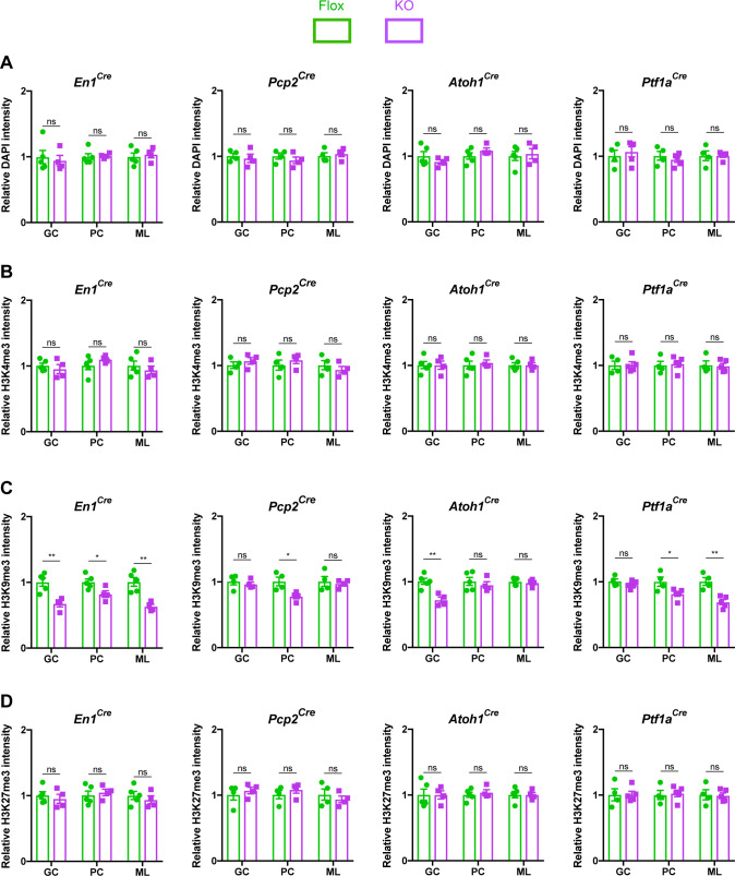
Figure 4—source data 1. Related to Figure 4.

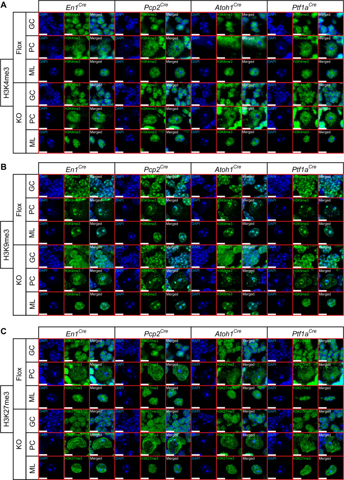
Figure 4.

Figure 4—figure supplement 1. Heterochromatin architecture in mice lacking Mecp2 in cerebellar neurons.

Figure 4—figure supplement 1.

(A) Representative images of cerebellar neurons in Flox and KO mice showing DAPI and H3K4me3. Scale bar, 5 µm. (B) Representative images of cerebellar neurons in Flox and KO mice showing DAPI and H3K9me3. Scale bar, 5 µm. (C) Representative images of cerebellar neurons in Flox and KO mice showing DAPI and H3K27me3. Scale bar, 5 µm.
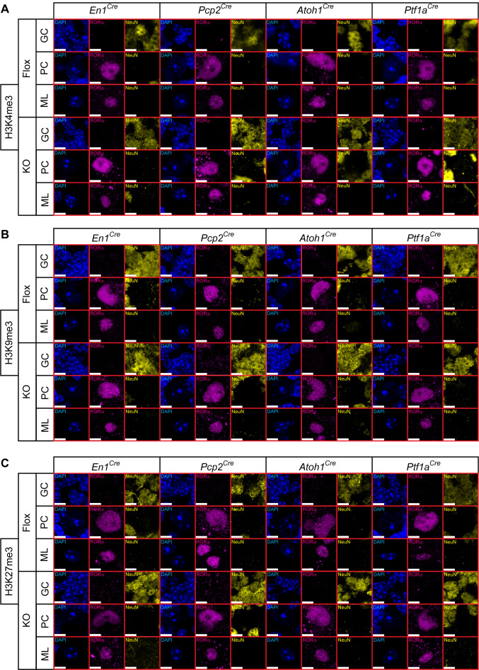
Figure 4—figure supplement 2. Cerebellar neurons were identified by the expression of RORα and NeuN.

Figure 4—figure supplement 2.

(A) Neurons stained for H3K4me3 from Figure 4—figure supplement 1 were co-stained for RORα and NeuN to identify molecular layer interneurons (RORα), Purkinje cells (RORα), and granule cells (NeuN). Scale bar, 5 µm. (B) Neurons stained for H3K9me3 from Figure 4—figure supplement 1 were co-stained for RORα and NeuN to identify molecular layer interneurons (RORα), Purkinje cells (RORα), and granule cells (NeuN). Scale bar, 5 µm. (C) Neurons stained for H3K27me3 from Figure 4—figure supplement 1 were co-stained for RORα and NeuN to identify molecular layer interneurons (RORα), Purkinje cells (RORα), and granule cells (NeuN). Scale bar, 5 µm.