Abstract
Objective
To investigate the three-dimensional forces created by clear aligners on mandibular teeth during differential activation with en-masse retraction and/or intrusion in vitro.
Methods
Six sets of clear aligners were designed for differential en-masse retraction and/or intrusion procedures in a first premolar extraction model. Group A0 was a control group with no activation. Groups A1–5 underwent different degrees of retractions and/or intrusions. Each group consisted of 10 aligners. Aligner forces were measured on a multi-axis force/torque transducer measurement system in real-time.
Results
In the en-masse retraction groups (A1 and A2), lingual and extrusive forces were observed on the incisors; the canines mainly received distal forces; intrusive forces were seen on the second premolars; and the molars received mesial forces. In the en-masse retraction and intrusion groups (A3, A4, and A5), incisors also received lingual and extrusive forces; canines received distal and intrusive forces; mesial and extrusive forces were seen on the second premolars; and the second molars received distal and intrusive forces. The vertical forces on the incisors did not differ significantly among groups A1, A3, and A5. However, the vertical forces on the second premolars reversed from intrusion in group A1 to extrusion in groups A3 and A5.
Conclusions
With clear aligners, the “bowing effect” is seen during en-masse anterior teeth retraction and can be partially relieved by performing en-masse retraction accompanied by anterior teeth intrusion. Vertical control of incisors remained unsolved during en-masse retraction, even when intrusive activation was added to the anterior teeth.
Keywords: Aligners, Tooth movement, Orthodontic treatment, Activation amount
INTRODUCTION
Clear aligner therapy (CAT) has been attracting increasing interest over the past two decades due to its improved aesthetics, comfort, and maintenance of oral hygiene. Early treatment indications for CAT include mild to moderate crowding or diastema, non-skeletal narrow arches, and mild relapse after fixed appliance therapy.1 With advances in materials and the use of creative adjuncts, the range of treatable malocclusions has expanded to include anterior-posterior, vertical, and transverse corrections.2-6
At present, increasing numbers of dentists are using the clear aligner in extraction cases, and some of them have reported satisfactory results in the literature.7-11 However, vertical control remains a problem in extraction cases treated by CAT. Clinicians have struggled with the “bowing effect” seen in orthodontic treatment by fixed appliances. This effect is especially relevant with lingual orthodontics that specifically involve extrusion of incisors and intrusion of second premolars.12 Bowman et al.7 found that control of intrusion with clear aligners was poor and that the mesial aspects of molars tend to intrude while the crowns tip forward. Baldwin et al.9 also observed excessive tipping movement adjacent to extraction sites in 24 patients who underwent extraction of at least one premolar. However, the underlying biomechanics of the “bowing effect” seen in CAT have not been investigated.
Innovations in micro-sensor technology have made it possible to measure orthodontic forces in vitro in real time.13-18 Hahn et al.13,14 constructed a measuring device with a separated maxillary central incisor fixed on the sensor. The results showed that the forces produced by thick appliances were generally higher than those produced by thin materials. Kohda et al.15 studied the correlation between orthodontic forces produced by thermoplastic appliances and the material, thickness, and amount of activation. They found that thicker materials and smaller activations generally deliver high forces. Simon et al.16 measured the three-dimensional (3D) forces and moments generated by a series of aligners using a measurement system with two force/moment (F/M) sensors. They found that the attachments had a significant impact on force transfer. Li et al.17 investigated the force changes on upper central incisors using aligners with different lingual activations (0.2, 0.3, 0.4, 0.5, and 0.6 mm) by using a micro-stress sensor system. Their results suggested that the activation should not exceed 0.5 mm without consideration of the periodontal ligament to maintain the greatest efficiency. Liu and Hu18 studied vertical forces associated with different intrusion strategies in non-extraction deep-bite cases using this system. However, none of the studies to date have investigated the biomechanics of extraction cases.
In this study, clear aligners that underwent different anterior teeth en-masse retraction and/or intrusion activations were investigated, and the corresponding 3D forces were measured in real time to determine the force changes, especially vertical force changes. The aim of this study was to identify a strategy to prevent or relieve the “bowing effect” from occurring in first premolar extraction treatment with clear aligners.
MATERIALS AND METHODS
Study protocol
Sixty removable thermoplastic aligners were divided into six groups based on movement strategies for lower anterior teeth. Each group included 10 aligners. Group A0 was set as the control group and did not undergo any activation, and the other five sets of clear aligners were designated as experimental groups with different degrees of anterior teeth en-masse activations for retraction and/or intrusion. Transverse rectangular attachments were bonded onto the second premolars, the first molars, and the second molars. More details of the force activations performed in each group are presented in Table 1.
Table 1.
Study protocol
| Group | Amount of retraction (mm) |
Amount of intrusion (mm) |
|---|---|---|
| A0 | 0 | 0 |
| A1 | 0.25 | 0 |
| A2 | 0.5 | 0 |
| A3 | 0.25 | 0.25 |
| A4 | 0.5 | 0.5 |
| A5 | 0.25 | 0.5 |
Group A0, control group with no activation; Group A1, underwent 0.25-mm retraction; Group A2, underwent 0.50-mm retraction; Group A3, underwent 0.25-mm retraction and 0.25-mm intrusion; Group A4, underwent 0.50-mm retraction and 0.50-mm intrusion; Group A5, underwent 0.25-mm retraction and 0.5-mm intrusion.
Aligner fabrication and test apparatus construction
OrthoDS software ver. 5.3 (Wuxi Angel Align Biotechnology Co., Ltd., Wuxi, China) was used to design aligners with 0.25-mm and 0.50-mm activations for retraction and/or intrusion of the mandibular anterior teeth on a digitized mandibular standard extraction model. The corresponding photosensitive resin models were printed by a 3D printer (Objet30 Pro; Stratasys Ltd., Rehovot, Israel) according to the digital models. Six sets of corresponding thermoplastic aligners were fabricated using thermoforming technology with 0.8-mm-thick thermoplastic material (Duran; Scheu-Dental, Iserlohn, Germany).
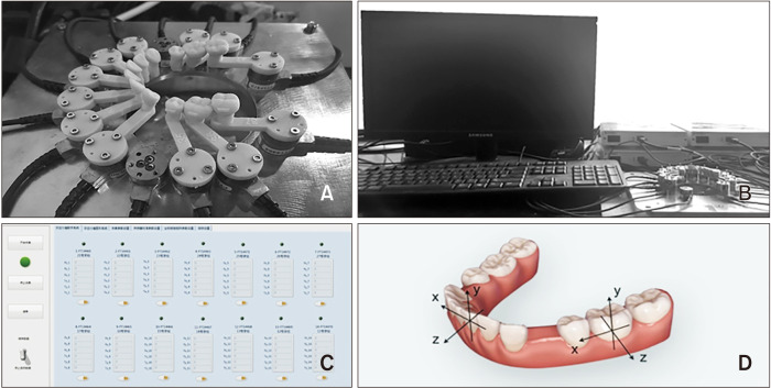
The test apparatus consisted of 12 multi-axis F/M transducers (IFPSMC3/4; ATI Industrial Automation, Apex, NC, USA) and corresponding 12 3D-printed resin teeth (Figure 1A). The resin teeth were printed by Object30 OrthoDesk (Stratasys Ltd.) and connected separately with the multi-axis F/M transducers by hexagonal screws.
Figure 1.
The force measurement system. A, Three-dimensional-printed resin teeth connected separately with the multi-axis force/moment transducer by hexagonal screws. B, The computer linked with the measurement system. C, The real-time visualization window of Angelalign Mechanical Measurement Software (Wuxi Angel Align Biotechnology Co., Ltd., Wuxi, China). D, The coordinate system for the forces and moments measured. The y-axis runs through the center of tooth and parallel to the long axis of this tooth. The x-axis is oriented parallel to the mesiodistal direction of teeth. The z-axis represents the labiolingual/buccolingual force.
Data collection
When each clear aligner was inserted, a computer (Figure 1B) connected to the transducer collected and recorded forces and moments every 1 second over a 40-second cycle. Each group contained 10 aligners, and this process was repeated for the six separate groups. The real-time visualization window of Angelalign Mechanical Measurement Software (Wuxi Angel Align Biotechnology Co.) was used to monitor changes in forces and moments and record real-time values (Figure 1C). The measured F/M values were transferred to the center of resistance of each tooth. To describe the forces the teeth received in 3D directions, we set up separate coordinate systems for each tooth to describe movements in all 3 spatial dimensions (Figure 1D). The y-axis ran through the center of resistance of each tooth and parallel to the long axis of this tooth, and positive values represented extrusive forces. The x-axis was oriented parallel to the mesiodistal direction of teeth, and positive values represented mesial forces. The z-axis represented the labiolingual/buccolingual direction, and positive values represented labial/buccal forces.
Each aligner in group A0 was inserted on the F/M measuring system to calibrate the influence of positioning errors before activated invisible aligners were inserted. The mean forces in the 3D direction of group A0 were obtained (Table 2). Five groups of active aligners were successively placed on the F/M measuring system, and the 3D forces generated by the different aligners were measured and recorded. After subtracting the corresponding data from group A0, the average 3D forces in the other five groups were obtained (Table 2). Histograms were used to describe the forces to directly observe the trend of distribution (Figures 2–5). All procedures were performed manually by a single operator to reduce variability. The intra-examiner reliability was good (intraclass correlation coefficient = 96.5%, 95% confidence interval: 0.958–0.971, p < 0.001).
Table 2.
Force distribution within clear aligner and comparisons of the forces in group A1–5
| Tooth number | Force direction | A0 | A1 | A2 | A3 | A4 | A5 |
|---|---|---|---|---|---|---|---|
| 37 | Fx¶ | 0.26 ± 0.35 | 1.00 ± 1.58‡‖ | 1.82 ± 0.54‡§‖ | −0.43 ± 0.71*† | −0.13 ± 0.92†‖ | −1.68 ± 0.96*†§ |
| Fy¶ | 1.37 ± 0.27 | 0.58 ± 0.98‡§‖ | 0.70 ± 0.81‡§‖ | −0.74 ± 0.44*† | −0.67 ± 1.10*†‖ | −1.80 ± 0.73*†§ | |
| Fz** | −0.94 ± 0.12 | 0.34 ± 0.17‡ | 0.65 ± 0.25 | 0.71 ± 0.18*§ | 0.44 ± 0.15‡ | 0.49 ± 0.24 | |
| 36 | Fx¶ | −1.11 ± 0.14 | 0.64 ± 0.39§‖ | 1.21 ± 0.56§ | 1.30 ± 0.39§ | 2.85 ± 0.76*†‡§ | 1.58 ± 0.47*§ |
| Fy¶ | −2.43 ± 0.24 | −0.33 ± 1.71‖ | −0.27 ± 0.59 | 0.12 ± 0.59 | 0.70 ± 2.12 | 1.38 ± 0.60* | |
| Fz¶ | 1.77 ± 0.12 | −0.63 ± 0.48 | −0.96 ± 0.33 | −0.78 ± 0.30 | −1.11 ± 0.57 | −1.11 ± 0.25 | |
| 35 | Fx¶ | 2.75 ± 0.37 | 0.03 ± 0.26§‖ | 0.47 ± 0.53§‖ | 0.62 ± 0.34§‖ | 1.78 ± 0.58*†‡ | 1.50 ± 0.58*†‡ |
| Fy¶ | 0.38 ± 0.36 | −0.74 ± 0.80‡§‖ | −0.93 ± 0.63‡§‖ | 0.82 ± 0.47*† | 0.88 ± 1.13*† | 1.61 ± 0.57*† | |
| Fz¶ | −1.74 ± 0.26 | 0.20 ± 0.80‡§‖ | −0.40 ± 0.45§‖ | −0.82 ± 0.31*§ | −1.63 ± 0.48*†‡ | −1.13 ± 0.41*† | |
| 33 | Fx¶ | −0.16 ± 0.12 | −1.19 ± 0.21†‡§‖ | −2.20 ± 0.17*‡§‖ | −0.63 ± 0.09*†§ | −1.79 ± 0.11*†‡‖ | −0.62 ± 0.13*†§ |
| Fy** | −0.30 ± 0.17 | 0.40 ± 0.14†‡§‖ | −0.65 ± 0.22*§‖ | −0.80 ± 0.11*§‖ | −2.39 ± 0.38*†‡ | −2.05 ± 0.34*†‡ | |
| Fz¶ | −0.34 ± 0.23 | −0.24 ± 0.72 ‡§‖ | 0.31 ± 0.46 §‖ | 0.87 ± 0.26 *§‖ | 1.68 ± 0.40*†‡ | 2.11 ± 0.47 *†‡ | |
| 32 | Fx¶ | 0.14 ± 0.05 | −0.51 ± 0.13†‡§‖ | −0.96 ± 0.09*‡§‖ | −0.23 ± 0.08*†§ | −0.72 ± 0.04*†‡‖ | −0.26 ± 0.08*†§ |
| Fy¶ | 0.01 ± 0.07 | 0.15 ± 0.18§‖ | 0.08 ± 0.13 | 0.03 ± 0.17 | −0.08 ± 0.09* | −0.03 ± 0.11* | |
| Fz¶ | −0.84 ± 0.12 | 0.05 ± 0.25†‡§ | −0.33 ± 0.19*§ | −0.27 ± 0.15*§ | −0.67 ± 0.21*†‡‖ | −0.18 ± 0.15§ | |
| 31 | Fx¶ | 0.14 ± 0.09 | −0.14 ± 0.12 | −0.25 ± 0.10 | −0.15 ± 0.05 | −0.17 ± 0.09 | −0.15 ± 0.16 |
| Fy¶ | −0.46 ± 0.15 | 0.18 ± 0.27†§‖ | 0.74 ± 0.20*‡ | 0.31 ± 0.29† | 0.55 ± 0.27* | 0.58 ± 0.22* | |
| Fz¶ | −0.72 ± 0.13 | −0.40 ± 0.30†§‖ | −1.53 ± 0.33*‡§ | −0.75 ± 0.18†§‖ | −2.55 ± 0.28*†‡‖ | −1.23 ± 0.22*‡§ | |
| 41 | Fx¶ | 1.12 ± 0.10 | −0.03 ± 0.11§ | −0.04 ± 0.09§ | 0.07 ± 0.07 | 0.14 ± 0.08*† | 0.08 ± 0.10 |
| Fy¶ | 0.23 ± 0.10 | −0.04 ± 0.27 | 0.03 ± 0.28 | 0.11 ± 0.29 | −0.14 ± 0.27 | 0.18 ± 0.20 | |
| Fz** | −1.26 ± 0.13 | −0.46 ± 0.34†§‖ | −1.30 ± 0.46*§ | −0.86 ± 0.14§‖ | −2.36 ± 0.30*†‡‖ | −1.12 ± 0.14*‡§ | |
| 42 | Fx** | 0.81 ± 0.12 | −0.34 ± 0.09†‖ | −0.59 ± 0.06*‡§‖ | −0.23 ± 0.08†§ | −0.42 ± 0.06†‡‖ | −0.20 ± 0.06*†§ |
| Fy** | 0.04 ± 0.03 | −0.23 ± 0.05†‖ | −0.40 ± 0.14* | −0.24 ± 0.05‖ | −0.38 ± 0.15 | −0.37 ± 0.09*‡ | |
| Fz** | 0.93 ± 0.19 | −0.03 ± 0.19 †‡§‖ | −0.28 ± 0.10*§‖ | −0.34 ± 0.12 *§‖ | −1.08 ± 0.19 *†‡‖ | −0.62 ± 0.09 *†‡§ | |
| 43 | Fx** | 1.58 ± 0.16 | −1.42 ± 0.13†‡§‖ | −2.39 ± 0.16*‡‖ | −0.83 ± 0.29*†§ | −2.20 ± 0.15*‡‖ | −0.88 ± 0.24*†§ |
| Fy¶ | 0.25 ± 0.15 | −0.02 ± 0.37†‡§‖ | −0.94 ± 0.19*§‖ | −1.21 ± 0.23*§‖ | −3.18 ± 0.30*†‡‖ | −2.59 ± 0.40*†‡§ | |
| Fz** | −0.10 ± 0.31 | 0.11 ± 0.34‡§‖ | 0.49 ± 0.36‡§‖ | 1.07 ± 0.37*†§‖ | 1.89 ± 0.46*†‡ | 2.11 ± 0.35 | |
| 45 | Fx** | 0.85 ± 0.41 | −0.18 ± 0.51‡§‖ | 0.20 ± 0.45§‖ | 1.06 ± 0.82*§ | 2.32 ± 0.39*†‡ | 1.84 ± 0.38*† |
| Fy** | 0.36 ± 0.41 | −0.86 ± 0.53‡§‖ | −1.12 ± 0.62‡§‖ | 0.73 ± 0.95*† | 1.13 ± 0.41*†‖ | 1.81 ± 0.46*†§ | |
| Fz** | −0.18 ± 0.13 | 0.08 ± 0.23‡§‖ | −0.04 ± 0.29‡§‖ | −0.77 ± 0.45*†§ | −1.43 ± 0.28*†‡‖ | −0.65 ± 0.28*†‡ | |
| 46 | Fx** | −1.55 ± 0.43 | 0.63 ± 0.53†§ | 1.75 ± 0.52*‡‖ | 0.23 ± 1.18†§ | 1.77 ± 0.56*‡‖ | 0.80 ± 0.57†§ |
| Fy** | −2.02 ± 0.47 | 0.07 ± 0.64 | 0.64 ± 0.43 | −0.17 ± 1.36 | 0.63 ± 0.66 | 0.83 ± 0.61 | |
| Fz¶ | 1.71 ± 0.21 | −0.46 ± 0.28 | −0.44 ± 0.22 | 0.04 ± 0.50 | −0.27 ± 0.23 | −0.02 ± 0.40*†§ | |
| 47 | Fx** | −0.21 ± 0.78 | 0.86 ± 0.93‖ | 1.32 ± 0.36‖ | 0.17 ± 0.93 | 0.76 ± 0.73‖ | −0.81 ± 0.73*†§ |
| Fy** | 1.43 ± 0.44 | 0.48 ± 0.56‖ | 0.64 ± 0.34§‖ | 0.08 ± 0.58‖ | 0.04 ± 0.42†‖ | −0.90 ± 0.40*†‡§ | |
| Fz** | −1.58 ± 0.18 | 0.06 ± 0.13‖ | −0.04 ± 0.12‖ | −0.04 ± 0.40 | 0.00 ± 0.15‖ | −0.34 ± 0.18*†§ |
Values are presented as mean ± standard deviation.
Group A0 was used as the reference data, and the figures from other groups were obtained by subtracting from the A0.
FDI tooth numbering system was used.
Fx, force along the x-axis: positive values represent mesial forces; Fy, force along the y-axis: positive values represent extrusive forces; Fz, force along the z-axis: positive values represent labial or buccal forces.
Significantly different from *group A1, †group A2, ‡group A3, §group A4, and ‖group A5 on the same tooth.
¶Bonferroni’s test, p < 0.05.
**Dunnett’s T3, p < 0.05.
See Table 1 for definitions of each group.
Figure 2.
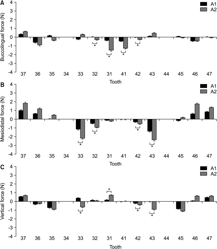
Comparisons of the three-dimensional forces in groups A1 and A2. A, Forces in the buccolingual direction. B, Forces in the mesiodistal direction. C, Forces in the vertical direction.
Group A1, underwent 0.25-mm retraction; Group A2, underwent 0.50-mm retraction.
*p < 0.05.
Figure 3.
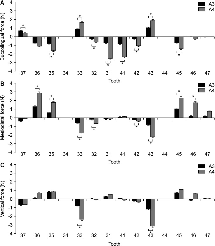
Comparisons of the three-dimensional forces in groups A3 and A4. A, Forces in the buccolingual direction. B, Forces in the mesiodistal direction. C, Forces in the vertical direction.
Group A3, underwent 0.25-mm retraction and 0.25-mm intrusion; Group A4, underwent 0.50-mm retraction and 0.50-mm intrusion.
*p < 0.05.
Figure 4.
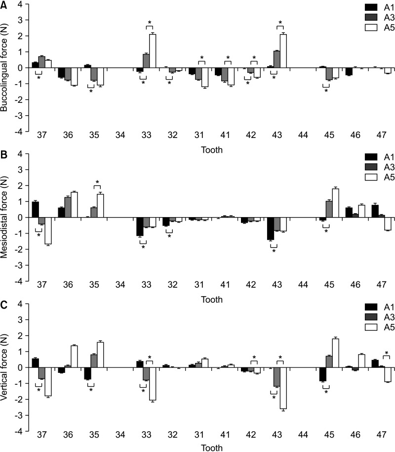
Comparisons of the three-dimensional forces in groups A1, A3, and A5. A, Forces in the buccolingual direction. B, Forces in the mesiodistal direction. C, Forces in the vertical direction.
Group A1, underwent 0.25-mm retraction; Group A3, underwent 0.25-mm retraction and 0.25-mm intrusion; Group A5, underwent 0.25-mm retraction and 0.5-mm intrusion.
*p < 0.05.
Figure 5.
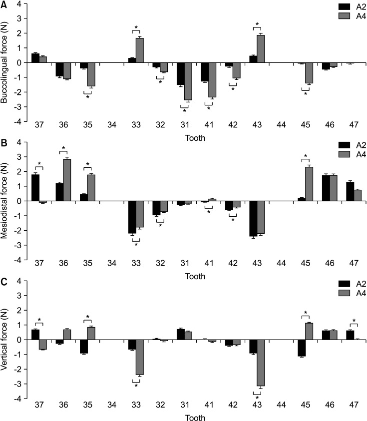
Comparisons of three-dimensional forces in groups A2 and A4. A, Forces in the buccolingual direction. B, Forces in the mesiodistal direction. C, Forces in the vertical direction.
Group A2, underwent 0.50-mm retraction; Group A4, underwent 0.50-mm retraction and 0.50-mm intrusion.
*p < 0.05.
Statistical analysis
To compare the differences among the five groups, all statistical analyses were performed using SPSS ver. 20.0 (IBM Co., Armonk, NY, USA). One-way analysis of variance was used to examine the differences among groups. The Bonferroni test was applied when homogeneity of variance assumptions was satisfied; otherwise, the equivalent Dunnettʼs T3 test was used. Both statistical analyses were conducted at α = 0.05 level of significance (Table 2).
RESULTS
In group A0, forces were produced in all 3 dimensions even when there was no designed activation (Table 2). Only anterior teeth retraction movement was observed in groups A1 and A2. The 3D force measurements for each group are shown in Table 2 and Figure 2. In group A1 with 0.25-mm en-masse retraction movement, the four incisors received mild to moderate lingual forces, while molars received moderate mesial forces as anchorage. The second molar received a more mesial force than the first molar. As a consequence of their location, the canines mainly received distal forces. The vertical direction mainly received mild to moderate forces in the lower anterior region. The canines received extrusive forces, while incisors received small extrusive or intrusive forces. The second premolars received moderate intrusive forces, and the second molars received moderate extrusive forces.
In group A2, the amount of en-masse retraction designed was 0.50 mm in the lower anterior area. In comparison with group A1, group A2 showed higher lingual forces on the incisors, distal forces on the canines, and mesial forces on the molars. The lingual forces on the incisors and distal forces on the canines showed statistically significant differences between these two groups (p < 0.05). However, there were no significant differences in the forces on the posterior teeth (p > 0.05). In the vertical direction, the canines received moderate intrusive forces. Most of the incisors received extrusive forces. The second premolars experienced intrusive forces and the second molars experienced an extrusive force. The extrusive forces on the left central incisors increased while those on the canines were reversed to intrusive forces with increasing amounts of en-masse retraction, indicating that the “bowing effect” had worsened. Vertical forces on the left central incisors, right lateral incisor, and bilateral canines showed statistically significant differences between the two groups (p < 0.05). However, there were no significant differences for the posterior teeth (p > 0.05).
Groups A3, A4, and A5 showed anterior teeth retraction and intrusion movements. In group A3 with a 0.25-mm en-masse retraction and intrusion activation in the lower anterior area, the aligners produced moderate lingual forces on the incisors and mesial forces on the second premolars and first molars (Figure 3). The aligners also exerted mild distal forces on the left second molar, while the canines received labial and distal forces. In the vertical direction, the incisors received limited vertical forces, while the canines received moderate intrusive forces. The vertical forces on the second premolars were extrusive forces.
In group A4, the amounts of en-masse retraction and intrusion designed were 0.5 mm each in the lower anterior area (Figure 3). In comparison with group A3, group A4 showed higher lingual forces on the incisors and mesial forces on the second premolars and first molars. The canines received more labial and distal forces. All the forces mentioned above showed statistically significant differences between these two groups (p < 0.05). However, the incisors received limited vertical forces as well. The canines received higher intrusive forces, and the forces on canines significantly differed between groups A3 and A4 (p < 0.05). The second premolars in both groups received similar extrusive forces but did not show a statistically significant difference (p > 0.05).
In group A5, aligners were designed for 0.25-mm anterior teeth retraction and 0.5-mm anterior teeth intrusion (Figure 4). In comparison with group A3, group A5 showed twice the amount of intrusion movement. The incisors received greater lingual forces, and the second premolars received greater mesial forces. Notably, the second molars received obvious distal forces, and the canines received greater labial forces. The labiolingual forces on the central incisors, right lateral incisor, and bilateral canines, as well as the mesial force on the left second premolar showed statistically significant differences between the two groups (p < 0.05). In the vertical direction, the aligners exerted more intrusive forces on canines and second molars and more extrusive forces on the second premolars. However, the incisors received limited vertical forces. Statistically significant differences in the vertical forces of bilateral canines and the right second molar were observed only between groups A3 and A5 (p < 0.05, Figure 5).
On comparing the differences in forces in the three en-masse retractions and/or intrusion groups (A1, A3, and A5, Figure 4), the lingual forces aligners exerted on incisors and the labial forces on canines increased, while the distal forces on canines decreased. The mesial forces on the second premolar increased, but the forces received by the second molars changed from mesial forces in group A1 to distal forces in group A5. Notably, the “bowing effect” seen in group A1 was partially alleviated in groups A3 and A5, especially in the posterior area. This was because anterior teeth retraction without intrusion was designed in group A1, while anterior teeth retraction and intrusion were designed in groups A3 and A5. The forces on second premolars changed from an intrusive force in group A1 to an extrusive force in groups A3 and A5. The forces on the second molars also changed from an extrusive force in group A1 to an intrusive force in groups A3 and A5. In addition, the vertical forces on incisors did not change significantly even when the intrusive activation on anterior teeth in group A5 was increased. Only the intrusive forces on the canines became greater, with the burden on the anchorage teeth being correspondingly aggravated. This tendency could also be seen between groups A2 and A4, in which the amount of activation was 0.5 mm (Figure 5).
DISCUSSION
The present study was performed to explore the distribution of 3D forces on mandibular teeth undergoing en-masse retraction and/or intrusion in extraction treatment using clear aligners.
Compared to conventional fixed appliances, clear thermoformed plastic aligners cover the entire dentition as an overlay appliance during teeth movement, making them significantly different in effectiveness and accuracy.19-21 The orthodontic performance of clear aligners in extraction cases remains inferior to that obtained with conventional fixed appliances.22,23 Many researchers observed the “bowing effect” when extraction spaces were closed with aligners in clinical settings.7,9,11 This effect was believed to result from the sagging of the plastic around the extraction sites.9,11 Therefore, it is crucial for orthodontists to clearly understand the biomechanics underlying CAT, especially in the vertical direction.
A thorough investigation of the exact biomechanical mechanisms involved in clear aligners is challenging. Photoelastic models24 and the finite element method25 were generally used to study the biomechanics of orthodontics. In a recent in vivo study, a pressure film was used to measure the pressure imparted by removable thermoplastic appliances on the surface of the upper first premolar during the buccal tipping movement.26 In another in vivo study, strain-gauge rosettes were attached to the external surface of aligners to obtain force values.27 Recent improvements in micro-sensor technology have made it possible to measure orthodontic forces and moments of appliances in vitro.13,16,17,28 Nevertheless, there is currently little research regarding the clear aligner force system. Therefore, we designed this system for real-time in vitro measurement of orthodontic forces.
The bowing effect exists in both the mandible and maxilla. However, we found that this effect is more severe on the mandibular dentition during the orthodontic treatment of tooth extraction cases using aligners. Extrusion of the lower anterior teeth and intrusion of the lower second premolars are more visible than the changes in the upper teeth. As a consequence, we only investigated forces in the lower jaw. The biomechanical characteristics of the upper dental arch will be studied in the future and compared with those of the lower dental arch. In our study, when exerting en-masse retraction activation from 0.25 mm in group A1 to 0.5 mm in group A2, apparent lingual forces and limited extrusive forces were exerted on the incisors, while moderate distal and intrusive forces were applied on the canines. The second premolars mainly experienced intrusive forces, while the second molars received mesial and extrusive forces. This was similar to the “bowing effect,” which was in agreement with observed clinical phenomena. In these cases, second molars would be extruded, while canines and second premolars would be intruded upon en-masse retraction with clear aligners, resulting in a deep curve of Spee with teeth tipping movement adjacent to extraction sites. Due to the reduction in aligner length, incisors received lingual forces, and posterior teeth as a whole received mesial forces. The molars were the main anchorage teeth.
A deep curve of Spee is related to a deep overbite. Effective intrusion of the anterior teeth is generally necessary during en-masse retraction. In the current study, intrusive activation was added in group A3 to prevent extrusion of anterior teeth and intrusion of posterior teeth while exerting en-masse retraction. In a comparison of groups A1 with A3, the changes in the vertical forces that the incisors received were not statistically significant, while the lingual force increased. The canines experienced relatively large intrusive forces but received lighter distal forces. In this situation, the incisors were still out of vertical control even when anterior teeth retraction was accompanied by anterior teeth intrusion. In fixed appliance treatment, a bite opening curve, which is often used to intrude anterior teeth, leads to labial crown torque and increases the length of the arch.29,30 In CAT, intrusion was parallel to the long axis of the tooth and went through the center of resistance of the tooth. In comparison to only en-masse retraction, the length of the aligner was further reduced during retraction accompanied by intrusion. As a result, lingual forces on the incisors were greater, and the second premolars became the main anchorage. The vertical forces on the second premolars changed from intrusive to extrusive forces, and moderate mesial forces were also observed. The extrusive forces on the second molars decreased or transformed into intrusive forces, and mesial forces decreased or transformed into distal forces. Therefore, the “bowing effect” that appeared in en-masse retraction was partially relieved on the posterior teeth. This is similar to the reverse curve of Spee, which is commonly used in the straight-wire treatment of deep-bite cases, and this finding suggests that anterior teeth intrusion should be added during en-masse retraction.
After doubling the amount of intrusion with en-masse retraction in group A5, the situation on the incisors remained the same as that in group A3. There were no obvious intrusive forces on incisors; in fact, some extrusive forces were measured. At the same time, more lingual forces were observed on the incisors. Only the canines received greater labial and intrusive forces, while the second premolars received increased mesial and extrusive forces. The second molars received intrusive and distal forces. This suggests that there is no additional benefit on the vertical control of anterior teeth during en-masse retraction resulting from increased anterior teeth intrusion with clear aligner treatment.
In addition, the retraction of anterior teeth using clear aligners is affected by the design of anterior tooth movement, activation time, and aligner material.14,31 Different amounts of activation are recommended by different invisible orthodontic suppliers. Li et al.17 suggested that lingual activation for upper central incisors should not exceed 0.5 mm. In the current study, 0.8-mm transparent thermoplastic material was used to fabricate aligners for 0.25- and 0.5-mm activations for retractive and/or intrusive movement of mandibular anterior teeth. In groups A2, A4, and A5, the vertical forces produced by 0.5-mm activation in the aligner were greater than the optimal translational forces (0.75–1.25 N).32 Thus, 0.25-mm activation is recommended because the forces in groups A1 and A3 did not exceed 1.42 N.
This study has several limitations. First, due to the lack of emulation of the intra-oral environment and periodontal ligaments, the simulated tooth movement produced by the clear aligners cannot completely predict results in vivo. Second, abrasion arising from mastication and changed mechanical properties was not taken into consideration. Third, en-masse retraction and/or intrusion of only mandibular anterior teeth were investigated.
The deep overbite caused by en-masse retraction in extraction cases treated with CAT remains unsolved. If anterior tooth retraction can be divided into two steps, with the first step being distal movement of the canines followed by retraction and intrusion of the incisors, the “bowing effect” may be alleviated and canine movement may be more similar to translation. Further research on this aspect will be conducted in the future.
CONCLUSION
In this study, the vertical force changes associated with different en-masse retraction and/or intrusion strategies in extraction cases treated with clear aligners were analyzed. The following conclusions can be drawn from this study. First, with CAT, the “bowing effect” appeared during en-masse retraction. Second, the “bowing effect” can be partially relieved during en-masse retraction when accompanied by anterior teeth intrusion. Third, the issue of vertical control of incisors remains unsolved during en-masse retraction in CAT, even when intrusive activation was added on the anterior teeth.
Footnotes
CONFLICTS OF INTEREST
No potential conflict of interest relevant to this article was reported.
REFERENCES
- 1.Turpin DL. Clinical trials needed to answer questions about Invisalign. Am J Orthod Dentofacial Orthop. 2005;127:157–8. doi: 10.1016/j.ajodo.2004.12.009. [DOI] [PubMed] [Google Scholar]
- 2.Shin K. The Invisalign appliance could be an effective modality for treating overbite malocclusions within a mild to moderate range. J Evid Based Dent Pract. 2017;17:278–80. doi: 10.1016/j.jebdp.2017.06.010. [DOI] [PubMed] [Google Scholar]
- 3.Bowman SJ, Celenza F, Sparaga J, Papadopoulos MA, Ojima K, Lin JC. Creative adjuncts for clear aligners, part 2: intrusion, rotation, and extrusion. J Clin Orthod. 2015;49:162–72. [PubMed] [Google Scholar]
- 4.Khosravi R, Cohanim B, Hujoel P, Daher S, Neal M, Liu W, et al. Management of overbite with the Invisalign appliance. Am J Orthod Dentofacial Orthop. 2017;151:691–9.e2. doi: 10.1016/j.ajodo.2016.09.022. [DOI] [PubMed] [Google Scholar]
- 5.Bowman SJ, Celenza F, Sparaga J, Papadopoulos MA, Ojima K, Lin JC. Creative adjuncts for clear aligners, part 1: class II treatment. J Clin Orthod. 2015;49:83–94. [PubMed] [Google Scholar]
- 6.Houle JP, Piedade L, Todescan R, Jr, Pinheiro FH. The predictability of transverse changes with Invisalign. Angle Orthod. 2017;87:19–24. doi: 10.2319/122115-875.1. [DOI] [PMC free article] [PubMed] [Google Scholar]
- 7.Bowman SJ, Celenza F, Sparaga J, Papadopoulos MA, Ojima K, Lin JC. Creative adjuncts for clear aligners, part 3: extraction and interdisciplinary treatment. J Clin Orthod. 2015;49:249–62. [PubMed] [Google Scholar]
- 8.Womack WR. Four-premolar extraction treatment with Invisalign. J Clin Orthod. 2006;40:493–500. [PubMed] [Google Scholar]
- 9.Baldwin DK, King G, Ramsay DS, Huang G, Bollen AM. Activation time and material stiffness of sequential removable orthodontic appliances. Part 3: premolar extraction patients. Am J Orthod Dentofacial Orthop. 2008;133:837–45. doi: 10.1016/j.ajodo.2006.06.025. [DOI] [PubMed] [Google Scholar]
- 10.Giancotti A, Greco M, Mampieri G. Extraction treatment using Invisalign Technique. Prog Orthod. 2006;7:32–43. [PubMed] [Google Scholar]
- 11.Ojima K, Dan C, Nishiyama R, Ohtsuka S, Schupp W. Accelerated extraction treatment with Invisalign. J Clin Orthod. 2014;48:487–99. [PubMed] [Google Scholar]
- 12.Liu D, Yan B, Lei F, Li J, Wang X, Rong Q, et al. Different sliding mechanics in space closure of lingual orthodontics: a translational study by three-dimensional finite element method. Am J Transl Res. 2019;11:120–30. [PMC free article] [PubMed] [Google Scholar]
- 13.Hahn W, Dathe H, Fialka-Fricke J, Fricke-Zech S, Zapf A, Kubein-Meesenburg D, et al. Influence of thermoplastic appliance thickness on the magnitude of force delivered to a maxillary central incisor during tipping. Am J Orthod Dentofacial Orthop. 2009;136:12.e1–7. discussion 12–3. doi: 10.1016/j.ajodo.2008.12.015. [DOI] [PubMed] [Google Scholar]
- 14.Hahn W, Zapf A, Dathe H, Fialka-Fricke J, Fricke-Zech S, Gruber R, et al. Torquing an upper central incisor with aligners--acting forces and biomechanical principles. Eur J Orthod. 2010;32:607–13. doi: 10.1093/ejo/cjq007. [DOI] [PubMed] [Google Scholar]
- 15.Kohda N, Iijima M, Muguruma T, Brantley WA, Ahluwalia KS, Mizoguchi I. Effects of mechanical properties of thermoplastic materials on the initial force of thermoplastic appliances. Angle Orthod. 2013;83:476–83. doi: 10.2319/052512-432.1. [DOI] [PMC free article] [PubMed] [Google Scholar]
- 16.Simon M, Keilig L, Schwarze J, Jung BA, Bourauel C. Forces and moments generated by removable thermoplastic aligners: incisor torque, premolar derotation, and molar distalization. Am J Orthod Dentofacial Orthop. 2014;145:728–36. doi: 10.1016/j.ajodo.2014.03.015. [DOI] [PubMed] [Google Scholar]
- 17.Li X, Ren C, Wang Z, Zhao P, Wang H, Bai Y. Changes in force associated with the amount of aligner activation and lingual bodily movement of the maxillary central incisor. Korean J Orthod. 2016;46:65–72. doi: 10.4041/kjod.2016.46.2.65. [DOI] [PMC free article] [PubMed] [Google Scholar]
- 18.Liu Y, Hu W. Force changes associated with different intrusion strategies for deep-bite correction by clear aligners. Angle Orthod. 2018;88:771–8. doi: 10.2319/121717-864.1. [DOI] [PMC free article] [PubMed] [Google Scholar]
- 19.Krieger E, Seiferth J, Saric I, Jung BA, Wehrbein H. Accuracy of invisalign® treatments in the anterior tooth region. First results. J Orofac Orthop. 2011;72:141–9. doi: 10.1007/s00056-011-0017-4. [DOI] [PubMed] [Google Scholar]
- 20.Gu J, Tang JS, Skulski B, Fields HW, Jr, Beck FM, Firestone AR, et al. Evaluation of Invisalign treatment effectiveness and efficiency compared with conventional fixed appliances using the Peer Assessment Rating index. Am J Orthod Dentofacial Orthop. 2017;151:259–66. doi: 10.1016/j.ajodo.2016.06.041. [DOI] [PubMed] [Google Scholar]
- 21.Kravitz ND, Kusnoto B, BeGole E, Obrez A, Agran B. How well does Invisalign work? A prospective clinical study evaluating the efficacy of tooth movement with Invisalign. Am J Orthod Dentofacial Orthop. 2009;135:27–35. doi: 10.1016/j.ajodo.2007.05.018. [DOI] [PubMed] [Google Scholar]
- 22.Li W, Wang S, Zhang Y. The effectiveness of the Invisalign appliance in extraction cases using the the ABO model grading system: a multicenter randomized controlled trial. Int J Clin Exp Med. 2014;8:8276–82. [PMC free article] [PubMed] [Google Scholar] [Retracted]
- 23.Bollen AM, Huang G, King G, Hujoel P, Ma T. Activation time and material stiffness of sequential removable orthodontic appliances. Part 1: ability to complete treatment. Am J Orthod Dentofacial Orthop. 2003;124:496–501. doi: 10.1016/S0889-5406(03)00576-6. [DOI] [PubMed] [Google Scholar]
- 24.Caputo AA, Chaconas SJ, Hayashi RK. Photoelastic visualization of orthodontic forces during canine retraction. Am J Orthod. 1974;65:250–9. doi: 10.1016/S0002-9416(74)90330-3. [DOI] [PubMed] [Google Scholar]
- 25.Knop L, Gandini LG, Jr, Shintcovsk RL, Gandini MR. Scientific use of the finite element method in orthodontics. Dental Press J Orthod. 2015;20:119–25. doi: 10.1590/2176-9451.20.2.119-125.sar. [DOI] [PMC free article] [PubMed] [Google Scholar]
- 26.Barbagallo LJ, Shen G, Jones AS, Swain MV, Petocz P, Darendeliler MA. A novel pressure film approach for determining the force imparted by clear removable thermoplastic appliances. Ann Biomed Eng. 2008;36:335–41. doi: 10.1007/s10439-007-9424-5. [DOI] [PubMed] [Google Scholar]
- 27.Vardimon AD, Robbins D, Brosh T. In-vivo von Mises strains during Invisalign treatment. Am J Orthod Dentofacial Orthop. 2010;138:399–409. doi: 10.1016/j.ajodo.2008.11.027. [DOI] [PubMed] [Google Scholar]
- 28.Badawi H, Major P. Three-dimensional orthodontic force measurements. Am J Orthod Dentofacial Orthop. 2010;137:299–300. doi: 10.1016/j.ajodo.2010.01.006. [DOI] [PubMed] [Google Scholar]
- 29.Ferguson JW. Lower incisor torque: the effects of rectangular archwires with a reverse curve of Spee. Br J Orthod. 1990;17:311–5. doi: 10.1179/bjo.17.4.311. [DOI] [PubMed] [Google Scholar]
- 30.Pandis N, Polychronopoulou A, Sifakakis I, Makou M, Eliades T. Effects of levelling of the curve of Spee on the proclination of mandibular incisors and expansion of dental arches: a prospective clinical trial. Aust Orthod J. 2010;26:61–5. [PubMed] [Google Scholar]
- 31.Clements KM, Bollen AM, Huang G, King G, Hujoel P, Ma T. Activation time and material stiffness of sequential removable orthodontic appliances. Part 2: Dental improvements. Am J Orthod Dentofacial Orthop. 2003;124:502–8. doi: 10.1016/S0889-5406(03)00577-8. [DOI] [PubMed] [Google Scholar]
- 32.Jacobson A. Contemporary orthodontics, 3rd edition. Am J Orthod Dentofacial Orthop. 2000;117:PA1. doi: 10.1016/S0889-5406(00)70018-7. [DOI] [Google Scholar]


