PLOS ONE logoLink to PLOS ONE
. 2021 Jan 27;16(1):e0246066. doi: 10.1371/journal.pone.0246066

Targeted temperature management at 33°C or 36℃ induces equivalent myocardial protection by inhibiting HMGB1 release in myocardial ischemia/reperfusion injury

Jin Ho Beom 1, Ju Hee Kim 1, Jeho Seo 1, Jung Ho Lee 2,3, Yong Eun Chung 4, Hyun Soo Chung 1, Sung Phil Chung 1, Chul Hoon Kim 2,*, Je Sung You 1,*
Editor: Federica Limana5
PMCID: PMC7840046  PMID: 33503060

Abstract

Acute myocardial infarction (AMI) is lethal and causes myocardial necrosis via time-dependent ischemia due to prolonged occlusion of the infarct-related artery. No effective therapy or potential therapeutic targets can prevent myocardial ischemia/reperfusion (I/R) injury. Targeted temperature management (TTM) may reduce peri-infarct regions by inhibiting the extracellular release of high mobility group box-1 (HMGB1) as a primary mediator of the innate immune response. We used a rat left anterior descending (LAD) coronary artery ligation model to determine if TTM at 33°C and 36°C had similar myocardial protective effects. Rats were divided into sham, LAD I/R+37°C normothermia, LAD I/R+33°C TTM, and LAD I/R+36°C TTM groups (n = 5 per group). To verify the cardioprotective effect of TTM by specifically inhibiting HMGB1, rats were assigned to sham, LAD I/R, and LAD I/R after pre-treatment with glycyrrhizin (known as a pharmacological inhibitor of HMGB1) groups (n = 5 per group). Different target temperatures of 33°C and 36°C caused equivalent reductions in infarct volume after myocardial I/R, inhibited the extracellular release of HMGB1 from infarct tissue, and suppressed the expression of inflammatory cytokines from peri-infarct regions. TTM at 33°C and 36°C significantly attenuated the elevation of cardiac troponin, a sensitive and specific marker of heart muscle damage, after injury. Similarly, glycyrrhizin alleviated myocardial damage by suppressing the extracellular release of HMGB1. TTM at 33°C and 36°C had equivalent myocardial protective effects by similar inhibiting HMGB1 release against myocardial I/R injury. This is the first study to suggest that a target core temperature of 36°C is applicable for cardioprotection.

Introduction

Coronary heart disease is the leading cause of death worldwide, and acute myocardial infarction (AMI) is the most severe manifestation of this disease [1, 2]. In AMI, prolonged occlusion of the infarct-related artery leads to high levels of myocardial necrosis as a time-dependent ischemic process. To minimize myocardial necrosis, blood flow to the infarct-related artery must be restored by mechanical reperfusion using a coronary artery stent and thrombolytic therapy as rapidly as possible [3, 4]. Although the door to balloon time has been significantly decreased, overall in-hospital mortality has not significantly declined in patients with ST elevation myocardial infarction (STEMI) undergoing primary percutaneous coronary intervention (PCI) [1, 5]. To achieve safe and effective therapeutic benefits, reperfusion therapy should be performed within 12 h of symptom onset as the therapeutic window [6]. Paradoxically, timely myocardial reperfusion is the cornerstone of therapy for acute STEMI [7]. However, this process leads to myocardial injury and cardiomyocyte death, known as myocardial reperfusion injury, which disrupts the therapeutic effects of reperfusion [7, 8]. Currently, no effective therapies or potential therapeutic targets are available for preventing reperfusion injury in STEMI [7, 9]. Therefore, the application of active adjunctive therapy to extend the critical therapeutic window and prevent reperfusion injury may improve clinical outcomes in patients with AMI. As the extent of myocardial salvage is an important determinant of the final infarct size in AMI, attenuation of ischemic/reperfusion (I/R) injury is critical for novel therapeutic strategies [10].

Targeted temperature management (TTM, which involves therapy hypothermia (TH) or prophylactic controlled normothermia) has been widely used as a gold standard treatment for minimizing secondary brain damage and improving neurologic outcomes in survivors of sudden cardiac arrest [1114]. Although mild therapeutic hypothermia of TTM at 32–34°C improves the survival and neurologic outcomes of patients who have been successfully resuscitated after cardiac arrest, a study comparing TTM at 33°C and 36°C after cardiac arrest showed that TTM at 33°C was not beneficial compared to TTM at 36°C in patients with out- of- hospital cardiac arrest of presumed cardiac aetiology [11, 12]. As a new concept regarding TTM, the 2015 American Heart Association Guidelines for Cardiopulmonary Resuscitation and Emergency Cardiovascular Care recommended selecting and maintaining a constant target temperature of 32–36°C over a duration of at least 24 h in patients with return of spontaneous circulation after cardiac arrest [14]. TTM may be a promising strategy for improving myocardial salvage and cardiac function [15]. Several studies showed that a core temperature of <35°C during reperfusion limits the infarct size. However, this goal core temperature is not always achieved [16]. Therapeutic hypothermia commonly induces harmful effects, including bradycardia, atrial and ventricular arrhythmia, decreased cardiac output, and mild diastolic dysfunction [17]. The optimal target temperature and duration are unknown in established post-cardiac arrest care [14]. Considering all of the expected benefits and disadvantages according to the target temperatures during TTM, determining the optimal target temperature that clinically improves the outcomes of patients with myocardial I/R injury remains challenging.

Although the pathophysiology of myocardial I/R injury is very complex and poorly understood, inflammatory response and apoptotic cell death are known to play an important role in the development of ischemic heart damage by myocardial I/R injury [18, 19]. Apoptosis is an important mechanism in I/R injury, and therapeutic hypothermia reduces apoptosis in myocytes. Therapeutic hypothermia-induced myocardial protection is significantly associated with beneficial modifications in apoptotic signal pathways [18]. High mobility group box-1 (HMGB1), which is involved in the structural organization of DNA in eukaryotic cells, serves as a primary mediator of the innate immune response after release by necrotic cells or active release during sterile injury [20]. HMGB1 is rapidly released upon I/R injury and is elevated after 30 min of ischemia [21]. Extracellular HMGB1 binding to Toll-like receptor 4 enhances the inflammatory response to myocardial damage after I/R and induces cardiomyocyte apoptosis [19, 20]. Therefore, synergistic interactions between HMGB1 and inflammatory factors amplify inflammatory responses and increase damage after I/R injury [19]. Plasma levels of HMGB1 are independently associated with increased mortality of STEMI patients treated with PCI [22]. Intravenous administration of glycyrrhizin, which attenuates extracellular release of HMGB1, significantly reduces the infarct size and decreased the levels of serum HMGB1, tumour necrosis factor (TNF)-α, and interleukin (IL)-6 [23]. In a previous study using a middle cerebral artery occlusion rat model, both glycyrrhizin-mediated inhibition of HMGB1 and intracerebroventricular neutralizing antibody treatment significantly reduced the infarct volume [24]. Thus, HMGB1 is a valuable molecular target for new adjunctive therapies that extend the critical therapeutic window by blocking sterile inflammation during early myocardial damage after I/R injury. The exact mechanism by which hypothermia attenuates myocardial damage due to ischemia and reperfusion remains unknown [25]. It is critical to understand the direct functional and mechanistic relationships between TTM and HMGB1 in a clinically relevant model of AMI.

We previously demonstrated that TTM at both 33°C and 36°C equivalently helped rescue ischemic penumbra from exacerbated ischemic injury by attenuating pro-inflammatory cytokine production via HMGB1 blockade in a clinically relevant middle cerebral artery occlusion rat model [24, 26]. Although TTM at 36°C is advantageous for ameliorating hypothermia-induced cardiac arrhythmia, shivering, and rewarming damage, whether core temperatures of 36°C and 33°C are equally effective in our preclinical model of left anterior descending (LAD) coronary artery ligation remains unclear.

We hypothesized that TTM could attenuate the inflammatory response in peri-infarct regions by inhibiting the extracellular release of HMGB1 using a rat LAD coronary artery ligation model and subsequently reduce the myocardial infarcted area, resulting in increased myocardial protection after I/R injury. We investigated whether TTM at 36°C has a myocardial protective effect via the same mechanism.

Methods

Preparation of experimental animals

Healthy, age-matched, adult male Wistar rats weighing 400–430 g were acquired from a single source breeder at Orientbio (Seongnam, Republic of Korea). All experiments and animal care were conducted in strict accordance with guidelines and protocols approved by the Institutional Animal Care and Use Committee of the Yonsei University Health System (2016–0043) and National Institutes of Health.

Experimental rat model of myocardial I/R injury

Before surgery of the experimental rat model, anaesthesia was induced with 5% isoflurane in a mixture of 0.7 L/min nitrous oxide and 0.3 L/min oxygen and maintained using 2% isoflurane in the same gas mixture. After anaesthesia, tracheostomy was conducted using a midline neck incision and intravenous catheter (4712-020-116. I.V Catheter 16G, Sewoon Medical Co., Cheon-An, Korea). Mechanical ventilation (tidal volume, 3.0 mL; respiratory rate, 50/min) was supported by a rodent ventilator (SAP-830/AP, CWE, Inc., Ardmore, PA). The heart was exposed by left vertical thoracotomy and pericardiectomy. Ligation of the LAD coronary artery was performed on rats as described previously [27]. An LAD coronary artery was ligated at the mid portion between the pulmonary artery and apex through a 6–0 ethilon suture. Immediately before ligation, the PE-10 tube (polyethylene tube, OD 0.61 mm) was placed between the LAD and suture. The suture was ligated with the PE-10 tube. Ischemia was confirmed, with cyanosis and dyskinesia of the myocardium supplied by LAD observed to be developed after ligation. Reperfusion was induced by removing the PE-10 tube after 30 min of LAD ligation and was sustained for 3 h 30 min. The skin was closed with 4–0 nylon sutures after reperfusion. The same surgical procedures were performed in sham animals except for ligation [27]. After 4 hours of LAD ligation, anaesthesia was performed with 5% isoflurane in a mixture of 0.7 L / min nitrous oxide and 0.3 L / min oxygen by inhalation. and euthanasia was carried out.

Experiment protocol

We divided the present study into two main experiments. To assess the effects of myocardial protection exerted by TTM at 33°C and 36°C, the rats were randomly divided into four experimental groups: sham + 37°C (n = 5), sham + 33°C TTM (n = 5) sham + 36°C TTM (n = 5), LAD I/R + 37°C normothermia (n = 5), LAD I/R + 33°C TTM (n = 5), and LAD I/R + 36°C TTM (n = 5). The target core temperature was monitored in the rectum of rats and maintained during all experiments using a feedback-controlled heating pad (HB 101, Harvard Apparatus, Holliston, MA, USA). In the sham and normothermic groups, the target core temperature temperatures were maintained at 37.0 ± 0.5°C. In the TTM groups with target temperatures of 33°C and 36°C, external surface cooling was started at 15 min after LAD coronary ligation by placing ice packs on the animal’s torso. The TTM target temperatures of 33°C and 36°C were maintained at 33.0 ± 0.5°C and 36.0± 0.5°C, respectively. To prevent shivering caused by TTM, vecuronium (0.9 mg/kg) was injected intramuscularly into all animals. Glycyrrhizin is a pharmacological inhibitor of HMGB1 and has been suggested to prevent HMGB1 release from cells by directly binding to HMGB1 [2729]. To verify the cardioprotective effect of TTM by specifically inhibiting HMGB1 in our animal model, rats were randomly assigned to three different experimental groups: sham (n = 5), LAD I/R (n = 5), and LAD I/R after pre-treatment with glycyrrhizin (n = 5). Glycyrrhizin (100 mg/kg) was injected intraperitoneally into the rats at 30 min before the ligation of the LAD coronary artery.

Assessment of infarct volume

To assess myocardial infarction, 2,3,5-triphenyltetrazolium chloride (TTC) (T8877, Sigma-Aldrich, St. Louis, MO, USA) staining was performed. The chest of anesthetized rats was re-opened at 4 h after sham treatment or LAD I/R surgery. The heart was quickly removed and sectioned into 2-mm-thick slices in a pre-chilled coronal matrix device (HSRA001-1, Zivic Instruments, Pittsburgh, PA, USA). Coronal sections were immersed for 30 min in a 1% TTC solution in sterile distilled water at 37°C and then fixed in 4% paraformaldehyde in phosphate-buffered saline for 48 h. Each stained section was scanned with a flatbed scanner (PERFECTION V800 PHOTO, EPSN, Nagano, Japan). To measure the infarct volume, heart tissue between 0 and 8 mm from the apex of the heart was used. We measured the infarcted area in the anterior and posterior sides of each 2-mm-thick slice using ImageJ 1.48v software. To determine the infarct volume in each slice, the average value of the infarct area on the anterior and posterior sides was multiplied by the thickness (2mm) [thickness × (top area + bottom area)/2]. In addition, the total infarct volume was calculated as the sum of the infarct volume per slice.

Immunohistochemistry analysis

For immunohistochemistry analysis, 2,3,5-TTC staining was performed to confirm the peri-infarct area in the left ventricle [28]. Next, 2-mm-thick slices between 4 and 6 mm from the apex of the rat heart were selected, fixed with a 4% paraformaldehyde solution and embedded in paraffin. Between 4 and 6 mm from the apex of the rat heart was chosen because the peri-infarcted region was easily observable given that it was properly mixed with normal and infarct tissue after TTC staining. Using a microtome (LEICA RM 2335, Wetzlar, Germany), the heart sections were cut at 4 μm thickness on New Silane III-coated microslides (Muto Pure Chemical, Tokyo, Japan) from a region including the infarct area. The sections were permeabilized and blocked with citrate buffer, 3% H2O2, and 5% bovine serum albumin in Tris-buffered saline (TBS) for 1 h at room temperature (RT). The sections were incubated in TBS containing Tween 20 and anti-HMGB1 polyclonal primary antibody overnight at 4°C (1:100, ab18256; Abcam, Cambridge, UK). The sections were washed three times with TBS for 5 min and incubated for 1 h at RT with fluorescent secondary antibodies conjugated to Alexa-fluor 594 (1:100, A11032; Invitrogen, Carlsbad, CA, USA). The sections were washed three times with TBS and mounted with ProLongDiamond Antifade Mountant containing DAPI (P36962, Invitrogen). The peri-ischemic areas of stained sections were observed with a confocal microscope (LSM 700; Carl Zeiss GmbH, Jena, Germany).

Enzyme-linked immunosorbent assay (ELISA) for cardiac troponin T (cTnT) and HMGB1

To obtain serum samples from rats, blood was drawn from the right atrium at 4 h after ligation of the LAD coronary artery with a 22-gauge needle. One millilitre of collected blood was transferred into a Z Serum Sep Clot Activator (Greiner Bioone, Kremsmunster, Austria), followed by centrifugation for 15 min at 3,000 rpm. The cTnT concentrations were determined using an cTnT ELISA kit (MBS2024997, MyBioSource, San Diego, CA, USA) and and HMGB1 concentrations were determined using the Rat HMGB1 ELISA kit (Solarbio, Beijing, China).

Real-time polymerase chain reaction (RT-PCR)

To prepare peri-infarcted myocardium tissue, 2,3,5-TTC staining was conducted to confirm the peri-infarct area in the left ventricle [29]. Tissue RNA was isolated using a Hybrid-R kit (305–010, GeneAll Biotechnology, Seoul, Korea). PrimerQuest (IDT, Skokie, IL, USA) was used to design primers for glyceraldehyde-3-phosphate dehydrogenase, TNF-α, IL-1β, and IL-6. Single-stranded cDNA was synthesized from 500 ng of total RNA using the PrimeScript 1st strand cDNA Synthesis Kit (6110A, Takara Bio, Shiga, Japan) (S1 Table). Quantitative PCR was performed using a 7500 ABI system (Applied Biosystems, Foster City, CA, USA) utilizing the SYBR-Green reagent (Q5602, Gendepot, Katy, TX, USA).

TUNEL assay

Apoptotic cells were detected by terminal deoxynucleotidyl transferase (TdT)-mediated dUTP nick end labeling (TUNEL) using DeadEndTM Fluorometric TUNEL system (Promega, WI, USA) according to the manufacturer’s instructions. A confocal microscope (LSM700, Carl Zeiss GmbG, Jena, Germany) was used to identify the stained sections. One slide from each animal was selected and stained. The two peri-ischemic areas of the stained sections were observed with a confocal microscope (LSM 700; Carl Zeiss GmbH, Jena, Germany). The average values of TUNEL-positive cells in the peri-infarct area were derived from two areas on the stained sections. Numbers of TUNEL-positive cells in the infarct area were normalised using the numbers from the hearts of sham animals.

Statistical analysis

All experimental results are expressed as the mean ± standard deviation of the mean. Statistical analyses were performed using unpaired t-test or by one-way analysis of variance (ANOVA) followed by Bonferroni post hoc tests for multiple comparisons between groups. Differences with P < 0.05 were considered as significant.

Results

Target temperatures of 33°C and 36°C equivalently reduce infarct volume in myocardial I/R injury

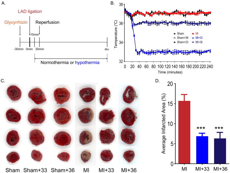
The core target temperatures of 33°C ± 0.5°C and 36°C ± 0.5°C were reached within 13 ± 0.80 and 5 ± 0.49 min after the onset of TTM. In the present study, the average values of the core temperature on reperfusion were 33.2°C ± 0.07°C in the 33°C group and 35.8°C ± 0.05°C in the 36°C groups (Fig 1A and 1B). Infarct volumes were assessed by TTC staining after 4 h of ischemic injury (Fig 1C and 1D). In the normothermic group, the mean ratio of the infarcted area after myocardial I/R was 15.7 ± 3.55% compared to the total area between 0 and 8 mm from the apex, whereas the mean ratio of the infarcted area at 33°C and 36°C of TTM was 6.9 ± 1.66% and 6.28 ± 3.05%, respectively. There was a significant difference between the normothermic group and TTM groups (P = 0.001).

Fig 1. Targeted temperature management at 33°C and 36°C similarly reduces infarct volume in myocardial I/R injury.

Fig 1

A. Experimental schedule. B. Changes in rat body temperature after LAD ligation (the number of animals: n = 5, respectively). C. Representative image of 2,3,5-triphenyltetrazolium chloride (TTC) staining. D. Volume of myocardial infarction stained with TTC (the number of animals: n = 5, respectively). ***P < 0.001, comparison of myocardial I/R with normothermia and hypothermia (33°C and 36°C), Statistical analyses were performed by one-way analysis of variance (ANOVA) followed by Bonferroni post hoc tests for multiple comparisons between groups.

To detect differences in myocardial protective effects at 33°C and 36°C in TTM after myocardial I/R injury, we compared the degree of reduction of the infarct volume in the 33°C and 36°C TTM groups. There was no significant difference between the infarct volumes of the 33°C TTM group and the 36°C TTM group (P = 0.999). These results suggest that application of TTM at both 33°C and 36°C has significant myocardial protective effects and that these temperatures lead to equivalent protection against myocardial I/R injury.

Different target temperatures of 33°C and 36°C TTM similarly suppress extracellular release of HMGB1 from peri-infarct tissue after myocardial I/R injury

When ischemic damage to the myocardium is induced by LAD ligation of the heart, HMGB1 is released from the nucleus of myocardial cells [30, 31].

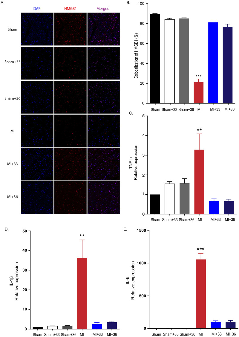
We found that the HMGB1 immunoreactivity was significantly decreased in the myocardium after ligation of LAD in rats. To investigate whether TTM at 33°C and 36°C significantly reduced the release of extracellular HMGB1 following I/R injury, we compared HMGB1 immunoreactivity between the normothermia and TTM groups after LAD ligation. We found that 21.15 ± 7.29% of 4,6-diamidino-2-phenylindole (DAPI)-positive cells in the peri-ischemic myocardium of LAD ligation rats were HMGB1-positive. However, we also found that target temperatures of 33°C and 36°C similarly restored the number of HMGB1-positive cells in post-infarct tissues. The percentages of HMGB1-positive cells were 81.28 ± 5.21% and 76.68 ± 6.27% for TTM at 33°C and 36°C, respectively. While significant increases for the proportion of HMGB1-positive cells were observed for TTM at 33°C and 36°C compared to the normothermic group (P < 0.001), there was no significant difference between the 33°C and 36°C groups (P = 0.999). This suggests that both 33°C and 36°C TTM cause similarly significant reductions in the extracellular release of HMGB1 after ischemic myocardial damage (Fig 2A and 2B).

Fig 2. Hypothermia suppresses extracellular release of HMGB1 after myocardial I/R injury and inflammatory cytokine expression in peri-infarct regions.

Fig 2

A. Representative immunohistochemistry results for 33°C and 36°C targeted temperature management after myocardial I/R injury. B. Immunohistochemistry results (the number of animals: n = 5, respectively), ***P < 0.001, comparing myocardial I/R with normothermia and hypothermia (33°C and 36°C), one-way analysis of variance (ANOVA), followed by Bonferroni post hoc test. C. Quantification of tumour necrosis factor-α (TNF-α) expression by RT-PCR (the number of animals: n = 5, respectively), **P < 0.01, comparison of myocardial I/R with normothermia and hypothermia (33°C and 36°C) by one-way analysis of variance (ANOVA) followed by Bonferroni post hoc test. D. Quantification of interleukin-1β (IL-1β) expression by RT-PCR (the number of animals: n = 5, respectively), **P < 0.01. Comparison of myocardial I/R with normothermia and hypothermia (33°C and 36°C) by ANOVA followed by Bonferroni post hoc test. E. Quantification of IL-6 expression by RT-PCR (the number of animals: n = 5, respectively), ***P < 0.01. Comparison of myocardial I/R with normothermia and hypothermia (33°C and 36°C) by ANOVA followed by Bonferroni post hoc test. All statistical analyses were performed by one-way analysis of variance (ANOVA) followed by Bonferroni post hoc tests for multiple comparisons between groups.

TTM at 33°C and 36°C similarly inhibited inflammatory cytokine expression from peri-infarct regions

Cardiac mRNA expression of three major inflammatory cytokines (i.e., TNF-α, IL-1β, and IL-6) was assessed by quantitative RT-PCR in the peri-infarcted myocardium 4 h after LAD ligation. In normothermic rats maintained at 37°C after myocardial I/R, the expression levels of TNF-α (3.28 ± 1.62, P = 0.001), IL-1β (36.15 ± 18.5, P < 0.001), and IL-6 (1055.89 ± 185.63, P < 0.001) were significantly increased. Compared to the normothermic I/R group, TTM treatment at 33°C was closely associated with lower expression of inflammatory cytokines in the peri-infarcted myocardium (TNF-α (0.67 ± 0.23, P = 0.001), IL-1β (2.67 ± 1.33, P < 0.001), IL-6 (98.43 ± 42.12, P < 0.001) (Fig 2C, 2D, and 2E)). TTM at 36°C also decreased the expression of these cytokines in the peri-infarcted myocardium (TNF-α (0.67 ± 0.19, P = 0.001), IL-1β (3.49 ± 1.31, P < 0.001), IL-6 (98.68 ± 50.89, P < 0.001)). First, there were no significant differences in the mRNA expression of three inflammatory cytokines between the 33°C and 36°C TTM groups (P < 0.999, P = 0.999, P < 0.999, respectively). Thus, the application of TTM prevents the aggravation of damage by suppressing the production of inflammatory cytokines in the peri-infarct area after myocardial I/R injury (Fig 2C, 2D, and 2E). This indicates that the different target temperatures of 33°C and 36°C TTM similarly attenuate inflammatory cytokine expression after cardiac I/R injury.

TUNEL assay

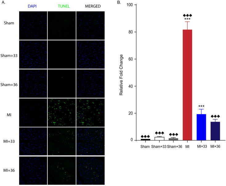
After myocardial I/R, TUNEL-positive apoptotic cells, which appear as light green dots under the confocal microscope, were significantly increased in the normothermic group (81.7 ± 16.11) compared to the 33°C (19.4 ± 7.19; P < 0.001) and 36°C (13.7 ± 5.12; P < 0.001) TTM groups. However, there was no significant difference between the number of TUNEL-positive cells of the two TTM groups (P = 0.676) (Fig 3). These results also imply that application of TTM at both temperatures has significant myocardial protective effects by reducing apoptosis and that these core temperatures lead to equivalent protection against myocardial I/R injury.

Fig 3. Quantitative analysis of apoptotic cell death by TUNEL assay.

Fig 3

A. Representative TUNEL assay results for 33°C and 36°C targeted temperature management after myocardial I/R injury. B. TUNEL assay results (the number of animals: n = 5, respectively), ♠♠♠ P < 0.001, comparing sham with normothermia and hypothermia (33°C and 36°C), ***P < 0.001, comparing myocardial I/R with normothermia and hypothermia (33°C and 36°C); Statistical analyses were performed by one-way analysis of variance (ANOVA) followed by Bonferroni post hoc tests for multiple comparisons between groups.

Glycyrrhizin alleviates myocardial damage by suppressing the extracellular release of HMGB1 in myocardial I/R injury

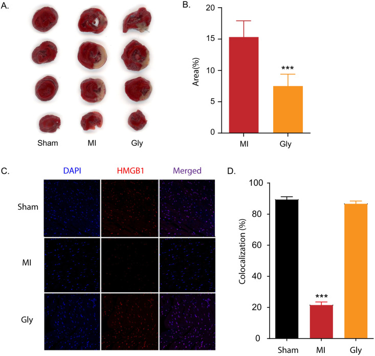
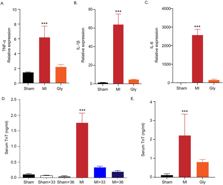
Glycyrrhizin is a pharmacological HMGB1 inhibitor that binds directly to HMGB1 and prevents the extracellular release of HMGB1 to block its cytokine function [3234]. We compared the effects of glycyrrhizin treatment on infarct volume, extracellular release of HMGB1, expression of inflammatory cytokines, and plasma level of cTnT in our animal model. In the myocardial I/R group treated with an intra-peritoneal injection of glycyrrhizin, the infarct volume was significantly decreased (7.5 ± 3.81%) compared to that in the normothermic myocardial I/R group (15.3 ± 5.17%, P = 0.04) ((Fig 4A and 4B). Glycyrrhizin also significantly increased the proportion of HMGB1-positive cells in the I/R injured myocardium (21.52 ± 3.94% in normothermic rats after myocardial I/R versus 86.61 ± 3.65% in glycyrrhizin-treated myocardial I/R rats, P < 0.001) (Fig 4C and 4D). In glycyrrhizin–treated AMI rats, TNF-α (2.18 ± 0.77, P < 0.001), IL-1β (4.59 ± 0.95, P < 0.001), and IL-6 (145.78 ± 107.26, P < 0.001) levels were decreased compared to those in the myocardial I/R group (TNF-α; 6.18 ± 3.42, IL-1β; 63.78 ± 25.36, and IL-6; 2565.74 ± 707.31, respectively) (Fig 5A, 5B, and 5C). Additionally, cTnT levels were significantly lower in the glycyrrhizin-treated myocardial I/R group (0.80 ± 0.12 ng/mL) than in the normothermic group after myocardial I/R (2.39 ± 0.83 ng/mL, P = 0.001) (Fig 5E).

Fig 4. Glycyrrhizin reduces infarct volume in myocardial I/R injury and glycyrrhizin suppresses extracellular release of HMGB1 in myocardial I/R injury.

Fig 4

To verify the cardioprotective effect of TTM by specifically inhibiting HMGB1, rats were assigned to sham, LAD I/R, and LAD I/R after pre-treatment with glycyrrhizin (known as a pharmacological inhibitor of HMGB1) groups. A. Representative image for 2,3,5-triphenyltetrazolium chloride (TTC) staining comparing myocardial I/R injury with normothermia and glycyrrhizin pre-treatment. B. Quantification of TTC staining results in A (the number of animals: n = 5, respectively). ***P < 0.001 comparing myocardial I/R injury with and without glycyrrhizin, unpaired t-test. C. Quantification of immunohistochemistry results in D (the number of animals: n = 5, respectively). D. Representative images showing HMGB1 immunoreactivity from myocardial I/R injury with and without glycyrrhizin B treatment. ***P < 0.001, comparing myocardial I/R injury with and without glycyrrhizin by one-way analysis of variance (ANOVA) followed by Bonferroni post hoc test.

Fig 5. Inflammatory cytokine expression in myocardial I/R injury with and without glycyrrhizin treatment and levels of cardiac troponin T (cTnT) in plasma.

Fig 5

A. Quantification of tumour necrosis factor-α (TNF-α) expression by RT-PCR comparing myocardial I/R injury with and without glycyrrhizin treatment (the number of animals: n = 5, respectively), ***P < 0.001, comparing myocardial I/R injury with and without glycyrrhizin by one-way analysis of variance (ANOVA) followed by Bonferroni post hoc test B. Quantification of interleukin-1β (IL-1β) expression by RT-PCR comparing myocardial I/R injury with and without glycyrrhizin (the number of animals: n = 5, respectively), ***P < 0.001, comparing myocardial I/R injury with and without glycyrrhizin treatment by one-way ANOVA followed by Bonferroni post hoc test C. Quantification of interleukin-6 (IL-6) expression by RT-PCR comparing myocardial I/R injury with and without glycyrrhizin treatment (the number of animals: n = 5, respectively), ***P < 0.001, comparing myocardial I/R injury with and without glycyrrhizin treatment by one-way ANOVA followed by Bonferroni post hoc test. D. Quantification of serum TnT level by ELISA comparing myocardial I/R with normothermia and hypothermia (33°C and 36°C) (number of animals: n = 5), ***P < 0.001, comparison of myocardial I/R with normothermia and hypothermia (33°C and 36°C) by one-way analysis of variance (ANOVA) followed by Bonferroni post hoc test. E. Quantification of serum TnT level by ELISA with comparing myocardial I/R injury with and without glycyrrhizin pre-treatment (number of animals: n = 5), ***P < 0.001, comparing myocardial I/R injury with and without glycyrrhizin pre-treatment by one-way ANOVA followed by Bonferroni post hoc test.

Effects of 33°C and 36°C TTM on cTnT and HMGB1 levels in the plasma

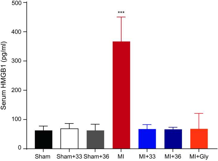
To examine the myocardial protective effects of 33°C and 36°C TTM on cTnT levels reflecting myocardial damage, we measured cTnT levels in the plasma. The levels of cTnT were higher in the normothermia group after LAD ligation compared to those in the sham-operated group (1.75 ± 0.53 and 0.10 ± 0.05 ng/mL, respectively; P < 0.001). However, rats subjected to either 33°C or 36°C TTM showed lower cTnT levels than those in the normothermia group (0.33 ± 0.08 in 33°C and 0.19 ± 0.07 ng/mL in 36°C TTM group). There was no significant difference in plasma cTnT at 33°C and 36°C TTM (P = 0.999), indicating that both target core temperatures for TTM equivalently reduced myocardial damage (Fig 5D) Next, we performed an ELISA to measure HMGB1 levels in serum samples obtained at 4 h after the onset of ischemia. As expected, the level of circulating of HMGB1 was increased after I/R injury, but this increase was significantly attenuated by TTM at 33°C and 36°C (normothermic group after myocardial I/R, 367.08 ± 83.58 pg/mL, 33°C TTM after myocardial I/R, 67.15 ± 15.55 pg/mL and 36°C TTM after myocardial I/R, 66.30 ± 7.43 pg/mL, P < 0.001) (Fig 6).

Fig 6. Quantification of levels of HMGB1 in plasma.

Fig 6

Quantification of serum HMGB1 level by ELISA comparing myocardial I/R with normothermia, hypothermia (33°C and 36°C), and glycyrrhizin pre-treatment (number of animals: n = 5), ***P < 0.001, Statistical analyses were performed by one-way analysis of variance (ANOVA) followed by Bonferroni post hoc tests for multiple comparisons between groups.

Discussion

Our results suggest that TTM at both 33°C and 36°C reduces myocardial injury following acute myocardial I/R injury by suppressing the extracellular release of HMGB1. We found that TTM attenuated the increase in infarct size, apoptosis, extracellular release of HMGB1, and pro-inflammatory effect against the propagation of injury in rats with AMI. We also showed that TTM at both 33°C and 36°C significantly attenuated the elevation of cardiac troponin, which is a sensitive and specific marker of heart muscle damage, after myocardial I/R injury. TTM at 36°C showed similar myocardial protective effects against myocardial I/R injury as TTM at 33°C in our rat AMI model.

The inflammatory cascade in myocardial injury and infarction is significantly associated with debris removal and scar formation [9]. Despite the fundamental role of inflammation in wound healing after AMI, an overwhelming inflammatory response can lead to devastating effects on cardiomyocytes [9]. The onset of cell death begins within 30 min to 1 h after the cessation of blood flow through a combination of necrosis and apoptosis. Inflammation, which contributes to lethal myocardial injury, is initiated during ischemia and is sustained over several hours after reperfusion [35]. Inhibiting the inflammatory process can provide a potential therapeutic window for cardioprotection [35]. HMGB1 is known to subsequently act as a pro-inflammatory cytokine that activates inflammatory cells by its extracellular release from necrotic cells in the pathophysiology of various diseases [20, 36, 37]. The serum concentration of HMGB1 is significantly associated with infarct size and residual left ventricular function in patients with STEMI [38]. Furthermore, extracellular HMGB1 mediates inflammation and enhances the regeneration of damaged tissues. Takahashi et al. reported that HMGB1 has beneficial effects at low concentrations and deleterious effects at high concentrations [36]. HMGB1 levels are significantly increased within 1 h and maintained for up to 24 h during I/R injury [39, 40]. Therefore, it is important to prevent the action of HMGB1 to alleviate ischemic injury of the myocardium [20, 41]. Nevertheless, HMGB1 plays dual roles in cardiac injury. In the initial stage of cerebrovascular and cardiovascular diseases, HMGB1 is released from the cell to participate in the cascade amplification reaction of inflammation, causing vasospasm and apoptosis [20, 41, 42]. In the recovery stage of disease, HMGB1 can promote tissue repair, regeneration, and remodelling [20, 41, 42]. It is necessary to investigate whether the newly generated HMGB1 plays a role in propagating inflammatory detriment or repairing damages over time after injury. The interaction between extracellular HMGB1 and Toll-like receptor 4 enhances the inflammatory response to myocardial damage after I/R by activating the release of pro-inflammatory cytokines, such as TNF-α from macrophage/monocytes [20]. TNF-α influences the production of other pro-inflammatory cytokines, such as IL-1 β and IL-6, resulting in a negative cycle of pro-inflammatory cytokine production and aggravation of injury after myocardial infarction [43, 44]. We also demonstrated that TTM attenuated myocardial I/R-induced apoptosis. Temperatures of 33°C or 36°C in TTM induced equivalent myocardial protection by attenuating apoptosis after I/R injury.

Our previous study demonstrated that therapeutic application of TTM helps alleviate peri-infarct damage from the propagation of ischemic injury in an ischemic stroke model by reducing inflammatory cytokines through the blockage of HMGB1 release [24, 26]. This is the first study to show a direct mechanistic and functional link between HMGB1 and TTM in a clinically relevant AMI animal model. Interestingly, myocardial I/R injured rats treated with glycyrrhizin showed equivalent myocardial protection as myocardial I/R rats that underwent TTM. This suggests that the extracellular release of HMGB1 is critical for the propagation of I/R injury by increasing the expression of proinflammatory cytokines in the peri-infarct myocardium and that TTM helps attenuate this propagation of I/R injury by inhibiting HMGB1 after I/R injury. In addition, we demonstrated that the serum HMGB1 level was lower in the TTM group than in the group without TTM, revealing a correlation between TTM and HMGB1. However, there is a lack of data regarding the link between HMGB1 and I/R injury and direct mechanisms in our study. In the case of the AMI animal model, we could not confirm the relationship between HMGB1 and I/R injury because HMGB1 neutralizing antibodies could not be injected locally. Instead, we indirectly revealed the association of HMGB1 with I/R injury by using glycyrrhizin as a pharmacological HMGB1 inhibitor and via experiments using our middle cerebral artery occlusion model [24, 26]. However, these analyses with glycyrrhizin and neutralizing antibodies only demonstrated that the extracellular release of HMGB1 is a key factor in the cardiac damage after I/R injury. There is no evidence that the blockade of the HMGB1 release is direct mechanism of the TTM-induced cardiac protection after I/R injury. Further studies are needed to identify the direct mechanisms underlying, and the link between, HMGB1 release and the action of TTM after I/R injury. In addition, Liu et al. identified several classes of agents that potently induce the nucleo-cytoplasmic relocation and subsequent cellular release of HMGB1 [45]. To clarify whether TTM can attenuate the inflammatory response in peri-infarct regions by inhibiting the extracellular release of HMGB1 after I/R injury, further studies are needed to verify ischemic damage using pharmacological agents that induce HMGB1 release during TTM after I/R injury in the AMI model.

Induced hypothermia may increase the rates of lethal arrhythmia, hypotension, shivering, infection, impaired coagulopathy, and rewarming injury and significantly alter the pharmacokinetics [46]. Its intrinsic adverse effects can significantly diminish the hypothermic benefits throughout the body [46]. TTM at 36°C may be preferred to TTM at 33°C in patients with cardiac arrest for several reasons [26]. In clinical practice, TTM consists of three phases: induction, maintenance, and rewarming [47]. TTM should be initiated as soon as possible according to international guidelines [46]. Moreover, rapidly induced hypothermia is important for modulating the efficacy of hypothermia in the clinical setting because minimizing the door to balloon time and reaching the target temperature within that at pre-reperfusion are critical for salvaging cardiac muscles [48]. As TTM at 36°C is close to the lower margin of the normal body temperature, it has the advantage of quickly achieving the target core temperature [46]. Clinical management to control shivering and minimize the risk of the devastating complications of TTM should be considered to maximize the benefits [47]. Shivering, as a major adverse effect of TTM, leads to extremely uncomfortable and massive increases in the metabolic demand and systemic energy consumption [47]. TTM at 36°C may be preferred to minimize the risk of shivering in the induction and rewarming phases because peripheral vasoconstriction and shivering are triggered at 36.5°C and 35.5°C in healthy humans [47]. In the rewarming phase, a small temperature change between the maintenance and rewarming periods can be beneficial for reducing the increased risk of secondary damage due to the adverse consequences of rewarming on the whole body. TTM at 33°C is generally recommended as the safer margin for the target core temperature in critically ill patients because temperatures below 32°C can induce serious cardiac arrhythmia. Application of TTM at 36°C to AMI patients also helps ameliorate the risk of several adverse effects. Unlike patients who are resuscitated after cardiac arrest, most patients with AMI remain awake and breathe spontaneously during acute management [47]. Simple and well-tolerated TTM at 36°C is more feasible during the acute period of AMI. In this experiment, we did not study the rewarming phase when applying 33°C and 36°C TTM. Rewarming treatment of the 33°C TTM group is likely to be more damaging than 36°C TTM group. However, due to the technical problems of our study, it was difficult to implement rewarming experiments. In future studies, additional rewarming treatments will be necessary because they can provide more meaningful interpretations compared to actual clinical practice.

TTM has been shown to be safe and feasible in clinical practice, and there were no differences in mortality or neurological outcomes between patients who underwent TTM at 33°C and those who underwent TTM 36°C after out-of-hospital cardiac arrest in recent multi-centre clinical trials [49]. Recently, the target temperature of TTM tends to change accordingly from 33–34°C to 36°C during post-resuscitation care [49]. However, previous animal studies demonstrated that rapid application of therapeutic hypothermia at 32–34°C prior to reperfusion significantly reduced the myocardial infarct size [25, 50, 51]. Although the infarct size in TTM at 35°C is decreased, Dash et al. demonstrated that TTM at 32°C is superior to TTM at 35°C and normothermic porcine after AMI [48]. Several clinical trials of TTM after AMI have shown inconsistent results with the major findings of many experimental studies.

This may be because of interspecies variability among animal models and differences in the immune response to I/R injury. Additionally, clinical outcomes may also be affected by disease- or organ-specific characteristics and molecular biological differences among organs [16, 17]. To reduce the gap between the TTM beneficial effects for cardiac arrest and AMI, we investigated whether TTM at 36°C has a potent myocardial protective effect. Our study showed that target temperatures of 33°C or 36°C in TTM similarly inhibited HMGB1 release and induced equivalent myocardial protection in terms of the infarct size in myocardial I/R injury in a rat model. This is the first study to suggest that a target core temperature of 36°C is applicable for cardioprotection in myocardial I/R injury. However, we compared the effects of two different temperatures in rats intubated with ventilation support, adequate sedation, and strict shivering control by vecuronium, mimicking the features of cardiac arrest. Therefore, further studies are needed to clarify the cardioprotective effects of both 33°C and 36°C TTM and the critical roles of HMGB1 in patients with AMI who are awake and exhibit spontaneous breathing. Finally, the importance of this study is that whilst there are many described pathways known to protect against I/R injury due to TTM, here we identified a novel pathway that inhibits the release of HMGB1.

Conclusion

We describe a new mechanistic and clinical link showing that TTM at 36°C is a therapeutic candidate that should be investigated in future clinical trials by reducing the propagation of myocardial damage to effectively inhibit the extracellular HMGB1 release after myocardial I/R injury.

Supporting information

S1 Table. Sequences.

(DOCX)

Data Availability

All relevant data are within the manuscript and its Supporting information files.

Funding Statement

This work was supported by the Basic Science Research Program of the National Research Foundation of Korea (NRF), funded by the Ministry of Science and ICT (grant numbers NRF-2015R1C1A1A01054641 and NRF-2018R1C1B6006159 to J.S.Y; NRF-2019R1A2C3002354 to C.H.K; and NRF-2019R1C1C1006332 to J.B.) and a faculty research grant from the Yonsei University College of Medicine (grant number 6-2017-0092 to J.B. and 6-2019-0188 to J.S.Y.). The funding bodies had no role in the design, collection, analysis, or interpretation of this study.

References

  • 1.Menees DS, Peterson ED, Wang Y, Curtis JP, Messenger JC, Rumsfeld JS, et al. Door-to-balloon time and mortality among patients undergoing primary PCI. N Engl J Med. 2013;369: 901–909. 10.1056/NEJMoa1208200 [DOI] [PubMed] [Google Scholar]
  • 2.Mozaffarian D, Benjamin EJ, Go AS, Arnett DK, Blaha MJ, Cushman M, et al. Heart disease and stroke statistics—2015 update: a report from the American Heart Association. Circulation. 2015;131: e29–322. 10.1161/CIR.0000000000000152 [DOI] [PubMed] [Google Scholar]
  • 3.Boersma E. Does time matter? A pooled analysis of randomized clinical trials comparing primary percutaneous coronary intervention and in-hospital fibrinolysis in acute myocardial infarction patients. Eur Heart J. 2006;27: 779–788. 10.1093/eurheartj/ehi810 [DOI] [PubMed] [Google Scholar]
  • 4.Greulich S, Mayr A, Gloekler S, Seitz A, Birkmeier S, Schaufele T, et al. Time-Dependent Myocardial Necrosis in Patients With ST-Segment-Elevation Myocardial Infarction Without Angiographic Collateral Flow Visualized by Cardiac Magnetic Resonance Imaging: Results From the Multicenter STEMI-SCAR Project. J Am Heart Assoc. 2019;8: e012429 10.1161/JAHA.119.012429 [DOI] [PMC free article] [PubMed] [Google Scholar]
  • 5.Heusch G, Gersh BJ. The pathophysiology of acute myocardial infarction and strategies of protection beyond reperfusion: a continual challenge. Eur Heart J. 2017;38: 774–784. 10.1093/eurheartj/ehw224 [DOI] [PubMed] [Google Scholar]
  • 6.Kushner FG, Hand M, Smith SC, King SB, Anderson JL, Antman EM, et al. 2009 Focused Updates: ACC/AHA Guidelines for the Management of Patients With ST-Elevation Myocardial Infarction (updating the 2004 Guideline and 2007 Focused Update) and ACC/AHA/SCAI Guidelines on Percutaneous Coronary Intervention (updating the 2005 Guideline and 2007 Focused Update): a report of the American College of Cardiology Foundation/American Heart Association Task Force on Practice Guidelines. Circulation. 2009;120: 2271–2306. 10.1161/CIRCULATIONAHA.109.192663 [DOI] [PubMed] [Google Scholar]
  • 7.Hausenloy DJ, Yellon DM. Targeting Myocardial Reperfusion Injury—The Search Continues. N Engl J Med. 2015;373: 1073–1075. 10.1056/NEJMe1509718 [DOI] [PubMed] [Google Scholar]
  • 8.Yellon DM, Hausenloy DJ. Myocardial reperfusion injury. N Engl J Med. 2007;357: 1121–1135. 10.1056/NEJMra071667 [DOI] [PubMed] [Google Scholar]
  • 9.Frohlich GM, Meier P, White SK, Yellon DM, Hausenloy DJ. Myocardial reperfusion injury: looking beyond primary PCI. Eur Heart J. 2013;34: 1714–1722. 10.1093/eurheartj/eht090 [DOI] [PubMed] [Google Scholar]
  • 10.Rochitte CE, Azevedo CF. The myocardial area at risk. Heart. 2012;98: 348–350. 10.1136/heartjnl-2011-301332 [DOI] [PubMed] [Google Scholar]
  • 11.Nielsen N, Wetterslev J, Cronberg T, Erlinge D, Gasche Y, Hassager C, et al. Targeted temperature management at 33 degrees C versus 36 degrees C after cardiac arrest. N Engl J Med. 2013;369: 2197–2206. 10.1056/NEJMoa1310519 [DOI] [PubMed] [Google Scholar]
  • 12.Mild therapeutic hypothermia to improve the neurologic outcome after cardiac arrest. N Engl J Med. 2002;346: 549–556. 10.1056/NEJMoa012689 [DOI] [PubMed] [Google Scholar]
  • 13.Schwartz BG, Kloner RA, Thomas JL, Bui Q, Mayeda GS, Burstein S, et al. Therapeutic hypothermia for acute myocardial infarction and cardiac arrest. Am J Cardiol. 2012;110: 461–466. 10.1016/j.amjcard.2012.03.048 [DOI] [PubMed] [Google Scholar]
  • 14.Callaway CW, Donnino MW, Fink EL, Geocadin RG, Golan E, Kern KB, et al. Part 8: Post-Cardiac Arrest Care: 2015 American Heart Association Guidelines Update for Cardiopulmonary Resuscitation and Emergency Cardiovascular Care. Circulation. 2015;132: S465–482. 10.1161/CIR.0000000000000262 [DOI] [PMC free article] [PubMed] [Google Scholar]
  • 15.Kohlhauer M, Berdeaux A, Ghaleh B, Tissier R. Therapeutic hypothermia to protect the heart against acute myocardial infarction. Arch Cardiovasc Dis. 2016;109: 716–722. 10.1016/j.acvd.2016.05.005 [DOI] [PubMed] [Google Scholar]
  • 16.Herring MJ, Hale SL, Dai W, Oskui PM, Kloner RA. Hypothermia in the setting of experimental acute myocardial infarction: a comprehensive review. Ther Hypothermia Temp Manag. 2014;4: 159–167. 10.1089/ther.2014.0016 [DOI] [PubMed] [Google Scholar]
  • 17.Kelly FE, Nolan JP. The effects of mild induced hypothermia on the myocardium: a systematic review. Anaesthesia. 2010;65: 505–515. 10.1111/j.1365-2044.2009.06237.x [DOI] [PubMed] [Google Scholar]
  • 18.Wang X, Guo Z, Ding Z, Mehta JL. Inflammation, Autophagy, and Apoptosis After Myocardial Infarction. J Am Heart Assoc. 2018;7 10.1161/jaha.117.008024 [DOI] [PMC free article] [PubMed] [Google Scholar]
  • 19.Ding HS, Yang J, Chen P, Yang J, Bo SQ, Ding JW, et al. The HMGB1-TLR4 axis contributes to myocardial ischemia/reperfusion injury via regulation of cardiomyocyte apoptosis. Gene. 2013;527: 389–393. 10.1016/j.gene.2013.05.041 [DOI] [PubMed] [Google Scholar]
  • 20.Andersson U, Tracey KJ. HMGB1 is a therapeutic target for sterile inflammation and infection. Annu Rev Immunol. 2011;29: 139–162. 10.1146/annurev-immunol-030409-101323 [DOI] [PMC free article] [PubMed] [Google Scholar]
  • 21.Lin Y, Chen L, Li W, Fang J. Role of high-mobility group box-1 in myocardial ischemia/reperfusion injury and the effect of ethyl pyruvate. Exp Ther Med. 2015;9: 1537–1541. 10.3892/etm.2015.2290 [DOI] [PMC free article] [PubMed] [Google Scholar]
  • 22.Sorensen MV, Pedersen S, Mogelvang R, Skov-Jensen J, Flyvbjerg A. Plasma high-mobility group box 1 levels predict mortality after ST-segment elevation myocardial infarction. JACC Cardiovasc Interv. 2011;4: 281–286. 10.1016/j.jcin.2010.10.015 [DOI] [PubMed] [Google Scholar]
  • 23.Zhai CL, Zhang MQ, Zhang Y, Xu HX, Wang JM, An GP, et al. Glycyrrhizin protects rat heart against ischemia-reperfusion injury through blockade of HMGB1-dependent phospho-JNK/Bax pathway. Acta Pharmacol Sin. 2012;33: 1477–1487. 10.1038/aps.2012.112 [DOI] [PMC free article] [PubMed] [Google Scholar]
  • 24.Lee JH, Yoon EJ, Seo J, Kavoussi A, Chung YE, Chung SP, et al. Hypothermia inhibits the propagation of acute ischemic injury by inhibiting HMGB1. Mol Brain. 2016;9: 81 10.1186/s13041-016-0260-0 [DOI] [PMC free article] [PubMed] [Google Scholar]
  • 25.Tissier R, Couvreur N, Ghaleh B, Bruneval P, Lidouren F, Morin D, et al. Rapid cooling preserves the ischaemic myocardium against mitochondrial damage and left ventricular dysfunction. Cardiovasc Res. 2009;83: 345–353. 10.1093/cvr/cvp046 [DOI] [PMC free article] [PubMed] [Google Scholar]
  • 26.Lee JH, Lim J, Chung YE, Chung SP, Park I, Kim CH, et al. Targeted Temperature Management at 33 degrees C or 36 degrees C Produces Equivalent Neuroprotective Effects in the Middle Cerebral Artery Occlusion Rat Model of Ischemic Stroke. Shock. 2018;50: 714–719. 10.1097/SHK.0000000000001106 [DOI] [PubMed] [Google Scholar]
  • 27.Wang WE, Yang D, Li L, Wang W, Peng Y, Chen C, et al. Prolyl hydroxylase domain protein 2 silencing enhances the survival and paracrine function of transplanted adipose-derived stem cells in infarcted myocardium. Circ Res. 2013;113: 288–300. 10.1161/CIRCRESAHA.113.300929 [DOI] [PMC free article] [PubMed] [Google Scholar]
  • 28.Li L, Yu Q, Liang W. Use of 2,3,5-triphenyltetrazolium chloride-stained brain tissues for immunofluorescence analyses after focal cerebral ischemia in rats. Pathol Res Pract. 2018;214: 174–179. 10.1016/j.prp.2017.11.016 [DOI] [PubMed] [Google Scholar]
  • 29.Reid E, Graham D, Lopez-Gonzalez MR, Holmes WM, Macrae IM, McCabe C. Penumbra detection using PWI/DWI mismatch MRI in a rat stroke model with and without comorbidity: comparison of methods. J Cereb Blood Flow Metab. 2012;32: 1765–1777. 10.1038/jcbfm.2012.69 [DOI] [PMC free article] [PubMed] [Google Scholar]
  • 30.Loukili N, Rosenblatt-Velin N, Li J, Clerc S, Pacher P, Feihl F, et al. Peroxynitrite induces HMGB1 release by cardiac cells in vitro and HMGB1 upregulation in the infarcted myocardium in vivo. Cardiovasc Res. 2011;89: 586–594. 10.1093/cvr/cvq373 [DOI] [PMC free article] [PubMed] [Google Scholar]
  • 31.Andrassy M, Volz HC, Igwe JC, Funke B, Eichberger SN, Kaya Z, et al. High-mobility group box-1 in ischemia-reperfusion injury of the heart. Circulation. 2008;117: 3216–3226. 10.1161/CIRCULATIONAHA.108.769331 [DOI] [PubMed] [Google Scholar]
  • 32.Kim SW, Jin Y, Shin JH, Kim ID, Lee HK, Park S, et al. Glycyrrhizic acid affords robust neuroprotection in the postischemic brain via anti-inflammatory effect by inhibiting HMGB1 phosphorylation and secretion. Neurobiol Dis. 2012;46: 147–156. 10.1016/j.nbd.2011.12.056 [DOI] [PubMed] [Google Scholar]
  • 33.Gong G, Xiang L, Yuan L, Hu L, Wu W, Cai L, et al. Protective effect of glycyrrhizin, a direct HMGB1 inhibitor, on focal cerebral ischemia/reperfusion-induced inflammation, oxidative stress, and apoptosis in rats. PLoS One. 2014;9: e89450 10.1371/journal.pone.0089450 [DOI] [PMC free article] [PubMed] [Google Scholar]
  • 34.Mollica L, De Marchis F, Spitaleri A, Dallacosta C, Pennacchini D, Zamai M, et al. Glycyrrhizin binds to high-mobility group box 1 protein and inhibits its cytokine activities. Chem Biol. 2007;14: 431–441. 10.1016/j.chembiol.2007.03.007 [DOI] [PubMed] [Google Scholar]
  • 35.Hausenloy DJ, Yellon DM. Myocardial ischemia-reperfusion injury: a neglected therapeutic target. J Clin Invest. 2013;123: 92–100. 10.1172/JCI62874 [DOI] [PMC free article] [PubMed] [Google Scholar]
  • 36.Takahashi M. High-mobility group box 1 protein (HMGB1) in ischaemic heart disease: beneficial or deleterious? Cardiovasc Res. 2008;80: 5–6. 10.1093/cvr/cvn212 [DOI] [PubMed] [Google Scholar]
  • 37.Takahashi M. High-Mobility Group Box 1 Protein in Myocardial Infarction: Should it be Stimulated or Inhibited? J Atheroscler Thromb. 2015;22: 553–554. 10.5551/jat.ED008 [DOI] [PubMed] [Google Scholar]
  • 38.Andrassy M, Volz HC, Riedle N, Gitsioudis G, Seidel C, Laohachewin D, et al. HMGB1 as a predictor of infarct transmurality and functional recovery in patients with myocardial infarction. J Intern Med. 2011;270: 245–253. 10.1111/j.1365-2796.2011.02369.x [DOI] [PubMed] [Google Scholar]
  • 39.Tsung A, Sahai R, Tanaka H, Nakao A, Fink MP, Lotze MT, et al. The nuclear factor HMGB1 mediates hepatic injury after murine liver ischemia-reperfusion. J Exp Med. 2005;201: 1135–1143. 10.1084/jem.20042614 [DOI] [PMC free article] [PubMed] [Google Scholar]
  • 40.Xu H, Yao Y, Su Z, Yang Y, Kao R, Martin CM, et al. Endogenous HMGB1 contributes to ischemia-reperfusion-induced myocardial apoptosis by potentiating the effect of TNF-&alpha;/JNK. Am J Physiol Heart Circ Physiol. 2011;300: H913–921. 10.1152/ajpheart.00703.2010 [DOI] [PMC free article] [PubMed] [Google Scholar]
  • 41.Lu H, Zhang Z, Barnie PA, Su Z. Dual faced HMGB1 plays multiple roles in cardiomyocyte senescence and cardiac inflammatory injury. Cytokine Growth Factor Rev. 2019;47: 74–82. 10.1016/j.cytogfr.2019.05.009 [DOI] [PubMed] [Google Scholar]
  • 42.Mu SW, Dang Y, Wang SS, Gu JJ. The role of high mobility group box 1 protein in acute cerebrovascular diseases. Biomed Rep. 2018;9: 191–197. 10.3892/br.2018.1127 [DOI] [PMC free article] [PubMed] [Google Scholar]
  • 43.Tian M, Yuan YC, Li JY, Gionfriddo MR, Huang RC. Tumor necrosis factor-alpha and its role as a mediator in myocardial infarction: A brief review. Chronic Dis Transl Med. 2015;1: 18–26. 10.1016/j.cdtm.2015.02.002 [DOI] [PMC free article] [PubMed] [Google Scholar]
  • 44.Yan W, Abu-El-Rub E, Saravanan S, Kirshenbaum LA, Arora RC, Dhingra S. Inflammation in myocardial injury- mesenchymal stem cells as potential immunomodulators. Am J Physiol Heart Circ Physiol. 2019. 10.1152/ajpheart.00065.20192019. [DOI] [PMC free article] [PubMed] [Google Scholar]
  • 45.Liu P, Zhao L, Loos F, Iribarren K, Lachkar S, Zhou H, et al. Identification of pharmacological agents that induce HMGB1 release. Sci Rep. 2017;7: 14915 10.1038/s41598-017-14848-1 [DOI] [PMC free article] [PubMed] [Google Scholar]
  • 46.Kirkegaard H, Taccone FS, Skrifvars M, Soreide E. Postresuscitation Care after Out-of-hospital Cardiac Arrest: Clinical Update and Focus on Targeted Temperature Management. Anesthesiology. 2019;131: 186–208. 10.1097/ALN.0000000000002700 [DOI] [PubMed] [Google Scholar]
  • 47.Choi HA, Badjatia N, Mayer SA. Hypothermia for acute brain injury—mechanisms and practical aspects. Nat Rev Neurol. 2012;8: 214–222. 10.1038/nrneurol.2012.21 [DOI] [PubMed] [Google Scholar]
  • 48.Dash R, Mitsutake Y, Pyun WB, Dawoud F, Lyons J, Tachibana A, et al. Dose-Dependent Cardioprotection of Moderate (32 degrees C) Versus Mild (35 degrees C) Therapeutic Hypothermia in Porcine Acute Myocardial Infarction. JACC Cardiovasc Interv. 2018;11: 195–205. 10.1016/j.jcin.2017.08.056 [DOI] [PMC free article] [PubMed] [Google Scholar]
  • 49.Arvidsson L, Lindgren S, Martinell L, Lundin S, Rylander C. Target temperature 34 vs. 36 degrees C after out-of-hospital cardiac arrest—a retrospective observational study. Acta Anaesthesiol Scand. 2017;61: 1176–1183. 10.1111/aas.12957 [DOI] [PubMed] [Google Scholar]
  • 50.Dai W, Herring MJ, Hale SL, Kloner RA. Rapid Surface Cooling by ThermoSuit System Dramatically Reduces Scar Size, Prevents Post-Infarction Adverse Left Ventricular Remodeling, and Improves Cardiac Function in Rats. J Am Heart Assoc. 2015;4 10.1161/jaha.115.002265 [DOI] [PMC free article] [PubMed] [Google Scholar]
  • 51.Herring MJ, Dai W, Hale SL, Kloner RA. Rapid Induction of Hypothermia by the ThermoSuit System Profoundly Reduces Infarct Size and Anatomic Zone of No Reflow Following Ischemia-Reperfusion in Rabbit and Rat Hearts. J Cardiovasc Pharmacol Ther. 2015;20: 193–202. 10.1177/1074248414535664 [DOI] [PubMed] [Google Scholar]

Decision Letter 0

Federica Limana

22 Sep 2020

PONE-D-20-25658

Targeted temperature management by inhibiting HMGB1 release in myocardial ischemia/reperfusion injury

PLOS ONE

Dear Dr. Sung You,

Thank you for submitting your manuscript to PLOS ONE. After careful consideration, we feel that it has merit but does not fully meet PLOS ONE’s publication criteria as it currently stands. Therefore, we invite you to submit a revised version of the manuscript that addresses the points raised during the review process.

Specifically, the reviewers raised criticisms concerning the quality of the images, asked for more detailed methods and for the necessity to perform an ELISA in order to quantify HMGB1 plasma levels. Further, they also raised doubts on the statistical analysis performed.

Please submit your revised manuscript by Nov 06 2020 11:59PM. If you will need more time than this to complete your revisions, please reply to this message or contact the journal office at plosone@plos.org. When you're ready to submit your revision, log on to https://www.editorialmanager.com/pone/ and select the 'Submissions Needing Revision' folder to locate your manuscript file.

Please include the following items when submitting your revised manuscript:

  • A rebuttal letter that responds to each point raised by the academic editor and reviewer(s). You should upload this letter as a separate file labeled 'Response to Reviewers'.

  • A marked-up copy of your manuscript that highlights changes made to the original version. You should upload this as a separate file labeled 'Revised Manuscript with Track Changes'.

  • An unmarked version of your revised paper without tracked changes. You should upload this as a separate file labeled 'Manuscript'.

If you would like to make changes to your financial disclosure, please include your updated statement in your cover letter. Guidelines for resubmitting your figure files are available below the reviewer comments at the end of this letter.

If applicable, we recommend that you deposit your laboratory protocols in protocols.io to enhance the reproducibility of your results. Protocols.io assigns your protocol its own identifier (DOI) so that it can be cited independently in the future. For instructions see: http://journals.plos.org/plosone/s/submission-guidelines#loc-laboratory-protocols

We look forward to receiving your revised manuscript.

Kind regards,

Federica Limana

Academic Editor

PLOS ONE

Journal Requirements:

When submitting your revision, we need you to address these additional requirements.

1. Please ensure that your manuscript meets PLOS ONE's style requirements, including those for file naming. The PLOS ONE style templates can be found at

https://journals.plos.org/plosone/s/file?id=wjVg/PLOSOne_formatting_sample_main_body.pdf and

https://journals.plos.org/plosone/s/file?id=ba62/PLOSOne_formatting_sample_title_authors_affiliations.pdf

2. PLOS requires an ORCID iD for the corresponding author in Editorial Manager on papers submitted after December 6th, 2016. Please ensure that you have an ORCID iD and that it is validated in Editorial Manager. To do this, go to ‘Update my Information’ (in the upper left-hand corner of the main menu), and click on the Fetch/Validate link next to the ORCID field. This will take you to the ORCID site and allow you to create a new iD or authenticate a pre-existing iD in Editorial Manager. Please see the following video for instructions on linking an ORCID iD to your Editorial Manager account: https://www.youtube.com/watch?v=_xcclfuvtxQ

3. Please amend your list of authors on the manuscript to ensure that each author is linked to an affiliation. Authors’ affiliations should reflect the institution where the work was done (if authors moved subsequently, you can also list the new affiliation stating “current affiliation:….” as necessary).

[Note: HTML markup is below. Please do not edit.]

Reviewers' comments:

Reviewer's Responses to Questions

Comments to the Author

1. Is the manuscript technically sound, and do the data support the conclusions?

The manuscript must describe a technically sound piece of scientific research with data that supports the conclusions. Experiments must have been conducted rigorously, with appropriate controls, replication, and sample sizes. The conclusions must be drawn appropriately based on the data presented.

Reviewer #1: Partly

Reviewer #2: Yes

**********

2. Has the statistical analysis been performed appropriately and rigorously?

Reviewer #1: I Don't Know

Reviewer #2: Yes

**********

3. Have the authors made all data underlying the findings in their manuscript fully available?

The PLOS Data policy requires authors to make all data underlying the findings described in their manuscript fully available without restriction, with rare exception (please refer to the Data Availability Statement in the manuscript PDF file). The data should be provided as part of the manuscript or its supporting information, or deposited to a public repository. For example, in addition to summary statistics, the data points behind means, medians and variance measures should be available. If there are restrictions on publicly sharing data—e.g. participant privacy or use of data from a third party—those must be specified.

Reviewer #1: No

Reviewer #2: Yes

**********

4. Is the manuscript presented in an intelligible fashion and written in standard English?

PLOS ONE does not copyedit accepted manuscripts, so the language in submitted articles must be clear, correct, and unambiguous. Any typographical or grammatical errors should be corrected at revision, so please note any specific errors here.

Reviewer #1: Yes

Reviewer #2: Yes

**********

5. Review Comments to the Author

Please use the space provided to explain your answers to the questions above. You may also include additional comments for the author, including concerns about dual publication, research ethics, or publication ethics. (Please upload your review as an attachment if it exceeds 20,000 characters)

Reviewer #1: Manuscript Number: PONE-D-20-25658

Title: Targeted temperature management by inhibiting HMGB1 release in myocardial ischemia/reperfusion injury

Plos One

Reviewer’s comments:

The authors investigated the expression and release of HMGB1 as a possible mediator of the innate immune response after acute myocardial ischemia/reperfusion at normothermia, with targeted temperature management at 33°C and 36°C, and with Glycyrrhizin, a pharmacological HMGB1 inhibitor, at normothermia. They observed similar cytoprotective effect by TTM at both temperatures and also by treatment with Glycyrrhizin instead of cooling, as shown in the attenuation of apoptosis (TUNEL) and necrosis (infracted area and serum TnT), relative to normoxic control. Moreover, they also showed an attenuation of the MI-induced innate immune response (IL-6, TnF-a, and IL-1ß expressions) by the various treatments, presumably due to the attenuation of HMGB1 release into the extracellular matrix. However, the attenuation of HMGB1 release by the different treatments was not shown in the date presented. Although the paper shows valuable and significant data, some major and minor issues clearly need to be addressed.

Major issues:

1. The authors only showed the intracellular expression of HMGB1 by IF staining and no data on HMGB1 serum concentration. Therefore, the only conclusion that can be drawn is MI-induced injury resulted in decreased intracellular HMGB1 expression, and no conclusion can be made if HMGB1 has been released to the extracellular matrix. Similar to their assessment of serum TnT by ELISA, data on serum HMGB1 would greatly support their conclusions.

2. The authors do not address the effect of their cooling treatment on MI-induced apoptosis, although it is clearly supported by their presented data.

3. The authors assess the infarct volume optically in 2 mm thick slices, which may or may not contain the entire infarct volume and does not clearly represent the apoptotic and necrotic areas. No indication of a z-stack was mentioned for this infarct volume analysis.

4. The title is grammatically incorrect and does not correspond with the presented findings, which is confusing for the readers.

5. Significant differences in the data illustrated in the graphs needs to be shown more clearly to better let the readers see which groups are being compared.

6. Data for experimental groups with n=5 should be shown as mean ± standard deviation and not as mean ± standard error of the mean.

7. It is unclear which statistical analysis was used for each group comparison.

Minor Issues:

• Figure Legends - Figure 1B: It is not clear what the authors mean by, “Traces of rat body temperature”, when they mention measuring the core temperature.

• Methods – TUNEL: How many slices per group were used?

• It is unclear how the heart tissue for the assessment of infarct volume is between 0 and 8 cm from the apex and in the same time the material for the immunohistochemistry analysis also comes from between 4 and 6 mm from the apex of the heart. It would be also interesting to reveal why only the apex of the heart was taken instead of its proximal parts.

• It would also be of interest to see how the immune response would change in a sham cooled group.

• Why were the TTM groups excluded from Figure 5A, 5B and 5C?

Reviewer #2: In the present paper, Beom and colleagues showed that targeted temperature management (TTM) at 33℃ and 36℃ had similar protective effects in a myocardial ischemia/reperfusion (I/R) model. Specifically, they demonstrated that TTM significantly reduced infarct volume and attenuated the elevation of cardiac troponin, a sensitive and specific marker of heart muscle damage, after injury. Moreover, they described that both TTM at 33℃ and 36℃ equally inhibited the extracellular release of high mobility group box-1(HMGB1) from necrotic cardiomyocytes in infarct tissue and suppressed the expression of inflammatory cytokines from peri-infarct regions. The exact mechanism by which hypothermia attenuates myocardial damage due to ischemia and reperfusion remains unknown. For this reason, the Authors hypothesized that the cardioprotective effect of TTM was dependent on the inhibition of HMGB1 release, basing on the fact that I/R injury after a pre-treatment with glycyrrhizin (known as a pharmacological inhibitor of HMGB1 release) showed similar protective effects as TTM.

As the Authors declared, this is the first study to suggest that a target core temperature of 36°C is applicable for cardioprotection: this becomes important from a therapeutic point of view, considering that therapeutic hypothermia set at lower temperature commonly induces several harmful effects.

I think the present paper is suitable to be published in PlosOne even if in my opinion there are some specific issues that need to be addressed.

- The Authors described that “in the TTM groups with target temperatures of 33°C and 36℃, external surface cooling was started at 15 min after LAD coronary ligation by placing ice packs on the animal’s torso” and that “the core target temperatures of 33 ± 0.5°C and 36 ± 0.5℃ were reached within 18 and 8 min after the onset of TTM, respectively, and then maintained for 4 h”. So, as well represented in Figure 1B, in the 33°C group the target temperature was reached after the reperfusion had already started and this was not the case of the 36℃, where the target temperature was reached before reperfusion. Don’t the Authors think that this could invalidate the comparison between the two groups?

- The Authors reported that “when ischemic damage to the myocardium is induced by LAD ligation of the heart, HMGB1 is released from the nucleus of myocardial cells [30,31]”. In my opinion, to unequivocally demonstrate the HMGB1 release in the plasma, the Authors should perform an ELISA assay for the quantification of HMGB1 in plasma, because with immunofluorescence they demonstrated only a decrease that could be potentially also a consequence of a reduced expression after MI.

Moreover, several studies report an increase of myocardial HMGB1 levels in experimental models of I/R [https://doi.org/10.1161/CIRCU LATIONAHA.108.769331; https://doi.org/10.1093/cvr/cvq373], beside the increase of circulating HMGB1 levels derived from necrotic cardiomyocytes and active secretion by hypoxic cardiac and infiltrating inflammatory cells: in fact, myocardial HMGB1 expression increases soon after ischemia and remains high several days after reperfusion [https://doi.org/10.1152/ajpheart.00703.2010]. Did the Authors observe a similar increase in myocardial HMGB1 expression? And if not, did it may depend on the too much short time of reperfusion (perhaps 4hours is too early to see an increase of HMGB1 expression)? Or did it maybe depend onthe myocardial area? In my opinion, the results presented in this paper could realistically describe the myocardial area interested by the infarct (in which nuclear HMGB1 is almost completely released), but the Authors talked about “peri-infarcted area” that means, I suppose, the area on the border between the infarcted area and border zone, in which cardiomyocytes don’t die and, as a consequence, don’t release HMGB1, but on the contrary increase HMGB1 expression. I suggest the Authors to deeper discuss this point.

- Figure 2a: A lot of background signal is present both in the nuclei staining (blue) and in HMGB1 staining (red) in SHAM, MI+33 and, even if less, in MI+36, making the comparison among different groups very complicated. I suggest the Authors to replace the pictures representing the groups mentioned above, to provide a more convincing figure and to better reflect the graph in Figure 2b.

- Figures 2c,d,e and 5a,b,c: In the graphs representing the RT-PCR results, it’s better to set the value of SHAM groups at 1 and, as a consequence, correlate the graph bars of the other groups.

- The Authors demonstrated that in MI+33 and MI+36 there was a very weak release of HMGB1 (even less than in SHAM) and expression of inflammatory cytokines (even less than in SHAM for TNFα). Given that other studies demonstrated a great increase of plasma concentrations of HMGB1 with a peak 12h after MI and a marked decrease of HMGB1 levels in the infarct zone by Western Blot 24h after MI [https://doi:10.1093/cvr/cvn163], I suggest the Authors to justify the choice to perform a reperfusion time of only 4hours, because it is possible that HMGB1 release and inflammatory cytokine production are just delayed by the TTM and some differences between MI+33 and MI+36 can become evident after a more long reperfusion time.

- Figure 3a:A lot of background signal is present both in the nuclei staining (blue) and in TUNEL positive nuclei staining (green) especially in MI group, making the comparison among different groups very complicated. I suggest the Authors to replace the pictures representing the MI group, to provide a more convincing figure and to better reflect the graph in Figure 3b.Moreover, it is really debatable that the number of nuclei present in MI peri-infarct region is higher than in SHAM left ventricle.

- Figure 4a: I think the Authors forgot to put the pictures of the SHAM hearts.

- Figures 4c and 4d:I suggest the Authors to switch the figure 4c and 4d with the immunofluorescence pictures before the quantificationas in figure 2.

- Figure 4d:A lot of background signal is present both in the nuclei staining (blue) and in HMGB1 staining (red) in SHAM and in Gly groups, making the comparison among different groups very complicated. I suggest the Authors to replace the pictures representing the groups mentioned above, to provide a more convincing figure and to better reflect the graph in Figure 4c.

- Figure 5d: Given that the Authors divided the present study into two main experiments, I believe that is more linear to provide two separate graphs also for the measurement of serum cTnT (one in figure 2 and one in figure 5). This becomes important also considering that the values showed for the other parameters (e.g. inflammatory cytokine expression) resulted a bit different between the different SHAM and MI groups of the two sets of experiments.

- Lastly, the major point of my revision. The Authors divided the present study into two main experiments: the first one to assess the effects of myocardial protection through TTM at 33°C and 36℃ and the second one to verify the cardioprotective effect of TTM by specifically inhibiting HMGB1 release from cells with Glycyrrhizin. But in this way the Authors cannot provide the direct demonstration that the cardioprotective effect of TTM is mediated by the blocking of HMGB1 release. They can only speculate about it on the basis of the similar results obtained in the two main experiments. The correct demonstration of this should be with the use of inducers of HMGB1 release in I/R damage in conditions of TTM. I suggest the Authors to take the cue from [http://doi:10.1038/s41598-017-14848-1] for new pharmacological agents that induce HMGB1 release. Otherwise it’s just a speculation and they have to underline that in their Discussion session!

**********

6. PLOS authors have the option to publish the peer review history of their article (what does this mean?). If published, this will include your full peer review and any attached files.

If you choose “no”, your identity will remain anonymous but your review may still be made public.

Do you want your identity to be public for this peer review? For information about this choice, including consent withdrawal, please see our Privacy Policy.

Reviewer #1: Yes: Giang Tong

Reviewer #2: No

[NOTE: If reviewer comments were submitted as an attachment file, they will be attached to this email and accessible via the submission site. Please log into your account, locate the manuscript record, and check for the action link "View Attachments". If this link does not appear, there are no attachment files.]

While revising your submission, please upload your figure files to the Preflight Analysis and Conversion Engine (PACE) digital diagnostic tool, https://pacev2.apexcovantage.com/. PACE helps ensure that figures meet PLOS requirements. To use PACE, you must first register as a user. Registration is free. Then, login and navigate to the UPLOAD tab, where you will find detailed instructions on how to use the tool. If you encounter any issues or have any questions when using PACE, please email PLOS at figures@plos.org. Please note that Supporting Information files do not need this step.

PLoS One. 2021 Jan 27;16(1):e0246066. doi: 10.1371/journal.pone.0246066.r002

Author response to Decision Letter 0


23 Nov 2020

please, check and review attached file - Reviewers' comments

Response to reviewers’ comments

Reviewer #1: Major issues:

Response: Dear reviewer, we appreciate your helpful comments and suggestions regarding our manuscript.

1. The authors only showed the intracellular expression of HMGB1 by IF staining and no data on HMGB1 serum concentration. Therefore, the only conclusion that can be drawn is MI-induced injury resulted in decreased intracellular HMGB1 expression, and no conclusion can be made if HMGB1 has been released to the extracellular matrix. Similar to their assessment of serum TnT by ELISA, data on serum HMGB1 would greatly support their conclusions.

Response: Thank you for this helpful comment. As per your comment, to identify extracellular release of HMGB1, we conducted an additional experiment to measure the serum HMGB1 level by HMGB1 ELISA. We have added the results to the manuscript and restructured the manuscript based on the reviewer’s comment.

Enzyme-linked immunosorbent assay (ELISA)

1. Enzyme-linked immunosorbent assay (ELISA) for cardiac troponin T (cTnT) and HMGB1

To obtain serum samples from rats, blood was drawn from the right atrium at 4 h after ligation of the LAD coronary artery with a 22-gauge needle. One millilitre of collected blood was transferred into a Z Serum Sep Clot Activator (Greiner Bioone, Kremsmunster, Austria), followed by centrifugation for 15 min at 3,000 rpm. The cTnT concentrations were determined using the cTnT ELISA kit (MBS2024997, MyBioSource, San Diego, CA, USA) and HMGB1 concentrations were determined using the Rat HMGB1 ELISA kit (Solarbio, Beijing, China).

Next, we performed an ELISA to measure HMGB1 levels in serum samples obtained at 4 h after the onset of ischemia. As expected, the level of circulating of HMGB1 was increased after I/R injury, but this increase was significantly attenuated by TTM at 33°C and 36°C.

(normothermic group after myocardial I/R, 367.08 ± 83.58, 33°C TTM after myocardial I/R, 67.15 ± 15.55 and 36°C TTM after myocardial I/R, 66.30 ± 7.43 pg/mL; P < 0.001)

(deletion) In addition, our previous study demonstrated that serum HMGB1 level was lower in the TTM group than the group without TTM, demonstrating the correlation between TTM and HMGB1

In addition, we demonstrated that the serum HMGB1 level was lower in the TTM group than in the group without TTM, revealing a correlation between TTM and HMGB1.

2. The authors do not address the effect of their cooling treatment on MI-induced apoptosis, although it is clearly supported by their presented data.

Response: We agree with this helpful comment. We have added this point to the Introduction and Discussion sections of the manuscript.

We found that TTM attenuated the increase in infarct size, apoptosis, extracellular release of HMGB1, and pro-inflammatory effect against the propagation of injury in rats with AMI.

The inflammatory response and apoptotic cell death are known to play important roles in the development of ischemic heart damage caused by myocardial I/R injury. Apoptosis is an important mechanism in I/R injury, and therapeutic hypothermia reduces apoptosis in myocytes. Therapeutic hypothermia-induced myocardial protection is significantly associated with beneficial modifications in apoptotic signal pathways.

We also demonstrated that TTM attenuated myocardial I/R-induced apoptosis. Temperatures of 33°C or 36℃ in TTM induced equivalent myocardial protection by attenuating apoptosis after I/R injury.

3. The authors assess the infarct volume optically in 2 mm thick slices, washich may or may not contain the entire infarct volume and does not clearly represent the apoptotic and necrotic areas. No indication of a z-stack was mentioned for this infarct volume analysis.

Response: Thank you for this helpful comment. To measure the total infarct volume, heart tissue between 0 and 8 mm from the apex of the heart was used to measure consistent areas in all tissues. Additionally, all heart tissues in the experiment included the site of LAD ligation and all injured areas below the ligation site.

According to your comment, we re-measured the entire infarct volume. We measured the infarcted area in the anterior and posterior sides of each 2-mm-thick slice using ImageJ 1.48v software. To determine the infarct volume in each slice, the average value of the infarct area on the anterior and posterior sides was multiplied by the thickness (2 mm) [thickness × (top area + bottom area)/2]. In addition, the total infarct volume was calculated as the sum of the infarct volume per slice.

4. The title is grammatically incorrect and does not correspond with the presented findings, which is confusing for the readers.

Response: According to your comment, we have revised the title.

Targeted temperature management at 33°C or 36℃ induces equivalent myocardial protection by inhibiting HMGB1 release in myocardial ischemia/reperfusion injury

5. Significant differences in the data illustrated in the graphs needs to be shown more clearly to better let the readers see which groups are being compared.

Response: Thank you for this helpful comment. We have revised the graphs based on your comment.

6. Data for experimental groups with n=5 should be shown as mean ± standard deviation and not as mean ± standard error of the mean.

Response: Thank you for this helpful comment. We have revised all data as the mean ± standard deviation based on your comment.

Statistical analysis

All experimental results are expressed as the mean ± standard error deviation of the mean. Statistical analyses were performed using unpaired t-test or by one-way analysis of variance (ANOVA) followed by Bonferroni post hoc tests for multiple comparisons between groups. Differences with P < 0.05 were considered as significant.

7. It is unclear which statistical analysis was used for each group comparison.

Response: We have discussed this point with our statisticians to confirm the statistical analysis. We have added this information to the manuscript and figure legends.

Statistical analysis

All experimental results are expressed as the mean ± standard deviation of the mean. Statistical analyses were performed using unpaired t-test or by one-way analysis of variance (ANOVA) followed by Bonferroni post hoc tests for multiple comparisons between groups. Differences with P < 0.05 were considered as significant.

Minor Issues:

• Figure Legends - Figure 1B: It is not clear what the authors mean by, “Traces of rat body temperature”, when they mention measuring the core temperature.

Response: Thank you for this helpful comment. We have revised figure legend as follows based on your comment.

Figure 1B. Changes in rat body temperature after LAD ligation

• Methods – TUNEL: How many slices per group were used?

Response: Thank you for this helpful comment. We have added this point to Methods as follows.

Methods

TUNEL assay

One slide from each animal was selected and stained. The two peri-ischemic areas of the stained sections were observed with a confocal microscope (LSM 700; Carl Zeiss GmbH, Jena, Germany). The average values of TUNEL-positive cells in the peri-infarct area were derived from two areas on the stained sections.

• It is unclear how the heart tissue for the assessment of infarct volume is between 0 and 8 cm from the apex and in the same time the material for the immunohistochemistry analysis also comes from between 4 and 6 mm from the apex of the heart. It would be also interesting to reveal why only the apex of the heart was taken instead of its proximal parts.

Response: Thank you for this helpful comment. We have added this point to Methods as follows.

To measure the total infarct volume, heart tissue between 0 and 8 mm from the apex of the heart was used to measure consistent areas in all tissues. Additionally, all heart tissues in the experiment included the site of LAD ligation and all injured areas below the ligation site. The proximal part of the heart has a complicated structure with several blood vessels, the right atrium, and the left atrium. Therefore, it was difficult to identify an accurate reference point for cutting the tissue, whereas the apex had a simple and consistent structure, making it a better point as a standard for accurate tissue cutting. After TTC staining, we confirmed the infarct area and performed immunostaining by selecting a slide containing normal tissue (peri-ischemic area) around the infarct. TTC staining showed that the infarcted and normal tissues (peri-ischemic area) were distributed in the tissue between 4 and 6 mm from the apex. We used this area for immunohistochemistry analysis.

Methods

Immunohistochemistry analysis

For immunohistochemistry analysis, 2,3,5-TTC staining was performed to confirm the peri-infarct area in the left ventricle. Next, 2-mm-thick slices between 4 and 6 mm from the apex of the rat heart were selected, fixed with 4% paraformaldehyde solution, and embedded in paraffin. Between 4 and 6 mm from the apex of the rat heart was chosen because the peri-infarcted region was easily observable given that it was properly mixed with normal and infarct tissue after TTC staining.

• It would also be of interest to see how the immune response would change in a sham cooled group.

Response: Thank you for this helpful comment. According to your recommendations, we conducted the experiments to identify changes in the sham at target temperatures of 33°C and 36°C. We have added the results to the manuscript and restructured the manuscript based on the reviewer’s comment.

To assess the effects of myocardial protection exerted by TTM at 33°C and 36℃, the rats were randomly divided into four experimental groups: sham + 37°C (n = 5), sham + 33°C TTM (n = 5), sham + 36°C TTM (n = 5), LAD I/R + 37°C normothermia (n = 5), LAD I/R + 33°C TTM (n = 5), and LAD I/R + 36°C TTM (n = 5).

• Why were the TTM groups excluded from Figure 5A, 5B and 5C?

Response: To verify the cardioprotective effect of TTM by specifically inhibiting HMGB1 in our animal model, we divided the present study into two main experiments: (1) examination of the cardioprotective effect of TTM and (2) analysis of the cardioprotective effect of a pharmacologic inhibitor against HMGB1.

We apologize for the confusion created by including the results of two experiments for the levels of cardiac troponin T (cTnT) in the plasma in Fig 5D to reduce the number of figures. We have separated the figure showing the measurement of serum cTnT into two figures.

Response to reviewers’ comments

Reviewer #2:

Response: Dear reviewer, we appreciate your helpful comments and suggestions regarding our manuscript.

- The Authors described that “in the TTM groups with target temperatures of 33°C and 36℃, external surface cooling was started at 15 min after LAD coronary ligation by placing ice packs on the animal’s torso” and that “the core target temperatures of 33 ± 0.5°C and 36 ± 0.5℃ were reached within 18 and 8 min after the onset of TTM, respectively, and then maintained for 4 h”. So, as well represented in Figure 1B, in the 33°C group the target temperature was reached after the reperfusion had already started and this was not the case of the 36℃, where the target temperature was reached before reperfusion. Don’t the Authors think that this could invalidate the comparison between the two groups?

Response: Thank you for this helpful comment. We apologize for the confusion created by our error. As we set the target core temperature to 33°C ± 0.5°C and 36°C ± 0.5℃, the time to reach the target core temperature is described as follows: “the core target temperatures of 33 ± 0.5°C and 36 ± 0.5℃ were reached within 18 and 8 min after the onset of TTM, respectively, and then maintained for 4 h”. We have corrected this text as follows: “the core target temperatures of 33°C and 36℃ were reached within 18 ± 1.41 and 8 ± 0.80 min after the onset of TTM, respectively, and then maintained for 4 h. The core target temperatures of 33°C ± 0.5°C and 36°C ± 0.5℃ were reached within 13 ± 0.80 and 5 ± 0.49 min after the onset of TTM. The average values of the core temperature upon reperfusion were 33.2°C ± 0.07°C in the 33°C group and 35.8°C ± 0.05°C in the 36°C groups.”

- The Authors reported that “when ischemic damage to the myocardium is induced by LAD ligation of the heart, HMGB1 is released from the nucleus of myocardial cells [30,31]”. In my opinion, to unequivocally demonstrate the HMGB1 release in the plasma, the Authors should perform an ELISA assay for the quantification of HMGB1 in plasma, because with immunofluorescence they demonstrated only a decrease that could be potentially also a consequence of a reduced expression after MI.

Response: Thank you for this helpful comment. To detect extracellular release of HMGB1, we measured the serum HMGB1 level by HMGB1 ELISA. We have added the results to the manuscript and restructured the manuscript based on your comments.

(normothermic group after myocardial I/R, 367.08 ± 83.58, 33°C TTM after myocardial I/R, 67.15 ± 15.55 and 36°C TTM after myocardial I/R, 66.30 ± 7.43 pg/mL; P < 0.001)

Enzyme-linked immunosorbent assay (ELISA)

2. Enzyme-linked immunosorbent assay (ELISA) for cardiac troponin T (cTnT) and HMGB1

2. To obtain serum samples from rats, blood was drawn from the right atrium at 4 h after ligation of the LAD coronary artery with a 22-gauge needle. One millilitre of collected blood was transferred into a Z Serum Sep Clot Activator (Greiner Bioone, Kremsmunster, Austria), followed by centrifugation for 15 min at 3,000 rpm. The cTnT concentrations were determined using the cTnT ELISA kit (MBS2024997, MyBioSource, San Diego, CA, USA) and HMGB1 concentrations were determined using the Rat HMGB1 ELISA kit (Solarbio, Beijing, China).

3. Next, we performed an ELISA to measure HMGB1 levels in serum samples obtained at 4 h after the onset of ischemia. As expected, the level of circulating of HMGB1 was increased after I/R injury, but this increase was significantly attenuated by TTM at 33°C and 36°C.

(normothermic group after myocardial I/R, 367.08 ± 83.58, 33°C TTM, 67.15 ± 15.55 and 36°C TTM, 66.30 ± 7.43 pg/mL, respectively; P < 0.001)

Moreover, several studies report an increase of myocardial HMGB1 levels in experimental models of I/R [https://doi.org/10.1161/CIRCU LATIONAHA.108.769331; https://doi.org/10.1093/cvr/cvq373], beside the increase of circulating HMGB1 levels derived from necrotic cardiomyocytes and active secretion by hypoxic cardiac and infiltrating inflammatory cells: in fact, myocardial HMGB1 expression increases soon after ischemia and remains high several days after reperfusion [https://doi.org/10.1152/ajpheart.00703.2010]. Did the Authors observe a similar increase in myocardial HMGB1 expression? And if not, did it may depend on the too much short time of reperfusion (perhaps 4hours is too early to see an increase of HMGB1 expression)? Or did it maybe depend on the myocardial area? In my opinion, the results presented in this paper could realistically describe the myocardial area interested by the infarct (in which nuclear HMGB1 is almost completely released), but the Authors talked about “peri-infarcted area” that means, I suppose, the area on the border between the infarcted area and border zone, in which cardiomyocytes don’t die and, as a consequence, don’t release HMGB1, but on the contrary increase HMGB1 expression. I suggest the Authors to deeper discuss this point.

Response: Thank you for this helpful comment. Firstly, we hypothesized that TTM could attenuate the inflammatory response in peri-infarct regions by inhibiting extracellular release of HMGB1 in a rat LAD coronary artery ligation model and subsequently reduce the myocardial infarcted area, resulting in increased myocardial protection after I/R injury. We investigated whether TTM at 36℃ has a myocardial protective effect via the same mechanism.

HMGB1 is a ubiquitously and abundantly expressed non-histone DNA binding protein with dual functions. When in the nucleus, HMGB1 functions to stabilize the DNA structure and modulates transcriptional activity. However, when cells are damaged or activated, HMGB1 is released into the extracellular environment where it acts as an inflammatory cytokine that mediates cytokine release, inflammation, and endothelial activation. HMGB1 is an early mediator of sterile injury and late mediator of infection. During ischemia and other forms of sterile cell injury, HMGB1 is released as an early mediator that in turn activates the later release of TNF and other cytokines. Studies of animal models revealed that HMGB1 levels are significantly increased during ischemia-reperfusion injury, increase within 1 h after reperfusion, and remain elevated for up to 24 h. Nuclear HMGB1 translocates from the neuronal cell nucleus into the cytoplasm within 1 h after the onset middle cerebral artery occlusion and is then exported from the cell.

As our study focused on HMGB1 already present in cells, we provide evidence that TTM inhibits the propagation of ischemic damage by inhibiting the extracellular release of HMGB1 already present in cells.

We conducted additional experiments according to the reviewers’ comments.

Upon LAD coronary artery ligation-induced ischemic injury, HMGB1 is released from the heart cell nuclei, reducing the number of HMGB1-positive cells in the peri-ischemic area. We next determined the serum HMGB1 level. As expected, the level of circulating of HMGB1 was increased after I/R injury, but this increase was significantly attenuated by TTM. We performed RT-PCR to examine the mRNA levels of HMGB1 in the peri-infarct region at 4 h after I/R injury and found no significant increase in the mRNA levels of HMGB1 of all groups by 4 h after I/R injury. Based on the mRNA expression of HMGB1, HMGB1 expression did not appear to be increased at 4 h after injury. It may take time for the new HMGB1 protein to be produced in the cell after the extracellular release of HMGB1 already present in cells.

To determine the specific inhibition effects of HMGB1 on ischemic injury, in a previous study, we injected a neutralizing antibody against HMGB1 into the intracerebroventricular space of rats. TTC staining after 4 h of ischemia showed that HMGB1 neutralizing antibody treatment reduced the MCAO-induced cortical infarct volume. This result supports our hypothesis that HMGB1 inhibition effectively protects the brain against the spread of ischemic injury. We also found that glycyrrhizin as a pharmacological HMGB1 inhibitor showed similar effects by inhibiting the extracellular release of HMGB1 compared with HMGB1 neutralizing antibody. Considering the characteristics of the heart where neutralizing antibodies cannot be injected locally and systemic injection of neutralizing antibodies causes problems, we could not evaluate the protective effects using neutralizing antibodies against HMGB1. Instead, we used glycyrrhizin rather than an HMGB1 neutralizing antibody. Both TTM and glycyrrhizin attenuate the spread of ischemic injury in peri-infarct regions by inhibiting the extracellular release of HMGB1 in a rat LAD coronary artery ligation model.

According to the reviewer’s comment, myocardial HMGB1 expression increases soon after ischemia and remains high for several days after reperfusion. However, we found s no significant increase in the mRNA levels of HMGB1 of all groups in 4 h after I/R injury.

(normothermic group after myocardial I/R, 1.33 ± 0.52, 33°C TTM after myocardial I/R, 1.06 ± 0.30, 36°C TTM after myocardial I/R, 0.63 ± 0.18, respectively; P = 0.20 (not significant))

HMGB1 plays dual roles in cardiac injury. In the initial stage of cerebrovascular and cardiovascular diseases, HMGB1 is released into the outside of the cell to participate in the cascade amplification reaction of inflammation, causing vasospasm and apoptosis. In the recovery stage of disease, HMGB1 can promote tissue repair, regeneration, and remodelling.

It would be appreciated if you could consider the role of extracellular release of HMGB1 that already exists in cells and role of HMGB1 that is newly generated after ischemic injury. Based on the role of HMGB1 already present in cells and released into the extracellular environment, 4 h appears to be an appropriate time for confirming the beneficial effect of TTM after injury.

We agree that identifying the role of newly generated HMGB1 after ischemic injury is an important topic of further study. Studies are needed to investigate whether newly generated HMGB1 is involved in inducing inflammatory reactions or repairs damage over time after injury.

Based on your comment, we have added this point to the Discussion section of the manuscript.

HMGB1 levels are significantly increased within 1 h and maintained for up to 24 h during I/R injury. Therefore, it is important to prevent the action of HMGB1 to alleviate ischemic injury of the myocardium. Nevertheless, HMGB1 plays dual roles in cardiac injury. In the initial stage of cerebrovascular and cardiovascular diseases, HMGB1 is released from the cell to participate in the cascade amplification reaction of inflammation, causing vasospasm and apoptosis. In the recovery stage of disease, HMGB1 can promote tissue repair, regeneration, and remodelling. It is necessary to investigate whether the newly generated HMGB1 plays a role in propagating inflammatory detriment or repairing damages over time after injury.

Additional References

1. Kim JB, Lim CM, Yu YM, Lee JK. Induction and subcellular localization of high-mobility group box-1 (HMGB1) in the postischemic rat brain. J Neurosci Res. 2008;86: 1125-1131. doi:10.1002/jnr.21555 PMID:17975839

2. Andersson U, Tracey KJ. HMGB1 is a therapeutic target for sterile inflammation and infection. Annu Rev Immunol. 2011;29: 139-162. doi:10.1146/annurev-immunol-030409-101323 PMID:21219181

3. Lee JH, Yoon EJ, Seo J, Kavoussi A, Chung YE, Chung SP, et al. Hypothermia inhibits the propagation of acute ischemic injury by inhibiting HMGB1. Mol Brain. 2016;9: 81. doi:10.1186/s13041-016-0260-0 PMID:27544687

4. Qiu J, Nishimura M, Wang Y, Sims JR, Qiu S, Savitz SI, et al. Early release of HMGB-1 from neurons after the onset of brain ischemia. J Cereb Blood Flow Metab. 2008;28: 927-938. doi:10.1038/sj.jcbfm.9600582 PMID:18000511

- Figure 2a: A lot of background signal is present both in the nuclei staining (blue) and in HMGB1 staining (red) in SHAM, MI+33 and, even if less, in MI+36, making the comparison among different groups very complicated. I suggest the Authors to replace the pictures representing the groups mentioned above, to provide a more convincing figure and to better reflect the graph in Figure 2b.

Response: Thank you for this helpful comment. Based on your recommendations, we have replaced the image representing the sham and MI+33°C groups with more convincing images and revised the graph in Fig 2b.

- Figures 2c,d,e and 5a,b,c: In the graphs representing the RT-PCR results, it’s better to set the value of SHAM groups at 1 and, as a consequence, correlate the graph bars of the other groups.

Response: Thank you for this helpful comment. We have revised the graphs in Figure 2 and 5.

- The Authors demonstrated that in MI+33 and MI+36 there was a very weak release of HMGB1 (even less than in SHAM) and expression of inflammatory cytokines (even less than in SHAM for TNFα). Given that other studies demonstrated a great increase of plasma concentrations of HMGB1 with a peak 12h after MI and a marked decrease of HMGB1 levels in the infarct zone by Western Blot 24h after MI [https://doi:10.1093/cvr/cvn163], I suggest the Authors to justify the choice to perform a reperfusion time of only 4hours, because it is possible that HMGB1 release and inflammatory cytokine production are just delayed by the TTM and some differences between MI+33 and MI+36 can become evident after a more long reperfusion time.

Response: Thank you for this helpful comment. In a study using HMGB1 Tg mice, Kitahara et al. demonstrated the first in vivo evidence that HMGB1 enhances angiogenesis, restores cardiac function, and improves survival after MI. Plasma HMGB1 levels after MI were significantly increased in both Wt and HMGB1-Tg mice, showing a peak at 12 h after MI. Particularly, the plasma HMGB1 level in HMGB1-Tg mice at 24 h after MI was significantly increased compared to that in Wt mice. These data suggest that large amounts of HMGB1 were released into the circulation from necrotic cardiomyocytes in HMGB1-Tg mice compared to in Wt mice. Our study demonstrated that TTM inhibits the propagation of ischemic damage by inhibiting the extracellular release of HMGB1 already present in cells.

HMGB1 plays dual roles in cardiac injury. In the initial stage of cardiovascular diseases, HMGB1 is released from the cell to participate in the cascade amplification reaction of inflammation, causing vasospasm and apoptosis. In the recovery stage of disease, HMGB1 can promote tissue repair, regeneration, and remodelling. It would be appreciated if you could consider the role of extracellular release of HMGB1 that already exists in nuclei of cells and the role of HMGB1 that is newly generated after ischemic injury. Based on the study by Kitahara et al., newly generated HMGB1 with a peak at 12 h after ischemic injury exerts effects on recovery. Our study focused on inflammatory aggravation in the initial stage after ischemic injury. Considering the role of extracellular release of HMGB1 that already exists in cells, 4 h appears to be an appropriate time for confirming the beneficial effect of TTM by extracellular release of HMGB1 that already exists in cells after injury. However, we agree that identifying the role of newly generated HMGB1 after ischemic injury is an important topic requiring further study. To identify the role of HMGB1 after a longer time following I/R, it is necessary to investigate whether newly generated HMGB1 plays a role in inducing the inflammatory reaction or repairing damages over time after injury.

- Figure 3a: A lot of background signal is present both in the nuclei staining (blue) and in TUNEL positive nuclei staining (green) especially in MI group, making the comparison among different groups very complicated. I suggest the Authors to replace the pictures representing the MI group, to provide a more convincing figure and to better reflect the graph in Figure 3b. Moreover, it is really debatable that the number of nuclei present in MI peri-infarct region is higher than in SHAM left ventricle.

Response: Thank you for this helpful comment. Based on your comment, we have replaced the image showing the MI group to a more convincing picture and revised the graph in Fig 3b. In addition, we have replaced the image of the sham group to the other image of the sham group according to your comment.

- Figure 4a: I think the Authors forgot to put the pictures of the SHAM hearts.

Response: Thank you for this helpful comment. We have added an image of the heart from the sham group to Fig 4a.

- Figures 4c and 4d: I suggest the Authors to switch the figure 4c and 4d with the immunofluorescence pictures before the quantifications in figure 2.

Response: Thank you for this helpful comment. We have changed the layout of Fig 4.

- Figure 4d: A lot of background signal is present both in the nuclei staining (blue) and in HMGB1 staining (red) in SHAM and in Gly groups, making the comparison among different groups very complicated. I suggest the Authors to replace the pictures representing the groups mentioned above, to provide a more convincing figure and to better reflect the graph in Figure 4c.

Response: Thank you for this helpful comment. We have replaced the image representing the sham and Gly groups to more convincing pictures and revised the graph in Fig 4d.

- Figure 5d: Given that the Authors divided the present study into two main experiments, I believe that is more linear to provide two separate graphs also for the measurement of serum cTnT (one in figure 2 and one in figure 5). This becomes important also considering that the values showed for the other parameters (e.g. inflammatory cytokine expression) resulted a bit different between the different SHAM and MI groups of the two sets of experiments.

Response: Thank you for this helpful comment. We have separated the figure of the measurement of serum cTnT into two figures.

- Lastly, the major point of my revision. The Authors divided the present study into two main experiments: the first one to assess the effects of myocardial protection through TTM at 33°C and 36℃ and the second one to verify the cardioprotective effect of TTM by specifically inhibiting HMGB1 release from cells with Glycyrrhizin. But in this way the Authors cannot provide the direct demonstration that the cardioprotective effect of TTM is mediated by the blocking of HMGB1 release. They can only speculate about it on the basis of the similar results obtained in the two main experiments. The correct demonstration of this should be with the use of inducers of HMGB1 release in I/R damage in conditions of TTM. I suggest the Authors to take the cue from [http://doi:10.1038/s41598-017-14848-1] for new pharmacological agents that induce HMGB1 release. Otherwise it’s just a speculation and they have to underline that in their Discussion session.

Response: Thank you for this helpful comment. This comment provides insight into the experimental methods used in our study. However, the study by Liu et al. is still in the screening stage of pharmacological agents that induce HMGB1 release, and it has been difficult to conduct experiments using the recommended agents because there are many factors that must be considered, such as the choice of various agents, effective dose, and interaction between TTM and agents. According to your comment, we have added this point to the Discussion as follows.

Liu et al. identified several classes of agents that potently induce the nucleo-cytoplasmic relocation and subsequent cellular release of HMGB1. To clarify whether TTM can attenuate the inflammatory response in peri-infarct regions by inhibiting the extracellular release of HMGB1 after I/R injury, further studies are needed to verify ischemic damage using pharmacological agents that induce HMGB1 release during TTM after I/R injury in the AMI model.

Attachment

Submitted filename: Response to reviewers.docx

Decision Letter 1

Federica Limana

23 Dec 2020

PONE-D-20-25658R1

Targeted temperature management at 33°C or 36℃ induces equivalent myocardial protection by inhibiting HMGB1 release in myocardial ischemia/reperfusion injury

PLOS ONE

Dear Dr. Je Sung You,

Thank you for submitting your manuscript to PLOS ONE. After careful consideration, we feel that it has merit but does not fully meet PLOS ONE’s publication criteria as it currently stands. Therefore, we invite you to submit a revised version of the manuscript that addresses the points raised during the review process.

In particular, the authors should revise the discussion as mentioned by one of the reviewer.

Please submit your revised manuscript by Feb 06 2021 11:59PM. If you will need more time than this to complete your revisions, please reply to this message or contact the journal office at plosone@plos.org. When you're ready to submit your revision, log on to https://www.editorialmanager.com/pone/ and select the 'Submissions Needing Revision' folder to locate your manuscript file.

Please include the following items when submitting your revised manuscript:

  • A rebuttal letter that responds to each point raised by the academic editor and reviewer(s). You should upload this letter as a separate file labeled 'Response to Reviewers'.

  • A marked-up copy of your manuscript that highlights changes made to the original version. You should upload this as a separate file labeled 'Revised Manuscript with Track Changes'.

  • An unmarked version of your revised paper without tracked changes. You should upload this as a separate file labeled 'Manuscript'.

If you would like to make changes to your financial disclosure, please include your updated statement in your cover letter. Guidelines for resubmitting your figure files are available below the reviewer comments at the end of this letter.

If applicable, we recommend that you deposit your laboratory protocols in protocols.io to enhance the reproducibility of your results. Protocols.io assigns your protocol its own identifier (DOI) so that it can be cited independently in the future. For instructions see: http://journals.plos.org/plosone/s/submission-guidelines#loc-laboratory-protocols

We look forward to receiving your revised manuscript.

Kind regards,

Federica Limana

Academic Editor

PLOS ONE

Journal Requirements:

Additional Editor Comments (if provided):

[Note: HTML markup is below. Please do not edit.]

Reviewers' comments:

Reviewer's Responses to Questions

Comments to the Author

1. If the authors have adequately addressed your comments raised in a previous round of review and you feel that this manuscript is now acceptable for publication, you may indicate that here to bypass the “Comments to the Author” section, enter your conflict of interest statement in the “Confidential to Editor” section, and submit your "Accept" recommendation.

Reviewer #2: (No Response)

**********

2. Is the manuscript technically sound, and do the data support the conclusions?

The manuscript must describe a technically sound piece of scientific research with data that supports the conclusions. Experiments must have been conducted rigorously, with appropriate controls, replication, and sample sizes. The conclusions must be drawn appropriately based on the data presented.

Reviewer #2: Partly

**********

3. Has the statistical analysis been performed appropriately and rigorously?

Reviewer #2: Yes

**********

4. Have the authors made all data underlying the findings in their manuscript fully available?

The PLOS Data policy requires authors to make all data underlying the findings described in their manuscript fully available without restriction, with rare exception (please refer to the Data Availability Statement in the manuscript PDF file). The data should be provided as part of the manuscript or its supporting information, or deposited to a public repository. For example, in addition to summary statistics, the data points behind means, medians and variance measures should be available. If there are restrictions on publicly sharing data—e.g. participant privacy or use of data from a third party—those must be specified.

Reviewer #2: Yes

**********

5. Is the manuscript presented in an intelligible fashion and written in standard English?

PLOS ONE does not copyedit accepted manuscripts, so the language in submitted articles must be clear, correct, and unambiguous. Any typographical or grammatical errors should be corrected at revision, so please note any specific errors here.

Reviewer #2: Yes

**********

6. Review Comments to the Author

Please use the space provided to explain your answers to the questions above. You may also include additional comments for the author, including concerns about dual publication, research ethics, or publication ethics. (Please upload your review as an attachment if it exceeds 20,000 characters)

Reviewer #2: Comments to Authors after paper revision

The Authors did their best to replay to all my comments on their article and I’m quite satisfied of their revision: new figures showed better quality, the results conserning missing experimental groups were provided, they performed ELISA assay for HMGB1 in serum, and I appreciated their care in clarifying my doubts about time of reperfusion and timing of HMGB1 release. I think that the present article had considerably improve its quality after the revision. For this reason, I agree for the publication, even if, in my opinion is still compulsory to better clarify the question of Glycyrrhizin. I understood the impossibility for the Authors to conduct experiments using pharmacological agents that induce HMGB1 release, but I think that is still not sufficiently clear in the paper that the second part of experiments (the part with Glycyrrhizin) did not provide the direct demonstration that the cardioprotective effect of TTM is mediated by the blocking of HMGB1 release. In fact, in my opinion, the blocking of HMGB1 release could be just a consequence and not be directly dependent on TTM. When the Authors compared Glycyrrhizin treatment with TTM, is just a speculation: the two treatments have the same final effect (myocardial protection during I/R), and present the same phenomenon (inhibition of HMGB1 release), but the mechanisms might be completely different. Glycyrrhizin impedes for sure HMGB1 release from cardiomyocytes because is a pharmacological inhibitor, TTM, on the contrary, might potentially act on some other factors that merge on the blocking of HMGB1 release. The effects are similar, but the mechanisms just could be. And the experiments with Glycyrrhizin did not provide any evidence on it; they only showed similarity between the two treatments and the Authors are just speculating, not demonstrating anything on this point. This has to be clear in order for the publication of the present paper.

**********

7. PLOS authors have the option to publish the peer review history of their article (what does this mean?). If published, this will include your full peer review and any attached files.

If you choose “no”, your identity will remain anonymous but your review may still be made public.

Do you want your identity to be public for this peer review? For information about this choice, including consent withdrawal, please see our Privacy Policy.

Reviewer #2: No

[NOTE: If reviewer comments were submitted as an attachment file, they will be attached to this email and accessible via the submission site. Please log into your account, locate the manuscript record, and check for the action link "View Attachments". If this link does not appear, there are no attachment files.]

While revising your submission, please upload your figure files to the Preflight Analysis and Conversion Engine (PACE) digital diagnostic tool, https://pacev2.apexcovantage.com/. PACE helps ensure that figures meet PLOS requirements. To use PACE, you must first register as a user. Registration is free. Then, login and navigate to the UPLOAD tab, where you will find detailed instructions on how to use the tool. If you encounter any issues or have any questions when using PACE, please email PLOS at figures@plos.org. Please note that Supporting Information files do not need this step.

PLoS One. 2021 Jan 27;16(1):e0246066. doi: 10.1371/journal.pone.0246066.r004

Author response to Decision Letter 1


2 Jan 2021

**please, check the attached file for Response to Reviewer’s Comment **

Response to Reviewer’s Comment

Response: Thank you for your helpful comment; we agree with it. Per your comment, we also think that the inhibition of HMGB1 release may be one of the mechanisms underlying the cardioprotective action of TTM and may result from several protective effects of TTM after I/R injury. Although the experiments involving glycyrrhizin and neutralizing antibodies only demonstrated that the extracellular release of HMGB1 is a key factor in the cardiac damage after I/R injury, there is no evidence that the blockade of the HMGB1 release is direct mechanism of the TTM-induced cardiac protection after I/R injury. We have mentioned this point in the Discussion section of the revised manuscript.

Discussion

We demonstrated that the serum HMGB1 level was lower in the TTM group than in the group without TTM, revealing a correlation between TTM and HMGB1. However, our study lacks data regarding the link between HMGB1 and I/R injury and on the direct mechanisms underlying this association. In the case of the AMI animal model, we could not confirm the relationship between HMGB1 and I/R injury because HMGB1-neutralizing antibodies could not be injected locally. Instead, we indirectly revealed the association of HMGB1 with I/R injury by using glycyrrhizin as a pharmacological HMGB1 inhibitor and via experiments using our middle cerebral artery occlusion model [24, 26]. However, these analyses with glycyrrhizin and neutralizing antibodies only demonstrated that the extracellular release of HMGB1 is a key factor in the cardiac damage after I/R injury. There is no evidence that the blockade of the HMGB1 release is direct mechanism of the TTM-induced cardiac protection after I/R injury. Further studies are needed to identify the direct mechanisms underlying, and the link between, HMGB1 release and the action of TTM after I/R injury. In addition, Liu et al. have identified several classes of agents that potently induce the nucleo-cytoplasmic relocation and subsequent cellular release of HMGB1 [45]. To clarify whether TTM can attenuate the inflammatory response in peri-infarct regions by inhibiting the extracellular release of HMGB1 after I/R injury, further studies are needed to verify the ischemic damage by using pharmacological agents that induce HMGB1 release during TTM after I/R injury in the AMI model.

Attachment

Submitted filename: Response to reviewers comment plos one TTM.docx

Decision Letter 2

Federica Limana

13 Jan 2021

Targeted temperature management at 33°C or 36℃ induces equivalent myocardial protection by inhibiting HMGB1 release in myocardial ischemia/reperfusion injury

PONE-D-20-25658R2

Dear Dr. Je Sung You,

We’re pleased to inform you that your manuscript has been judged scientifically suitable for publication and will be formally accepted for publication once it meets all outstanding technical requirements.

Within one week, you’ll receive an e-mail detailing the required amendments. When these have been addressed, you’ll receive a formal acceptance letter and your manuscript will be scheduled for publication.

An invoice for payment will follow shortly after the formal acceptance. To ensure an efficient process, please log into Editorial Manager at http://www.editorialmanager.com/pone/, click the 'Update My Information' link at the top of the page, and double check that your user information is up-to-date. If you have any billing related questions, please contact our Author Billing department directly at authorbilling@plos.org.

If your institution or institutions have a press office, please notify them about your upcoming paper to help maximize its impact. If they’ll be preparing press materials, please inform our press team as soon as possible -- no later than 48 hours after receiving the formal acceptance. Your manuscript will remain under strict press embargo until 2 pm Eastern Time on the date of publication. For more information, please contact onepress@plos.org.

Kind regards,

Federica Limana

Academic Editor

PLOS ONE

Additional Editor Comments (optional):

Reviewers' comments:

Reviewer's Responses to Questions

Comments to the Author

1. If the authors have adequately addressed your comments raised in a previous round of review and you feel that this manuscript is now acceptable for publication, you may indicate that here to bypass the “Comments to the Author” section, enter your conflict of interest statement in the “Confidential to Editor” section, and submit your "Accept" recommendation.

Reviewer #2: All comments have been addressed

**********

2. Is the manuscript technically sound, and do the data support the conclusions?

The manuscript must describe a technically sound piece of scientific research with data that supports the conclusions. Experiments must have been conducted rigorously, with appropriate controls, replication, and sample sizes. The conclusions must be drawn appropriately based on the data presented.

Reviewer #2: Yes

**********

3. Has the statistical analysis been performed appropriately and rigorously?

Reviewer #2: Yes

**********

4. Have the authors made all data underlying the findings in their manuscript fully available?

The PLOS Data policy requires authors to make all data underlying the findings described in their manuscript fully available without restriction, with rare exception (please refer to the Data Availability Statement in the manuscript PDF file). The data should be provided as part of the manuscript or its supporting information, or deposited to a public repository. For example, in addition to summary statistics, the data points behind means, medians and variance measures should be available. If there are restrictions on publicly sharing data—e.g. participant privacy or use of data from a third party—those must be specified.

Reviewer #2: Yes

**********

5. Is the manuscript presented in an intelligible fashion and written in standard English?

PLOS ONE does not copyedit accepted manuscripts, so the language in submitted articles must be clear, correct, and unambiguous. Any typographical or grammatical errors should be corrected at revision, so please note any specific errors here.

Reviewer #2: Yes

**********

6. Review Comments to the Author

Please use the space provided to explain your answers to the questions above. You may also include additional comments for the author, including concerns about dual publication, research ethics, or publication ethics. (Please upload your review as an attachment if it exceeds 20,000 characters)

Reviewer #2: In my opinion, with the update that Authors introduced in the Discussion Section, the present paper is now suitable for the publication in PLOS ONE.

**********

7. PLOS authors have the option to publish the peer review history of their article (what does this mean?). If published, this will include your full peer review and any attached files.

If you choose “no”, your identity will remain anonymous but your review may still be made public.

Do you want your identity to be public for this peer review? For information about this choice, including consent withdrawal, please see our Privacy Policy.

Reviewer #2: No

Acceptance letter

Federica Limana

18 Jan 2021

PONE-D-20-25658R2

Targeted temperature management at 33°C or 36℃ induces equivalent myocardial protection by inhibiting HMGB1 release in myocardial ischemia/reperfusion injury

Dear Dr. You:

I'm pleased to inform you that your manuscript has been deemed suitable for publication in PLOS ONE. Congratulations! Your manuscript is now with our production department.

If your institution or institutions have a press office, please let them know about your upcoming paper now to help maximize its impact. If they'll be preparing press materials, please inform our press team within the next 48 hours. Your manuscript will remain under strict press embargo until 2 pm Eastern Time on the date of publication. For more information please contact onepress@plos.org.

If we can help with anything else, please email us at plosone@plos.org.

Thank you for submitting your work to PLOS ONE and supporting open access.

Kind regards,

PLOS ONE Editorial Office Staff

on behalf of

Dr. Federica Limana

Academic Editor

PLOS ONE

Associated Data

    This section collects any data citations, data availability statements, or supplementary materials included in this article.

    Supplementary Materials

    S1 Table. Sequences.

    (DOCX)

    Attachment

    Submitted filename: Response to reviewers.docx

    Attachment

    Submitted filename: Response to reviewers comment plos one TTM.docx

    Data Availability Statement

    All relevant data are within the manuscript and its Supporting information files.


    Articles from PLoS ONE are provided here courtesy of PLOS

    RESOURCES