Abstract
This article provides an update on tinnitus for audiologists and other clinicians who provide tinnitus-specific services. Tinnitus can be attributable to hearing loss, somatosensory system dysfunction, or auditory cortex dysfunction, with hearing loss being the most common cause and serious underlying pathologies being rare. Hearing loss does not always lead to tinnitus, and patients with tinnitus do not always suffer from hearing loss. The first scenario is explained by a so-called inhibitory gating mechanism, whereas the second assumes that all tinnitus sufferers have some degree of hearing impairment, which might not be detected in standard audiological examinations. The treatments should aim at symptomatic relief and management of associated distress. Current treatment options include pharmacotherapy, education, counseling, cognitive behavioral therapy, and sound therapy.
Keywords: tinnitus, hearing loss, treatment
INTRODUCTION
Tinnitus is defined as the phantom perception of sound without corresponding acoustic or mechanical correlates in the cochlea.1 The term tinnitus is derived from the Latin word tinnire, meaning “to ring.”2 The prevalence of tinnitus varies between countries, which is usually due to differences in the definition of tinnitus as well as technical difficulties in quantifying its severity.3,4 The largest and most scientifically reliable study was performed as part of the national study of hearing in England (n=48,313), which revealed that 10.1% of the adult population is suffering from tinnitus and that its prevalence increases with age.5 The prevalence of any type of tinnitus in South Korean adults is estimated to be 19.7%, with 29.3% of tinnitus sufferers experiencing annoying tinnitus that affects the quality of their daily lives.6 The prevalence of tinnitus might be somewhat higher in females than in males.7,8,9
Tinnitus represents one of the most common and distressing otological problems,8,9 and it sometimes manifests with accompanying symptoms that lower the quality of life, such as anxiety, depression, insomnia, hearing loss, and hyperacusis.9,10 The severity of tinnitus is not necessarily related to its loudness or psychoacoustic characteristics.10 Most patients with tinnitus are not significantly bothered by it, but some of them experience anxiety, depression, and extreme life changes.11,12 The numerous published studies concerning tinnitus causes, mechanisms, and treatments can be overwhelming and difficult to understand for both clinicians and patients. The purpose of this article is to provide a brief updated summary of tinnitus, including its etiologies, causes, risk factors, triggering factors, mechanisms, natural course, and treatments.
CLASSIFICATION OF TINNITUS
Tinnitus has been classified in several ways, such as subjective or objective, pulsatile or nonpulsatile, tonal or nontonal, and acute or chronic. The classification into subjective and objective tinnitus is common, with subjective tinnitus only being audible to the affected individuals, whereas objective tinnitus can also be heard by an external observer.11,12,13 For mechanically induced objective tinnitus to be heard by anyone, the sound intensity needs to exceed a certain threshold. This means that “objective tinnitus” is a misleading term, and we instead prefer to call it “somatosound.”13,14 While the causes of tinnitus are benign in most cases, the clinical significance of somatosound covers a wide variety that clinicians should be aware of. Somatosound is often generated in various parts of the body, including the ear, head, neck, and other vascular structures such as arteriovenous malformations or fistulas, cavernous hemangiomas, aneurysms, and vascular stenosis.15,16 Sometimes muscular structures are responsible for somatosound, and symptoms of tinnitus that are abrupt, unilateral, and pulsatile with or without a neurological deficit could be considered as red-flag signs. For example, muscular tremor can be heard in patients with a lesion in the Guillain-Mollaret triangle that could be caused by ischemic, neoplastic, demyelinating, traumatic, inflammatory, or rare neurodegenerative processes such as progressive supranuclear palsy, multiple system atrophy, or amyotrophic lateral sclerosis (Table 1).12,17
Table 1. Red flag signs for tinnitus.
| Pulsatile tinnitus |
|---|
| Unilateral tinnitus with abrupt onset |
| Tinnitus with acute hearing difficulty |
| Any tinnitus with concomitant neurologic deficit |
| Any tinnitus with audible bruit or hums |
Tinnitus can also be categorized based on the sound being either pulsatile or nonpulsatile.12,13,18,19 This kind of clinical categorization is helpful in the differential diagnosis.19 Nonpulsatile tinnitus is often associated with age-related hearing loss and noise exposure.2,19,20 Neurological disorders such as brainstem infarction, cerebellopontine-angle tumor, and multiple sclerosis are known to cause unilateral nonpulsatile tinnitus.2 On the other hand, pulsatile tinnitus can be either subjective or objective depending on its cause and severity.12 Intracranial hypertension is one of the most common causes of subjective pulsatile tinnitus.21 Pulsatile tinnitus often has vascular origins and so is associated with the pulse, leading to so-called vascular tinnitus caused by conditions such as arterial bruits, high jugular bulb with or without diverticulum, systemic hypertension, venous hums, arteriovenous malformation, and vascular tumors.22,23 The relatively common condition of a high jugular bulb often results in vascular tinnitus that is audible to an examiner.23 The advent of high-resolution computed tomography (CT) scanning in the 1980s led to temporal-bone computed tomography (TBCT), which offers the highest structural resolution of the currently available imaging modalities and enables the identification of many vital structures in the inner ear, temporomandibular joint (TMJ), and adjacent vascular cavities including the carotid canal and jugular bulb.24 A high jugular bulb often causes vascular tinnitus, and TBCT is the first choice for detecting venous anomalies.23 One study found that a high jugular bulb was responsible for 47.4% of vascular tinnitus following venous hum (17.5%).
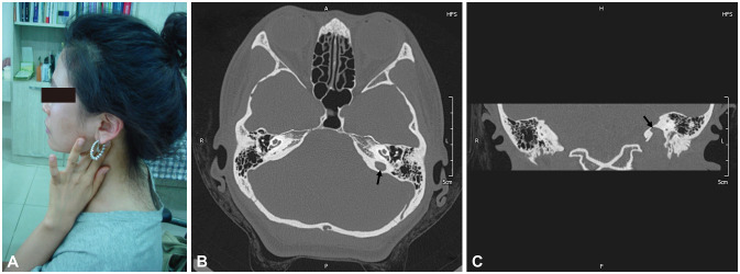
Tinnitus can be auscultated objectively on the affected side of the posterior neck and be louder when the head is turned to the unaffected side.25 Patients might describe the tinnitus as being attenuated or even disappearing after pressing the area where it is heard (Fig. 1). A diagnosis of a jugular-bulb anomaly might remove the need for further imaging investigations.23 However, magnetic resonance venography can be considered for excluding other vascular or intracranial pathologies.23 On the other hand, when a patient presents with a red-flag sign without any otoscopic abnormalities, vascular tinnitus of arterial origin should be considered. In such cases, transcranial Doppler sonography might be the first choice along with magnetic resonance angiography or brain CT angiography to search for conditions such as a dural arteriovenous fistula, atherosclerotic carotid artery disease, and cerebral aneurysm.23 Other imaging modalities such as magnetic resonance imaging (MRI) with enhancement would be needed to look for tumorous lesions in the brain, and diffusion-weighted imaging along with perfusion scan would be helpful for also identifying stroke lesions.23 Asynchronous pulsatile tinnitus may have a mechanical origin such as palatal muscle contraction, eustachian tube contraction, or myoclonus of the middle-ear muscle (Table 2).26,27,28
Fig. 1. A case of a high jugular bulb. (A) A 33-year-old female presented at our clinic with pulsatile tinnitus on her left ear with a 2-year history. The tinnitus was diminished by pressing just posterior to the left mandibular angle with her two fingers. There were no significant findings in blood tests for anemia, thyroid dysfunction, and hematological malignancy. Axial (B) and coronal (C) images of temporal-bone computed tomography revealed jugular-bulb diverticulum (arrows) at the level of the oval window without bony dehiscence, suggesting a high jugular bulb on the left side. Ten years had passed since her first visit to our clinic. In a recent follow-up call on June 25, 2020, the patient reported that her tinnitus was continuing but that it did not disturb her daily life or sleep. Written consent was obtained from the patient.
Table 2. Classification of tinnitus and its underlying etiology.
| Types | Causes |
|---|---|
| Tonal tinnitus | |
| Bilateral | |
| Chronic noise exposure | |
| Ototoxic medication | |
| Head trauma with/without acoustic trauma | |
| Meningitis | |
| Neurosyphilis | |
| Unilateral | |
| Neurologic signs | Brainstem lesion such as stroke, tumor, demyelinating disease, etc. |
| No neurologic signs | Chronic noise exposure on one ear |
| Ear infection | |
| Ear drum lesion | |
| Meniere disease | |
| Semicircular canal dehiscence | |
| Somatosound | |
| Bilateral or unilateral | |
| Pulse synchronous | Systemic hypertension |
| Arterial bruits | |
| High jugular bulb | |
| Arteriovenous malformation | |
| Intracranial aneurysm | |
| Vascular tumors | |
| Idiopathic intracranial hypertension | |
| Pulse asynchronous | Middle ear muscle myoclonus |
| Palatal muscle contraction | |
| Eustachian tube contraction | |
Another way to classify tinnitus is according to whether the sound is tonal or nontonal.14 Many clinicians use the term “tinnitus” when referring to tonal nonpulsatile tinnitus. Tinnitus can also be classified into acute or chronic depending on the duration of symptoms. Tinnitus that is temporary (lasting for up to 3 months) is referred to as acute tinnitus, while chronic or ongoing tinnitus refers to the condition lasting for longer than 3 months.29,30 This kind of duration-related classification is neither helpful nor reliable because the description of symptoms is solely based on the patient's history. Nonetheless, timely management is required when acute-onset tinnitus is accompanied with an unexplained balance problem and focal neurological symptoms, which is one of the red-flag signs prompting diagnostic workups such as TBCT or MRI.31,32 The present article uses the term tinnitus to refer to tonal nonpulsatile tinnitus.
TINNITUS MECHANISMS
The earliest speculations regarding the site of tinnitus generation described a cochlear origin.33 This view has shifted over time to tinnitus originating in the central auditory system rather than to mechanisms in the peripheral auditory system such as due to cochlear damage;13 that is, abnormal auditory signals activate neural plasticity within central auditory structures and manifest as tinnitus.33 However, it turns out that the pathophysiology of tinnitus is not limited to the auditory system,34 with the somatosensory system also provoking or modulating tinnitus,35 and the limbic system being necessary for its maintenance.34 Previous theories on tinnitus have assumed that it can play a reactive role for limbic structures, which might explain the acquired distress response.1,36 The present model assigns a more-central role to limbic and paralimbic structures in and around the subcallosal area, where they participate in a self-regulating gating process that can prevent the tinnitus signal from being perceived.36 Functional and structural abnormalities in the limbic and auditory areas can also contribute to tinnitus.36
Cause and etiology of tinnitus
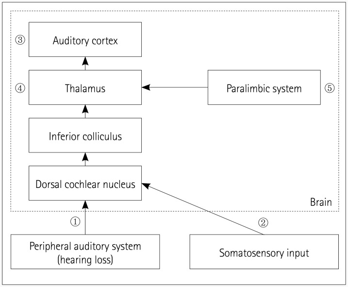
The term “cause” refers to biological or structural changes, while “etiology” refers only to the events associated with tinnitus onset, and not to the underlying mechanisms.13 Tinnitus does not represent a disease itself, but rather is a symptom of a variety of underlying diseases.8 The etiologies are known to include noise trauma, administration of ototoxic drugs (e.g., aminoglycosides, cisplatin, and salicylates), and head and neck injuries.37 Regardless of the causes, the three main locations where tinnitus is initiated are the peripheral auditory system,36 somatosensory system,35 and auditory cortex (Fig. 2).38,39 Lesions involving the dorsal cochlear nucleus (DCN),40 inferior colliculus (IC),13 and thalamus13 may also cause tinnitus. Furthermore, serotonin36 and the N-methyl-D-aspartate (NMDA) receptor41 are assumed to be potential therapeutic targets for blocking the initiation of tinnitus.
Fig. 2. Causes and mechanisms of tinnitus. The main causes of tinnitus are hearing loss, somatosensory system dysfunction, and auditory cortex lesions. 1) The most common cause of tinnitus is hearing loss. 2) Somatosensory stimuli disinhibit the ipsilateral cochlear nucleus, producing excitatory neuronal activity within the auditory pathway that results in tinnitus. 3) Damage to the brain cortex related to auditory perception alone can lead to tinnitus without contributions from peripheral influences such as hearing loss. 4) The socalled inhibitory gating mechanism blocks the tinnitus signal at the level of the thalamus. 5) An inhibitory feedback loop originates in paralimbic structures.
Anatomy of tinnitus
Cochlea
The most common cause of tinnitus is noise-induced hearing loss, followed by changes in the central auditory pathway.1,36 However, some people with tinnitus exhibit normal hearing ability,37 which is probably due to their degree of hearing impairment not being detectable in a standard audiological examination.36 On the other hand, many people with hearing loss never develop tinnitus.37
Somatosensory system
Tinnitus may be provoked or modulated by stimulation from the somatosensorial system.35 Somatosensory stimuli disinhibit the ipsilateral cochlear nucleus to produce excitatory neuronal activity within the auditory pathway that results in tinnitus. However, tinnitus without hearing loss following trauma makes the association more tenuous and raises the possibility of trauma only indirectly causing tinnitus, perhaps via a somatic mechanism.42
Brain cortex
Damage to the brain cortex related to auditory perception alone can lead to tinnitus without contributions from peripheral influences. Examples include acute hemorrhage of a small cavernous angioma located adjacent to the contralateral primary auditory cortex,38 and a lesion involving the superior temporal gyrus and the inferior portion of the supramarginal gyrus.39
DCN
Several studies have speculated that altered neuronal activity at the level of the DCN is primarily involved in tinnitus induction, due to an interplay between nonauditory brainstem structures and that nucleus.40 However, while the DCN may be necessary for tinnitus initiation, it is not necessary for the maintenance of chronic tinnitus.13
IC
Hyperactivity of IC neurons might be associated with chronic tinnitus but not with acute tinnitus.13
Thalamus
The thalamus is a structure that gaits sensory signals to the cortex to which the limbic structures of the brain are connected.13 The pathway from the medial geniculate body to the amygdala provides a possible key connection between the perception of tinnitus and the associated emotional component.13
Molecular mechanisms of tinnitus
Serotonin
Serotonin is considered to play a role in subjective tinnitus, such as that caused by the effect of certain antidepressant drugs.43 Tinnitus might be one of the symptoms of the overall depletion of serotonin, and patients with intermittent tinnitus might simply have fluctuating levels of serotonin.36 Serotonergic neurons in the nucleus accumbens and subcallosal networks can mediate the sensory gating mechanism.36 Serotonin depletion is assumed to cause tinnitus because a serotonin-depleted status as seen in hypersensitivity to noise, and reduced rapid-eye-movement sleep and depression can co-occur with tinnitus. Patients with tinnitus may have an independent systemic vulnerability in one or more limbicrelevant neurotransmitter systems, such as that involving serotonin. This is one of the reasons for these individuals being more susceptible to tinnitus.36 The underlying neurotransmitter levels may decline faster over time or with age in affected individuals than in unaffected individuals.36
NMDA receptor antagonist
Neuroprotective drugs such as NMDA receptor antagonists might aid in preventing or reversing chronic tinnitus.36 Trauma to the cochlea has been shown to initially trigger excessive production of glutamate that leads to the activation of NMDA receptors. Activation of cochlear NMDA receptors and related auditory nerve excitation has been suggested as the origin of cochlear tinnitus. Accordingly, NMDA receptor inhibition has been proposed for the pharmacological treatment of tinnitus,41 and the local administration of an NMDA antagonist to the rat cochlea was found to decrease tinnitus.44,45 The systemic administration of memantine (5 mg/kg), an NMDA antagonist, also significantly reduced acoustic-trauma-induced tinnitus in a rat model.46 In human trials, the administration of high-dose acamprosate, glutamate antagonist, and GABA agonist led to a significant decrease in the subjective loudness of tinnitus.47,48 However, memantine at 20 mg produced no significant improvement in tinnitus.49
Central gain enhancement
Cochlear damage would result in decreased excitatory input to the brain regions associated with the frequency range of the hearing loss and also decrease inhibition at nearby frequencies. Such a loss of sensory input from the cochlea would result in a compensatory increase in gain or neural amplification in the central auditory system. This is called “central gain enhancement,” which refers to a long-term alteration of inhibitory synapses at various levels of the auditory system that occurs after cochlear damage. Central gain enhancement may initially occur in the auditory cortex, and could subsequently be transmitted to the IC. Central gain enhancement occurs in a frequency-dependent manner and can be modulated by the amygdala (Fig. 3).50
Fig. 3. Central gain enhancement. A: Intact cochlear input. An intact cochlea modulates the activity of the corresponding auditory cortex. B: Damaged cochlear input. A cochlear lesion (C6) alters inhibitory synapses in the corresponding auditory cortex area (AC6), which in turn increases spontaneous activity that results in tinnitus. This occurs in a frequency-dependent manner modulated by amygdala. AC: auditory cortex, C: cochlea.
Inhibitory gating mechanism
The “inhibitory gating mechanism” or “noise cancellation system” is a system that blocks the tinnitus signal from arriving at the auditory cortex, which results in the tinnitus not being perceived.36 Under normal circumstances, the tinnitus signal is canceled out at the level of the thalamus by an inhibitory feedback loop that originates in the paralimbic structures.36 However, compromise of this gating mechanism may stop inhibition of the signal at the thalamic level, resulting in the signal being relayed to the auditory cortex to be perceived as tinnitus.34 In the long term this will lead to reorganization of the auditory cortex and sustained chronic tinnitus,34 whereas temporary dysfunction may result in intermittent tinnitus.36 Paralimbic dysfunction could be a consequence of overload of the paralimbic system attempting to compensate for the tinnitus signal.36 Disparities between the presence of hearing loss and tinnitus depend on individual differences in the effectiveness of the noise cancellation system supported by nonauditory regions.51
Nonauditory brain networks
Many patients with tinnitus experience emotions such as distress and other mood changes, which indicates that nonauditory brain areas also contribute to tinnitus in various ways. One study obtained quantitative electroencephalography data that demonstrated the involvement of these regions, and showed that various areas including the anterior cingulate cortex, dorsal lateral prefrontal cortex, insula, supplementary motor area, orbitofrontal cortex, parahippocampus, posterior cingulate cortex, and precuneus are involved in different aspects of tinnitus.52 Another study showed enhanced connectivity in both auditory and nonauditory regions in the tinnitus-affected brain based on functional near-infrared spectroscopy.53 The present study verified that connectivity can be strengthened between the auditory and frontotemporal, frontoparietal, temporal, occipitotemporal, and occipital cortices, suggesting that the central auditory and nonauditory brain regions are modified in tinnitus patients. Among those nonauditory brain networks involved in tinnitus, the limbic system is one of the important ones because about 50% of tinnitus patients have at least one psychiatric comorbidity.54 Brain structural changes in both the auditory and limbic systems were recently revealed with the help of high-resolution structural MRI, reiterating the importance of the connection between the auditory and nonauditory brain networks in tinnitus.55
RISK FACTORS
The heterogeneity of tinnitus has results in a scarcity of well-designed studies, including those of its risk factors. However, it is clear that hearing impairment is associated with tinnitus, and other well-known risk factors are noise exposure, autoimmune disorders, depression, lack of sleep, dyslipidemia, and smoking.56,57,58 Hypertension and sleep apnea are also closely related to tinnitus.59,60 One observation study showed that the presence of vertigo, otalgia, otorrhea, or TMJ disease can be related to tinnitus.61 That study also determined that tinnitus was more prevalent in patients with dental pain (21.1%), TMJ disease (22.5%), and combined dental pain and TMJ disorder (31.2%), compared to it being present in 19.6% of individuals with neither dental pain nor TMJ disease.61 A recently published clinic-based, cross-sectional questionnaire study and a population-based, a cross-sectional study showed that patients with glaucoma are more likely to have tinnitus.62 Many drugs are linked to tinnitus,63 with salicylate—the active component of the nonsteroidal anti-inflammatory drug aspirin—being one of the most-well-known drugs. High doses of aspirin can cause temporary hearing loss and reversible tinnitus by directly affecting cochlear function.64
TRIGGERING FACTORS
The onset of tinnitus occurs long after hearing loss in many patients,33 and it may be associated with negative life events such as unwanted retirement, redundancy, divorce,65 emotional stress, psychological factors, bereavement, unemployment, various physical or mental illnesses,33,66 noise exposure, and other somatic factors.67 The events that lead to tinnitus perception are referred to as “triggering factors.”67 Therefore, cochlear damage alone is not sufficient for the perception of tinnitus, with additional factors such as emotion or memory also making contributions.36,51
NATURAL COURSE
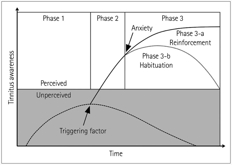
Tinnitus is usually initiated at the level of the cochlea and then remains below the perception threshold while it is masked by an inhibitory gating mechanism at the paralimbic level, and therefore is not perceived at the auditory cortex.34,35,36 The absence of triggering factors will mean that tinnitus does not occur, but the occurrence of a triggering factor such as a stressful event will unmask tinnitus so that it becomes perceived, which in turn will trigger more stressful events that will reinforce the tinnitus. Finally, tinnitus undergoes a phase of long-term maintenance that involves the central auditory and nonauditory neural networks.44
The natural course of tinnitus is depicted in Fig. 4. In phase 1, the tinnitus intensity is below the perception threshold. In phase 2, the occurrence of a stressful event triggers tinnitus perception. In phase 3-a, tinnitus triggers anxiety, which in turn reinforces the perception of tinnitus, leading to a vicious cycle; whereas in phase 3-b, tinnitus does not trigger anxiety, which results in habituation occurring and the perception of tinnitus decreasing.44
Fig. 4. The natural course of tinnitus. In phase 1, the tinnitus intensity is below the perception threshold. In phase 2, the occurrence of a stressful event triggers tinnitus perception. In phase 3-a, tinnitus triggers anxiety, which in turn reinforces the perception of tinnitus, leading to a vicious cycle; whereas in phase 3-b, tinnitus does not trigger anxiety, which results in habituation occurring and the perception of tinnitus decreasing. Modified from Guitton. Front Syst Neurosci 2012;6:12.44.
A longitudinal study found that approximately 40% and 20% of patients with mild and severe tinnitus at baseline, respectively, reported that it had resolved 5 years later.68 A recent well-designed systematic review and meta-analysis of controlled trials revealed changes in the effects of tinnitus over time on global tinnitus, depression, generalized anxiety, and quality of life, and in the loudness of the tinnitus.69 None of the patients in that review reported any significant changes in their global tinnitus during the nonintervention period, and it is difficult to draw conclusions due to the heterogeneity of the data and its self-reported nature in that study.
Some studies have suggested that tinnitus worsens depression, generalized anxiety, and the quality of life over time, while others have suggested that these aspects improve.69 Many studies have found that the loudness of tinnitus reduces over time, although some data were not subjected to meta-analysis. This finding provides statistical evidence that tinnitus generally improves over time without any intervention, although the effects vary markedly between individuals and their clinical meaning remain unclear.69
TREATMENTS
Tinnitus treatments must aim at resolving the underlying causes and their symptoms. For patients with mild tinnitus, simply providing the diagnosis followed by counseling about its natural history and progression should suffice. However, various interventions should be considered in patients with bothersome and persistent tinnitus.
Education and counseling
Education about tinnitus should emphasize that the condition itself is a symptom rather than a dangerous disease, and a comprehensive assessment can exclude any associated medical conditions that might require prompt treatment.70 Counseling should include providing information on the association between tinnitus and hearing loss. Lifestyle factors that can affect tinnitus either negatively or positively must also be discussed.70 Part of the counseling involves listening to the experiences of the patient and answering questions based on the neurophysiological model.66 The border between ordinary clinical counseling and formal psychotherapy is indistinct,71 and much of what the clinician says to the patient equates to educational counseling. Counseling is the only intervention offered in many cases of tinnitus.72
Cognitive-behavioral therapy
Cognitive-behavioral therapy is a relatively brief psychological treatment modality directed at identifying and modifying maladaptive behaviors and thoughts through behavioral changes and cognitive restructuring.73 Cognitive-behavioral therapy teaches skills to identify the negative thoughts that result in distress and restructure them such that the thoughts are more accurate or helpful.70 Although cognitive-behavioral therapy does not reduce the subjectively reported loudness of tinnitus, it improves the well-being of affected patients.70
Tinnitus retraining therapy
Tinnitus retraining therapy involves “retraining” the brain to become accustomed to the tinnitus sound and thereby encourage the patient to experience the tinnitus as a neutral stimulus.74 Tinnitus retraining therapy has two main components: 1) an educational counseling approach and 2) sound therapy in the form of masking or white noise, with the sound intensity of the device set slightly below that of the tinnitus perceived by the patient.74
Sound therapy
Sound therapy is a tinnitus retraining therapy that involves applying external sound to suppress the perception of tinnitus or the reaction to tinnitus for clinical benefit.70 The intensity of the external sound can be increased to the point where it does not interfere with the environment sounds that a patient needs to hear.75 Sound therapy can be applied using sound (noise) generators, hearing aids, or a combination of a hearing aid and a noise generator.76 The sound generators produce a broadband sound that can reduce (partial masking) or even eliminate (complete masking) the perception of tinnitus.33 Patients undergoing masking are also advised to use various sound sources for the masking, such as a CD player, radio, or tabletop sound generator.72 Sound therapy should be combined with some form of counseling, even if this simply involves providing information.77 A systemic review found that the efficacy of sound therapy against tinnitus was inconclusive, and currently, there is still no reliable evidence for the efficacy of sound-therapy devices.78
Hearing aids
A hearing aid is one of the therapeutic options for sound therapy and would be the first treatment option for patients with tinnitus who also have hearing difficulties. However, a study found that tinnitus was eliminated or attenuated in only about 50% of patients wearing hearing aids.79 That study demonstrated that the improvement in tinnitus was greater among those who used of ear-level devices as both hearing aids and sound generators (sound maskers). Moreover, hearing aids can also improve the quality of life by both treating hearing loss and making the tinnitus less noticeable.70 High-frequency amplification may be beneficial to and is readily accepted by patients who are only marginal hearing-aid candidates.33
Brain stimulation
One model for tinnitus is abnormal spontaneous neuronal activity in distinct regions of the central auditory system,80,81 which has promoted brain stimulation being proposed as a way to decrease neuronal activity. Although repetitive transcranial magnetic stimulation has been shown to reduce tinnitus in some studies, randomized controlled trials have failed to show its benefit.82,83 Patients with tinnitus who underwent deep brain stimulation (DBS) for movement disorders reported a reduction in the tinnitus intensity when their implants were switched on, even when there was no effect on their hearing.84 Though the implants were located in part of the classic auditory pathway, this method has recently become a viable treatment option. A recent phase I trial of caudate DBS for treatment-resistant tinnitus produced very encouraging results, and so this approach warrants further investigation.85
Pharmacotherapy
Currently, no drugs have been unanimously proven effective despite many having been investigated.4 Antidepressants are frequently prescribed in clinical practice for treating tinnitus, but the results have falling short of expectations, with no favorable outcomes based on seven randomized controlled trials and one cochrane review, although they may have a role in the management of any concomitant psychological distress.69,86 Anticonvulsants have also failed to demonstrate effectiveness. However, some drugs have been reported to provide moderate symptom relief in some patients.87 Among frequently prescribed medications, acamprosate, antidepressants, alprazolam, and clonazepam have been demonstrated to be effective in some studies. Acamprosate (333 mg, thrice daily) is effective in treating the severity of sensorineural tinnitus without causing marked side effects.48 Patients with tinnitus and concomitant sleep disturbance or major depression should consider antidepressant therapy.71 Anxiolytics such as alprazolam and clonazepam can be used to improve coping in patients who experience extreme stress due to their tinnitus.37,86,87,88 However, clinicians should not routinely recommend anxiolytics as the primary indication for treating persistent and bothersome tinnitus.70
The importance of providing education and counseling about the natural history and progression of tinnitus must also be emphasized. Furthermore, the above-mentioned medications must be tailored to the tinnitus symptoms experienced by individual patients, because most investigated drugs have been found to be ineffective, or have even induced adverse effects.
CONCLUSION
The recently introduced inhibitory gating mechanism is an impressive finding since it explains that 1) hearing loss does not always lead to tinnitus and 2) cochlear damage can elicit a tinnitus signal that remains unperceived until trigger factors such as emotion or memory-related events make the tinnitus signal arrive at the auditory cortex.
The complexities of the anatomical locations of tinnitus pathophysiology as well as the chronological gap between the initiation and triggers indicate that tinnitus exhibits unique features. First, because the onset of the tinnitus is correlated with stressful events rather than with the onset of hearing loss, it is difficult to determine the onset time of tinnitus.44 Second, because the origin and maintenance mechanisms of tinnitus are not yet clearly understood, tinnitus is treated in an indirect rather than an intuitive manner. Although tinnitus is thought to be initiated in the cochlea and generated or even made chronic in the brain, therapeutic trials that focus on either the cochlea or brain can be unsuccessful. Instead, therapeutic strategies should perhaps mainly focus on the limbic system and autonomic nervous system. Third, because the loudness and pitch of tinnitus do not accurately reflect the degree of patient discomfort,71 methods for weakening the intensity of the tinnitus sound do not always provide relief. Also, silencing external sounds by muffling both ear canals generally makes tinnitus worse.
Given the complexity and heterogeneity of tinnitus, single-factor strategies are likely to fail.44 Although there is no cure, patients with clinically significant tinnitus can be taught about stress management and provided with information about the therapeutic use of sound techniques and lifestyle modifications that will minimize its detrimental effects.89
Acknowledgements
None
Footnotes
- Conceptualization: Byung In Han.
- Investigation: all authors.
- Writing—original draft: Byung In Han, Sanghyo Ryu.
- Writing—review & editing: all authors.
Conflicts of Interest: The authors have no potential conflicts of interest to disclose.
References
- 1.Jastreboff PJ. Phantom auditory perception (tinnitus): mechanisms of generation and perception. Neurosci Res. 1990;8:221–254. doi: 10.1016/0168-0102(90)90031-9. [DOI] [PubMed] [Google Scholar]
- 2.Crummer RW, Hassan GA. Diagnostic approach to tinnitus. Am Fam Physician. 2004;69:120–126. [PubMed] [Google Scholar]
- 3.Bhatt JM, Lin HW, Bhattacharyya N. Prevalence, severity, exposures, and treatment patterns of tinnitus in the United States. JAMA Otolaryngol Head Neck Surg. 2016;142:959–965. doi: 10.1001/jamaoto.2016.1700. [DOI] [PMC free article] [PubMed] [Google Scholar]
- 4.Baguley D, McFerran D, Hall D. Tinnitus. Lancet. 2013;382:1600–1607. doi: 10.1016/S0140-6736(13)60142-7. [DOI] [PubMed] [Google Scholar]
- 5.Davis A, El Refaie A. Epidemiology of tinnitus. In: Tyler RS, editor. Tinnitus handbook. Clifton Park, NY: Delmar Cengage Learning, Inc.; 2000. pp. 1–23. [Google Scholar]
- 6.Park KH, Lee SH, Koo JW, Park HY, Lee KY, Choi YS, et al. Prevalence and associated factors of tinnitus: data from the Korean National Health and Nutrition Examination Survey 2009-2011. J Epidemiol. 2014;24:417–426. doi: 10.2188/jea.JE20140024. [DOI] [PMC free article] [PubMed] [Google Scholar]
- 7.Pinto PCL, Sanchez TG, Tomita S. The impact of gender, age and hearing loss on tinnitus severity. Braz J Otorhinolaryngol. 2010;76:18–24. doi: 10.1590/S1808-86942010000100004. [DOI] [PMC free article] [PubMed] [Google Scholar]
- 8.Lockwood AH, Salvi RJ, Burkard RF. Tinnitus. N Engl J Med. 2002;347:904–910. doi: 10.1056/NEJMra013395. [DOI] [PubMed] [Google Scholar]
- 9.Seo JH, Kang JM, Hwang SH, Han KD, Joo YH. Relationship between tinnitus and suicidal behaviour in Korean men and women: a cross-sectional study. Clin Otolaryngol. 2016;41:222–227. doi: 10.1111/coa.12500. [DOI] [PubMed] [Google Scholar]
- 10.Henry JA, Meikle MB. Psychoacoustic measures of tinnitus. J Am Acad Audiol. 2000;11:138–155. [PubMed] [Google Scholar]
- 11.Dobie RA. Overview: suffering from tinnitus. In: Snow JB Jr, editor. Tinnitus: theory and management. Hamilton, ON: BC Decker Inc.; 2004. pp. 1–7. [Google Scholar]
- 12.Chari DA, Limb CJ. Tinnitus. Med Clin North Am. 2018;102:1081–1093. doi: 10.1016/j.mcna.2018.06.014. [DOI] [PubMed] [Google Scholar]
- 13.Henry JA, Roberts LE, Caspary DM, Theodoroff SM, Salvi RJ. Underlying mechanisms of tinnitus: review and clinical implications. J Am Acad Audiol. 2014;25:5–22. doi: 10.3766/jaaa.25.1.2. [DOI] [PMC free article] [PubMed] [Google Scholar]
- 14.Han BI, Lee HW, Kim TY, Lim JS, Shin KS. Tinnitus: characteristics, causes, mechanisms, and treatments. J Clin Neurol. 2009;5:11–19. doi: 10.3988/jcn.2009.5.1.11. [DOI] [PMC free article] [PubMed] [Google Scholar]
- 15.Ahmad N, Seidman M. Tinnitus in the older adult: epidemiology, pathophysiology and treatment options. Drugs Aging. 2004;21:297–305. doi: 10.2165/00002512-200421050-00002. [DOI] [PubMed] [Google Scholar]
- 16.Lockwood AH, Salvi RJ, Burkard RF, Galantowicz PJ, Coad ML, Wack DS. Neuroanatomy of tinnitus. Scand Audiol Suppl. 1999;51:47–52. [PubMed] [Google Scholar]
- 17.Maghzi AH, Sahai S, Zimnowodzki S, Baloh RH. Palatal tremor as a presenting symptom of amyotrophic lateral sclerosis. Neurology. 2018;90:801–802. doi: 10.1212/WNL.0000000000005363. [DOI] [PMC free article] [PubMed] [Google Scholar]
- 18.Ward J, Vella C, Hoare DJ, Hall DA. Subtyping somatic tinnitus: a cross-sectional UK cohort study of demographic, clinical and audiological characteristics. PLoS One. 2015;10:e0126254. doi: 10.1371/journal.pone.0126254. [DOI] [PMC free article] [PubMed] [Google Scholar]
- 19.Wu V, Cooke B, Eitutis S, Simpson MTW, Beyea JA. Approach to tinnitus management. Can Fam Physician. 2018;64:491–495. [PMC free article] [PubMed] [Google Scholar]
- 20.Schleuning AJ., 2nd Management of the patient with tinnitus. Med Clin North Am. 1991;75:1225–1237. doi: 10.1016/s0025-7125(16)30383-2. [DOI] [PubMed] [Google Scholar]
- 21.Wall M. Idiopathic intracranial hypertension. Neurol Clin. 2010;28:593–617. doi: 10.1016/j.ncl.2010.03.003. [DOI] [PMC free article] [PubMed] [Google Scholar]
- 22.Park SN, Bae SC, Lee GH, Song JN, Park KH, Jeon EJ, et al. Clinical characteristics and therapeutic response of objective tinnitus due to middle ear myoclonus: a large case series. Laryngoscope. 2013;123:2516–2520. doi: 10.1002/lary.23854. [DOI] [PubMed] [Google Scholar]
- 23.Bae SC, Kim DK, Yeo SW, Park SY, Park SN. Single-center 10-year experience in treating patients with vascular tinnitus: diagnostic approaches and treatment outcomes. Clin Exp Otorhinolaryngol. 2015;8:7–12. doi: 10.3342/ceo.2015.8.1.7. [DOI] [PMC free article] [PubMed] [Google Scholar]
- 24.Fujii N, Inui Y, Katada K. Temporal bone anatomy: correlation of multiplanar reconstruction sections and three-dimensional computed tomography images. Jpn J Radiol. 2010;28:637–648. doi: 10.1007/s11604-010-0479-0. [DOI] [PubMed] [Google Scholar]
- 25.Jung H, Kim JR, Kim SH, Song JJ. Pulsatile tinnitus related with prominent venous plexus: case report and literature review. Korean J Otorhinolaryngol-Head Neck Surg. 2017;60:38–43. [Google Scholar]
- 26.Böhmer A. Hydrostatic pressure in the inner ear fluid compartments and its effects on inner ear function. Acta Otolaryngol Suppl. 1993;507:3–24. [PubMed] [Google Scholar]
- 27.Hofmann E, Behr R, Neumann-Haefelin T, Schwager K. Pulsatile tinnitus: imaging and differential diagnosis. Dtsch Arztebl Int. 2013;110:451–458. doi: 10.3238/arztebl.2013.0451. [DOI] [PMC free article] [PubMed] [Google Scholar]
- 28.Fortune DS, Haynes DS, Hall JW., 3rd Tinnitus. Current evaluation and management. Med Clin North Am. 1999;83:153–162. doi: 10.1016/s0025-7125(05)70094-8. [DOI] [PubMed] [Google Scholar]
- 29.Wallhäusser-Franke E, D'Amelio R, Glauner A, Delb W, Servais JJ, Hörmann K, et al. Transition from acute to chronic tinnitus: predictors for the development of chronic distressing tinnitus. Front Neurol. 2017;8:605. doi: 10.3389/fneur.2017.00605. [DOI] [PMC free article] [PubMed] [Google Scholar]
- 30.Swan AA, Nelson JT, Swiger B, Jaramillo CA, Eapen BC, Packer M, et al. Prevalence of hearing loss and tinnitus in Iraq and Afghanistan veterans: a Chronic Effects of Neurotrauma Consortium study. Hear Res. 2017;349:4–12. doi: 10.1016/j.heares.2017.01.013. [DOI] [PubMed] [Google Scholar]
- 31.Beukes EW, Manchaiah V, Baguley DM, Allen PM, Andersson G. Internet-based interventions for adults with hearing loss, tinnitus and vestibular disorders: a protocol for a systematic review. Syst Rev. 2018;7:205. doi: 10.1186/s13643-018-0880-9. [DOI] [PMC free article] [PubMed] [Google Scholar]
- 32.Bauer CA. Tinnitus. N Engl J Med. 2018;378:1224–1231. doi: 10.1056/NEJMcp1506631. [DOI] [PubMed] [Google Scholar]
- 33.Henry JA, Dennis KC, Schechter MA. General review of tinnitus: prevalence, mechanisms, effects, and management. J Speech Lang Hear Res. 2005;48:1204–1235. doi: 10.1044/1092-4388(2005/084). [DOI] [PubMed] [Google Scholar]
- 34.Adjamian P, Hall DA, Palmer AR, Allan TW, Langers DR. Neuroanatomical abnormalities in chronic tinnitus in the human brain. Neurosci Biobehav Rev. 2014;45:119–133. doi: 10.1016/j.neubiorev.2014.05.013. [DOI] [PMC free article] [PubMed] [Google Scholar]
- 35.Sanchez TG, Rocha CB. Diagnosis and management of somatosensory tinnitus: review article. Clinics (Sao Paulo) 2011;66:1089–1094. doi: 10.1590/S1807-59322011000600028. [DOI] [PMC free article] [PubMed] [Google Scholar]
- 36.Rauschecker JP, Leaver AM, Mühlau M. Tuning out the noise: limbic-auditory interactions in tinnitus. Neuron. 2010;66:819–826. doi: 10.1016/j.neuron.2010.04.032. [DOI] [PMC free article] [PubMed] [Google Scholar]
- 37.Elgoyhen AB, Langguth B. Pharmacological approaches to the treatment of tinnitus. Drug Discov Today. 2010;15:300–305. doi: 10.1016/j.drudis.2009.11.003. [DOI] [PubMed] [Google Scholar]
- 38.Yoneoka Y, Fujii Y, Nakada T. Central tinnitus: a case report. Ear Nose Throat J. 2001;80:864–866. [PubMed] [Google Scholar]
- 39.Hurst RW, Lee SI. Ictal tinnitus. Epilepsia. 1986;27:769–772. doi: 10.1111/j.1528-1157.1986.tb03608.x. [DOI] [PubMed] [Google Scholar]
- 40.Knipper M, Zimmermann U, Müller M. Molecular aspects of tinnitus. Hear Res. 2010;266:60–69. doi: 10.1016/j.heares.2009.07.013. [DOI] [PubMed] [Google Scholar]
- 41.Bing D, Lee SC, Campanelli D, Xiong H, Matsumoto M, Panford-Walsh R, et al. Cochlear NMDA receptors as a therapeutic target of noise-induced tinnitus. Cell Physiol Biochem. 2015;35:1905–1923. doi: 10.1159/000374000. [DOI] [PubMed] [Google Scholar]
- 42.Levine RA. Somatic (craniocervical) tinnitus and the dorsal cochlear nucleus hypothesis. Am J Otolaryngol. 1999;20:351–362. doi: 10.1016/s0196-0709(99)90074-1. [DOI] [PubMed] [Google Scholar]
- 43.Clewes J. A case report of onset of tinnitus following discontinuation of antidepressant and a review of the literature. Prim Care Companion CNS Disord. 2012;14:PCC. doi: 10.4088/PCC.11br01218. [DOI] [PMC free article] [PubMed] [Google Scholar]
- 44.Guitton MJ. Tinnitus: pathology of synaptic plasticity at the cellular and system levels. Front Syst Neurosci. 2012;6:12. doi: 10.3389/fnsys.2012.00012. [DOI] [PMC free article] [PubMed] [Google Scholar]
- 45.Brozoski TJ, Wisner KW, Odintsov B, Bauer CA. Local NMDA receptor blockade attenuates chronic tinnitus and associated brain activity in an animal model. PLoS One. 2013;8:e77674. doi: 10.1371/journal.pone.0077674. [DOI] [PMC free article] [PubMed] [Google Scholar]
- 46.Zheng Y, McNamara E, Stiles L, Darlington CL, Smith PF. Evidence that memantine reduces chronic tinnitus caused by acoustic trauma in rats. Front Neurol. 2012;3:127. doi: 10.3389/fneur.2012.00127. [DOI] [PMC free article] [PubMed] [Google Scholar]
- 47.Azevedo AA, Figueiredo RR. Tinnitus treatment with acamprosate: double-blind study. Braz J Otorhinolaryngol. 2005;71:618–623. doi: 10.1016/S1808-8694(15)31266-0. [DOI] [PMC free article] [PubMed] [Google Scholar]
- 48.Sharma DK, Kaur S, Singh J, Kaur I. Role of acamprosate in sensorineural tinnitus. Indian J Pharmacol. 2012;44:93–96. doi: 10.4103/0253-7613.91876. [DOI] [PMC free article] [PubMed] [Google Scholar]
- 49.Figueiredo RR, Langguth B, Mello de, Mello de Oliveira P. Tinnitus treatment with memantine. Otolaryngol Head Neck Surg. 2008;138:492–496. doi: 10.1016/j.otohns.2007.11.027. [DOI] [PubMed] [Google Scholar]
- 50.Auerbach BD, Rodrigues PV, Salvi RJ. Central gain control in tinnitus and hyperacusis. Front Neurol. 2014;5:206. doi: 10.3389/fneur.2014.00206. [DOI] [PMC free article] [PubMed] [Google Scholar]
- 51.Eggermont JJ, Roberts LE. Tinnitus: animal models and findings in humans. Cell Tissue Res. 2015;361:311–336. doi: 10.1007/s00441-014-1992-8. [DOI] [PMC free article] [PubMed] [Google Scholar]
- 52.Vanneste S, De Ridder D. The auditory and non-auditory brain areas involved in tinnitus. An emergent property of multiple parallel overlapping subnetworks. Front Syst Neurosci. 2012;6:31. doi: 10.3389/fnsys.2012.00031. [DOI] [PMC free article] [PubMed] [Google Scholar]
- 53.San Juan J, Hu XS, Issa M, Bisconti S, Kovelman I, Kileny P, et al. Tinnitus alters resting state functional connectivity (RSFC) in human auditory and non-auditory brain regions as measured by functional near-infrared spectroscopy (fNIRS) PLoS One. 2017;12:e0179150. doi: 10.1371/journal.pone.0179150. [DOI] [PMC free article] [PubMed] [Google Scholar]
- 54.Salviati M, Bersani FS, Terlizzi S, Melcore C, Panico R, Romano GF, et al. Tinnitus: clinical experience of the psychosomatic connection. Neuropsychiatr Dis Treat. 2014;10:267–275. doi: 10.2147/NDT.S49425. [DOI] [PMC free article] [PubMed] [Google Scholar]
- 55.Besteher B, Gaser C, Ivanšić D, Guntinas-Lichius O, Dobel C, Nenadić I. Chronic tinnitus and the limbic system: reappraising brain structural effects of distress and affective symptoms. Neuroimage Clin. 2019;24:101976. doi: 10.1016/j.nicl.2019.101976. [DOI] [PMC free article] [PubMed] [Google Scholar]
- 56.Kim HJ, Lee HJ, An SY, Sim S, Park B, Kim SW, et al. Analysis of the prevalence and associated risk factors of tinnitus in adults. PLoS One. 2015;10:e0127578. doi: 10.1371/journal.pone.0127578. [DOI] [PMC free article] [PubMed] [Google Scholar]
- 57.Bhatt JM, Bhattacharyya N, Lin HW. Relationships between tinnitus and the prevalence of anxiety and depression. Laryngoscope. 2017;127:466–469. doi: 10.1002/lary.26107. [DOI] [PMC free article] [PubMed] [Google Scholar]
- 58.Veile A, Zimmermann H, Lorenz E, Becher H. Is smoking a risk factor for tinnitus? A systematic review, meta-analysis and estimation of the population attributable risk in Germany. BMJ Open. 2018;8:e016589. doi: 10.1136/bmjopen-2017-016589. [DOI] [PMC free article] [PubMed] [Google Scholar]
- 59.Yang P, Ma W, Zheng Y, Yang H, Lin H. A systematic review and meta-analysis on the association between hypertension and tinnitus. Int J Hypertens. 2015;2015:583493. doi: 10.1155/2015/583493. [DOI] [PMC free article] [PubMed] [Google Scholar]
- 60.Koo M, Hwang JH. Risk of tinnitus in patients with sleep apnea: a nationwide, population-based, case-control study. Laryngoscope. 2017;127:2171–2175. doi: 10.1002/lary.26323. [DOI] [PubMed] [Google Scholar]
- 61.Kim YH, Park YG, Han KD, Vu D, Cho KH, Lee SY. Prevalence of tinnitus according to temporomandibular joint disorders and dental pain: the Korean national population-based study. J Oral Rehabil. 2018;45:198–203. doi: 10.1111/joor.12604. [DOI] [PubMed] [Google Scholar]
- 62.Loiselle AR, Neustaeter A, de Kleine E, van Dijk P, Jansonius NM. Associations between tinnitus and glaucoma suggest a common mechanism: a clinical and population-based study. Hear Res. 2020;386:107862. doi: 10.1016/j.heares.2019.107862. [DOI] [PubMed] [Google Scholar]
- 63.Bisht M, Bist SS. Ototoxicity: the hidden menace. Indian J Otolaryngol Head Neck Surg. 2011;63:255–259. doi: 10.1007/s12070-011-0151-8. [DOI] [PMC free article] [PubMed] [Google Scholar]
- 64.Stolzberg D, Salvi RJ, Allman BL. Salicylate toxicity model of tinnitus. Front Syst Neurosci. 2012;6:28. doi: 10.3389/fnsys.2012.00028. [DOI] [PMC free article] [PubMed] [Google Scholar]
- 65.Luxon LM. Tinnitus: its causes, diagnosis, and treatment. BMJ. 1993;306:1490–1491. doi: 10.1136/bmj.306.6891.1490. [DOI] [PMC free article] [PubMed] [Google Scholar]
- 66.Jastreboff PJ, Hazell JWP. Tinnitus retraining therapy (TRT): clinical implementation of the model. In: Jastreboff PJ, Hazell JWP, editors. Tinnitus retraining therapy: implementing the neurophysiological model. New York, NY: Cambridge University Press; 2004. pp. 64–144. [Google Scholar]
- 67.Dobie RA. Overview: suffering from tinnitus. In: Snow JB, editor. Tinnitus: theory and management. Lewiston, NY: BC Decker; 2004. pp. 1–7. [Google Scholar]
- 68.Nondahl DM, Cruickshanks KJ, Wiley TL, Klein R, Klein BE, Tweed TS. Prevalence and 5-year incidence of tinnitus among older adults: the epidemiology of hearing loss study. J Am Acad Audiol. 2002;13:323–331. [PubMed] [Google Scholar]
- 69.Phillips JS, McFerran DJ, Hall DA, Hoare DJ. The natural history of subjective tinnitus in adults: a systematic review and meta-analysis of no-intervention periods in controlled trials. Laryngoscope. 2018;128:217–227. doi: 10.1002/lary.26607. [DOI] [PubMed] [Google Scholar]
- 70.Tunkel DE, Bauer CA, Sun GH, Rosenfeld RM, Chandrasekhar SS, Cunningham ER, Jr, et al. Clinical practice guideline: tinnitus. Otolaryngol Head Neck Surg. 2014;151:S1–S40. doi: 10.1177/0194599814545325. [DOI] [PubMed] [Google Scholar]
- 71.Dobie RA. A review of randomized clinical trials in tinnitus. Laryngoscope. 1999;109:1202–1211. doi: 10.1097/00005537-199908000-00004. [DOI] [PubMed] [Google Scholar]
- 72.Henry JA, Schechter MA, Loovis CL, Zaugg TL, Kaelin C, Montero M. Clinical management of tinnitus using a “progressive intervention” approach. J Rehabil Res Dev. 2005;42:95–116. doi: 10.1682/jrrd.2005.01.0005. [DOI] [PubMed] [Google Scholar]
- 73.Andersson G, Kaldo V. Cognitive-behavioral therapy with applied relaxation. In: Tyler RS, editor. Tinnitus treatment: clinical protocols. New York, NY: Thieme; 2006. pp. 96–115. [Google Scholar]
- 74.Jastreboff PJ, Hazell JW. A neurophysiological approach to tinnitus: clinical implications. Br J Audiol. 1993;27:7–17. doi: 10.3109/03005369309077884. [DOI] [PubMed] [Google Scholar]
- 75.Folmer RL, Martin WH, Shi Y, Edlefsen LL. Tinnitus sound therapy. In: Tyler RS, editor. Tinnitus treatment: clinical protocols. New York, NY: Thieme; 2006. pp. 176–186. [Google Scholar]
- 76.Henry JA, Schechter MA, Nagler SM, Fausti SA. Comparison of tinnitus masking and tinnitus retraining therapy. J Am Acad Audiol. 2002;13:559–581. [PubMed] [Google Scholar]
- 77.Tyler RS. Neurophysiological models, psychological models, and treatments for tinnitus. In: Tyler RS, editor. Tinnitus treatment: clinical protocols. New York, NY: Thieme; 2006. pp. 1–22. [Google Scholar]
- 78.Hobson J, Chisholm E, El Refaie A. Sound therapy (masking) in the management of tinnitus in adults. Cochrane Database Syst Rev. 2012;11:CD006371. doi: 10.1002/14651858.CD006371.pub3. [DOI] [PMC free article] [PubMed] [Google Scholar]
- 79.Folmer RL, Carroll JR. Long-term effectiveness of ear-level devices for tinnitus. Otolaryngol Head Neck Surg. 2006;134:132–137. doi: 10.1016/j.otohns.2005.09.030. [DOI] [PubMed] [Google Scholar]
- 80.Eggermont JJ, Roberts LE. The neuroscience of tinnitus. Trends Neurosci. 2004;27:676–682. doi: 10.1016/j.tins.2004.08.010. [DOI] [PubMed] [Google Scholar]
- 81.Noreña AJ. An integrative model of tinnitus based on a central gain controlling neural sensitivity. Neurosci Biobehav Rev. 2011;35:1089–1109. doi: 10.1016/j.neubiorev.2010.11.003. [DOI] [PubMed] [Google Scholar]
- 82.Piccirillo JF, Garcia KS, Nicklaus J, Pierce K, Burton H, Vlassenko AG, et al. Low-frequency repetitive transcranial magnetic stimulation to the temporoparietal junction for tinnitus. Arch Otolaryngol Head Neck Surg. 2011;137:221–228. doi: 10.1001/archoto.2011.3. [DOI] [PMC free article] [PubMed] [Google Scholar]
- 83.Meng Z, Liu S, Zheng Y, Phillips JS. Repetitive transcranial magnetic stimulation for tinnitus. Cochrane Database Syst Rev. 2011:CD007946. doi: 10.1002/14651858.CD007946.pub2. [DOI] [PubMed] [Google Scholar]
- 84.Cheung SW, Larson PS. Tinnitus modulation by deep brain stimulation in locus of caudate neurons (area LC) Neuroscience. 2010;169:1768–1778. doi: 10.1016/j.neuroscience.2010.06.007. [DOI] [PubMed] [Google Scholar]
- 85.Cheung SW, Racine CA, Henderson-Sabes J, Demopoulos C, Molinaro AM, Heath S, et al. Phase I trial of caudate deep brain stimulation for treatment-resistant tinnitus. J Neurosurg. 2019;133:992–1001. doi: 10.3171/2019.4.JNS19347. [DOI] [PMC free article] [PubMed] [Google Scholar]
- 86.Johnson RM, Brummett R, Schleuning A. Use of alprazolam for relief of tinnitus. A double-blind study. Arch Otolaryngol Head Neck Surg. 1993;119:842–845. doi: 10.1001/archotol.1993.01880200042006. [DOI] [PubMed] [Google Scholar]
- 87.Salvi R, Lobarinas E, Sun W. Pharmacological treatments for tinnitus: new and old. Drugs Future. 2009;34:381–400. doi: 10.1358/dof.2009.034.05.1362442. [DOI] [PMC free article] [PubMed] [Google Scholar]
- 88.Ganança MM, Caovilla HH, Ganança FF, Ganança CF, Munhoz MS, da Silva ML, et al. Clonazepam in the pharmacological treatment of vertigo and tinnitus. Int Tinnitus J. 2002;8:50–53. [PubMed] [Google Scholar]
- 89.Henry JA, Zaugg TL, Myers PJ, Kendall CJ, Michaelides EM. A triage guide for tinnitus. J Fam Pract. 2010;59:389–393. [PubMed] [Google Scholar]


